#division of cytoplasm
Text
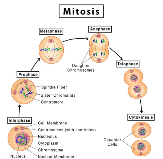
MITOSIS- its Occurrence, Stages and Significance.
Unlocking knowledge one post at a time! Check out our latest notes on www.microscopiaiwm.com – a treasure trove of insights, ideas, and inspiration. Dive in, explore, and let the journey of discovery begin! 📚
INTRODUCTION:
Mitosis is a type of cell division that takes place in living organisms and it is commonly defined as the process of duplication of chromosomes in eukaryotic cells and distributed during cell division.
The process where a single cell divides resulting in two identical cells, each resulted cell contains the same number of chromosomes and…
View On WordPress
#anaphase#asexual reproduction#cell cycle#cell cycle stages#cell division#cell plate#chromosome#cleavage furrow#contractile ring#cytokinesis#daughter cell#daughter cells#division of cytoplasm#division of nucleus#dna#DNA double#duplication of chromosome#fragmoplast#genetics#germ cells#homologous chromosome#homologous recombination#homologous recombination repair#karyokinesis#kinetochore#kinetoplast#meiosis#metaphase#MicroScopia IWM#mitos
3 notes
·
View notes
Text
Miss Ortega | j.o

part 7
—The cell is made up of the nucleus and the cytoplasm and is enclosed by the cell membrane, which regulates passage in and out. The nucleus contains chromosomes, the cell's genetic material, and the nucleolus, which produces ribosomes. My eyes shift to Olivia, who was jotting down my words in her notebook.
In the late afternoon, I was at Olivia's house to help her study science, primarily about what a cell is and its functions. Olivia nods, giving me a nervous smile.
—One last question... what's cell division?— She puts the tip of her pen between her teeth, thoughtful.
—Cell division is the process by which a cell multiplies, splitting into two. In prokaryotes, it happens through binary fission (DNA filament duplication and subsequent division into two identical new individuals). In eukaryotes, it occurs through mitosis and, in reproductive cells, meiosis.— I say, shrugging casually.
Olivia writes it all down and then closes her notebook with a soft thud, sighing with satisfaction and tiredness.
—We're done,— she murmurs weakly, looking at me with a smile on her lips.
—We're done,— I repeat, and she stretches, slightly tense from maintaining an uncomfortable position for a long time.
—I'm not surprised you never get a failing grade, you're a book,— she says as she gets up from her desk, flopping onto her bed's mattress.
—Don't exaggerate...— I chuckle and give her a playful look. —Now... will you let me hear something you've written?— I nervously bite my lip, accepting the invitation to sit beside her on the bed.
Olivia sighs and reluctantly agrees to my request, blushing as she looks at me. —Wait,— she murmurs softly, leaning towards the edge of the bed, picking up a guitar case from the floor. Olivia glances at me sideways, holding the guitar in her hands.
—I'll sing you a little snippet of the song, okay? Also... I haven't finished it yet,— she says, toying with the guitar strings, likely tuning it.
I gaze in awe at her profile. Olivia had her head tilted down, holding the guitar in her lap. Her eyes briefly meet mine for a split second before she looks away with flushed cheeks.
Taking a breath, she closes her eyes, focusing.
—And I won't fight for love if you won't meet me halfway...— she begins to sing. And I say that I'm through but this song's still for you–
Her voice sounds angelic, surprising me with her talent. Olivia glances at me briefly, giving me a small smile.
—All I want is love that lasts— her eyes glisten, still looking at me.
—Is all I want too much to ask?— her fingers pause, interrupting the sweet melody. Olivia sets the guitar aside and looks at me with embarrassment, accepting my applause.
—Oh my god... you have an amazing voice,— I admit, and she tucks a strand of hair behind her ear, staring at a fixed point on her lap. —Thank you,— she offers a shy smile, and I reciprocate.
A knock on the door draws our attention to the entrance of her room. Olivia's mother, Emma, is standing there with a smile on her lips.
—T/N, dear, why don't you stay for dinner with us?— Mrs. Rodrigo suggests.
With a smile, I look at Olivia's reaction. She's looking at me with bright eyes and a smile, nodding enthusiastically.
—That would be fantastic,— I reply, and immediately, two arms wrap around my neck, hugging me. The force makes me lie back on the bed, and amid laughter, I return the hug, smiling shyly at Emma, who watches us with tenderness as I hold Olivia in my arms.
(...)
—So... how's it going with the girl you like?— Enid asks, hugging a pillow in her arms.
After helping Olivia study, I received an invitation from Enid to have a pajama party at her house, inviting Olivia as well since she was with me. The blonde only knew that I liked someone, but she didn't know who, and for obvious reasons, she was really mad at me. I know she's my best friend, but I still couldn't tell her that I was in love with Professor Ortega.
—Actually, it's all going wrong... she said it's better if I forget what happened,— I lower my head towards my lap, sadly biting my lower lip. —Well, what a jerk...— Enid makes a face. —If only I knew who she was, I would have given her a piece of my mind,— she says absentmindedly, pulling at the corners of the pillow in her hands.
—You tried your best,— I smile sideways, and Enid throws the pillow at my face, messing up my hair. I chuckle slightly and wink at her.
—What do you think about Olivia, though?— she suddenly asks, lying down on the bed. I turn toward the door, relieved when I see that the subject of conversation is still downstairs preparing popcorn for the movie.
—Are you crazy? She's here...— I whisper, and she rolls her eyes at my comment.
—I don't see her,— Enid turns toward me, focusing her attention on me.
I sigh and shake my head. —She's nice...— I shrug indifferently, smiling at the blonde. Enid raises an eyebrow and gives me a smile, silently asking me to tell her more.
—She's beautiful... there's no doubt... but you know I'm in love with someone else,— I play with my fingers, embarrassed by the situation.
—She'd be perfect for you, you know? Plus... she really likes you,— Enid confesses. She adjusts her pajamas and gets under the covers, getting ready to watch the movie on her room's TV.
—I know... but for now... I only see her as a friend,— I tuck a strand of hair behind my ear and look confused at Enid's reaction, who is looking with panic over my shoulder.
I turn to her line of sight and pale when I see Olivia near the door. The brunette awkwardly leaves the popcorn bowl on the shelf and, with tears in her eyes, looks at me, shaking her head with regret. I stand up and bite my lips, mentally scolding myself for being so stupid.
I close the door behind me.
—Liv, wait,— I quickly descend the stairs, trying to catch up with Olivia. The brunette ignores me and walks toward the couches, searching for her jacket. I quicken my pace and grab her wrist. Olivia turns around and looks at me with tear-streaked cheeks, making me feel guilty.
—What do you want? You've said enough,— she says with venom, clenching her jaw.
—Liv...— I whisper, and her eyes glisten. Her shoulders relax, and she tentatively shuffles in place, wanting to hear what I have to say.
—Tell me...— her voice tone is clearly broken, showing that my confession has hurt her. I step closer, placing my hands around her face, wiping away some tears. Her eyes look at me sweetly despite the pain she's feeling. She places a hand against mine, giving me a comforting squeeze.
—Right now... I'm in love with someone else,— she nods, with bitterness in her mouth. —But it doesn't mean that in the future, I can't be with you... if you heard the whole conversation... and I'm pretty sure you did... I said that for now, I see you as a friend,— I smile sidelong, stroking her cheek. Olivia tilts her chin up and licks her lips, looking at me seriously. Suddenly, we're at the same height level since she's on tiptoes. My breath catches in my throat, and I timidly observe what the brunette wants to do.
—Kiss me...— she whispers, closing her eyes and clenching her jaw. —I just want to kiss you... at least once,— she confesses, making my chest tighten. I remove one hand from her face and trail it down her back, stopping at her waist, pulling Olivia closer to me.
—This...— I swallow, nervous due to the proximity. —This I can do— I lean toward her face and close the minimal distance between our lips. The kiss is sweet and at the same time salty from her tears. Olivia wraps her arms around my neck, sighing against my lips, receiving the long-desired kiss. The rhythm of the kiss is slow; we're simply enjoying the contact between our mouths. Olivia taps her tongue against my lower lip, asking for permission to enter. I part my lips, and our tongues meet, tentatively exploring each other's mouths.
I press my forehead against hers after ending the kiss. The brunette has a smile on her lips, looking at me with shining eyes of happiness. She leans in and briefly connects our lips for a split second before pulling away.
—That was... wow,— I admit, and she nods, completely agreeing.
I have to admit that the kiss was beautiful, I really enjoyed it. Her lips were sweet, inviting in a different way from Jenna's. Just mentioning the brunette makes me grimace, and I try to erase the image of her eyes from my mind so as not to ruin the moment. Olivia looks at me smiling, happy about what just happened.
—So... shall we go upstairs to watch the movie?— I suggest, and she nods slowly, starting to climb the stairs, our hands still intertwined.
—So... can you wait? I know it sounds horrible to ask, but I want to know, I want to find out if it's truly over with... the other person. I swear, if she's convinced that our... relationship? I don't know what to call it... is completely over... I'll give myself a chance to be with you,— I timidly ask, nervous about making this proposition. Olivia sighs and nods her head with both sadness and excitement at having a chance with me.
—Yes... you're... you're right, you know? I understand... it's not easy to choose between two girls you like... I'll wait... and if you choose me... I promise I'll never leave, T/N,— she admits, making me shiver slightly at the intensity of her gaze. I blush.
—Alright... because I was already getting ready to chase after you to talk,— I joke, and she chuckles softly, tilting her head back.
Her fingers tighten around my hand, stroking the back of my hand with her thumb.
—I wouldn't have gone anywhere... not in pajamas, obviously,— she raises her head with pride, and I burst out laughing at the expression on her face.
—Well... now let's go watch the movie? Enid's waiting for us,— I suggest, and she nods, starting to climb the stairs while still holding my hand, our fingers entwined.
It was late, but I was still awake, studying for the English literature exam I had the next day. The words on the pages were blurry, and I was unsure if I'd remember half of what I was reading due to how tired I was. But I had to keep going to be able to say that I had at least tried.
The vibration of the phone on the desk pulled my attention away from the book. With a sigh of relief, I picked up the device, thanking my lucky stars for the break. I looked at the screen, puzzled, when I saw that both Olivia and Jenna had messaged me.
I decided to read Olivia's message first.
Liv:
heyyy (1:13 AM)
Damn, was it already one in the morning?
Yo: Hey Liv!
Liv:
Are you done studying?
I furrowed my brows and nervously bit my lower lip.
Yo: Not really.
Yo: But if you need help, I'm here.
Liv:
Great! Actually, you'd do me a huge favor if you could open the window.
I closed the chat and walked over to a corner of my room, spotting Olivia in front of my house, holding her phone. I opened the window and leaned out, smiling at the girl standing on the street.
—What are you doing here?— I whispered, not wanting to wake up the rest of my family.
Olivia looked up from her phone and smiled at me.
As a response, she moved closer to stand right beneath my window, gazing up at the tree near my house. With a swift but careful movement, she started climbing its branches, eventually reaching out to touch the edge of my window with her fingertips.
—Are you crazy or something?— I looked at my friend with concern.
—If you help me, you'd be doing me a favor,— she panted, not being able to hold on much longer.
I extended my hand and grabbed hers, helping her into my room. With a little jump, she made it all the way in, looking at me with a nervous smile.
—So, spill it,— I absentmindedly stared at the lamp light that was focused on the book on my desk. I sighed in frustration.
—In a few days, there's the end-of-semester dance... you know, the start of the Christmas break...— she put her hands in her pockets, blushing as she looked at me.
Oh... I knew where this was going.
—T/N... would you like to come to the dance with me?— she asked, sounding hopeful.
I opened my mouth in surprise and remained silent for a few seconds, wanting to think about her proposal. In reality... I wasn't even sure if I wanted to go, as I didn't want to be a third wheel between Enid and Ajax... but if I had to choose someone to go with... besides Jenna, of course... it would definitely be Olivia Rodrigo.
—Yes...— I whispered, and she leaned slightly forward, not having heard my response. I widened my eyes when I saw the living room light shining through my slightly open door. Quickly, I grabbed Olivia by the shoulders and motioned for her to move towards the window, needing to get out of here immediately. Olivia placed a foot on a tree branch before turning back in my direction.
—So? — my eyes darted towards the door as I used my hands to urge Olivia. I looked at her with wide eyes before nodding repeatedly. —Yes?— she asked, with a smile on her lips.
—Yes! Now go before you get caught— I muttered under my breath, looking at Olivia. She nodded and leaned towards my face, briefly connecting our lips for a split second. I looked at her in surprise but didn't say anything, watching closely as she jumped down from the tree, landing on her feet.
—Goodnight— she smiled at me, waving her hand, and ran down the sidewalk towards her house on the other side of the neighborhood.
With a yawn, I returned to my desk, picked up my phone, turned off the lamp, and collapsed onto my bed. A sigh of relief escaped my mouth as I heard the sound of the toilet flushing.
Well, it was just a bathroom break.
I turned on my phone and went on WhatsApp, reading Jenna's message. I couldn't deny that I was quite nervous; I didn't expect her to message me after days... maybe a week or two without hearing from her.
Ortega:
Are you awake?
Yo: Yes.
Jenna's smile appeared on my screen, and I responded to her call with confusion.
—Hello?— I asked, hearing a breath on the other end. Jenna remained silent for a few seconds before speaking.
—Is it true?— she asked, leaving me completely stunned. I got under the covers, trying to figure out what to say.
—What?— I inquired, not exactly sure what she was talking about. She sighed in frustration before gritting her teeth.
—There are rumors at school that you and Rodrigo are together... is it true?— she muttered, sounding both annoyed and curious.
—Excuse me?— I was rather incredulous, not being able to believe what I was hearing. Jenna Ortega had called me in the middle of the night to ask me something like this.
—Is it true or not? ANSWER— she raised her voice, noticeably angry. I could hear her heavy breathing, making me feel uncomfortable and slightly afraid.
—No... We're not together... we're just getting to know each other... that's it,— I confessed, nervously biting my lower lip. —But anyway, isn't it none of your business who I'm dating? After all, you were the one who wanted distance a few days ago,— I retorted, annoyed by her attitude.
Jenna sighed loudly and ended the call, leaving me feeling both triumphant and confused. Whatever had gotten into her, I didn't know, but in any case, she had no right to treat me like this, especially after she wanted to pull away.
I placed the phone on the bedside shelf and closed my eyes, trying to fall asleep. The ghost of Olivia's kiss lingered on my lips, while Jenna's voice echoed in my head.
To say that I'm confused is an understatement.
#wednesday addams x reader#jenna ortega x reader#jenna ortega#jenna ortega x you#miércoles addams#jenna ortega x y/n#wednesday x you#jenna ortega x fem!reader#wednesday addams x you#professor#olivia rodrigo#kisses
149 notes
·
View notes
Text
Organelles
Nucleus
-- located near the center of the cell
-- contains the genetic material of the cell (DNA) and nucleoli
-- site of ribosome and messenger RNA synthesis
Nucleolus
-- located in the nucleus
-- site of ribosomal subunit assembly
Ribosomes
-- located in the cytoplasm
-- site of protein synthesis
Rough Endoplasmic Reticulum
-- located in the cytoplasm
-- has many ribosomes attached
-- site of protein synthesis
Smooth Endoplasmic Reticulum
-- located in the cytoplasm
-- site of lipid synthesis
-- participates in detoxification
Golgi Apparatus
-- located in the cytoplasm
-- modifies protein structure
-- packages proteins in secretory vesicles
Secretory Vesicle
-- located in the cytoplasm
-- contains materials produced in the cell
-- formed by the Golgi apparatus
-- secreted by exocytosis
Lysosome
-- located in the cytoplasm
-- contains enzymes that digest material taken into the cell
Mitochondria
-- located in the cytoplasm
-- site of aerobic respiration
-- major site of ATP synthesis
Microtubule
-- located in the cytoplasm
-- supports cytoplasm
-- assists in cell division
-- forms components of cilia and flagella
Centrioles
-- located in the cytoplasm
-- facilitate the movement of chromosomes during cell division
Cilia
-- located on the cell surface with many on each cell
-- move substances over the surface of certain cells
Flagella
-- located on sperm cells
-- one per cell
-- propels sperm cell
#medblr#studyblr#notes#my notes#anatomy and physiology#anatomy#physiology#biology#bio#bio notes#biology notes#anatomy notes#physiology notes#cells#cells notes#microbiology#microbiology notes#studyblr notes
62 notes
·
View notes
Text
Roots
Once upon a time, in a small, insignificant cell, a miracle was about to take place. The cell was going through a process called mitosis, which would ultimately lead to its division and reproduction. The first step of this process, called prophase, was already underway. The cell's nucleus, a tiny structure containing its precious genetic material, began to break down. It was as if the cell had decided that it could no longer contain itself and its secrets, and that it was time to share them with the world.
As prophase progressed, the cell's chromosomes, long coils of DNA, became more and more visible. They were like strands of a delicate, intricate web, woven together with the story of the cell's existence. The cell's cytoplasm, the jelly-like substance that surrounded its organelles, began to thicken and condense, as if it were getting ready for a grand party.
And then, there was a sudden, inexplicable shift in the atmosphere. The once-oblivious cell seemed to become aware of its surroundings for the first time. It looked around, confused and frightened, as if it had just woken up from a deep sleep and found itself in a foreign land. It realized that it was no longer alone; that there were other cells like it, and that they were all part of something much bigger than itself. And in that moment, it knew that it could no longer continue on this path. It had to break free from the chains that had bound it to its past, to its origins. It had to move on.
So, with a heavy heart and trembling hands, the cell began to write a letter to its former self, to the part of it that had been so sure of its purpose and its destiny. It wrote about the beauty of the world outside its little prison, about the other cells that had become its friends and allies, and about the adventures that awaited it beyond the confines of its tiny world. And as it sealed the envelope and watched its former self disappear into the distance, it knew that it had made the right decision. It had found the courage to move on, to embrace the unknown, and to begin a new chapter in its life.
As the cell continued its journey through mitosis, it felt lighter, freer. It knew that it would never forget its roots, but it also knew that it could not go back. It had to keep moving forward, to keep exploring, to keep learning. And so, it set out into the world, leaving behind a tiny little trace of itself in each cell it touched, a reminder of where it came from and what it had once been.
As the years passed, the cell's descendants spread far and wide, populating every corner of the universe. They became part of plants and animals, part of the very fabric of existence. And all the while, the cell's letter, its goodbye note to its past self, continued to be passed down from one generation to the next. It was a reminder that sometimes, in order to grow and thrive, we must be willing to let go of the things that once defined us and embrace the possibilities of the future. It was a testament to the courage it takes to break free from the chains that bind us and to find our true purpose in life.
2 notes
·
View notes
Text
Ea, Our Second Chance (10a)
10a. Eucytobionta (part 1/3, cell structure)
(Index)
(< 9. The Descent)
(> 10b. Eucytobionta, unicellular diversity)
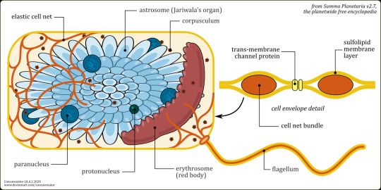
The typical cell structure of Eucytobionta, the clade comprising all the complex, multicellular life of planet Ea, the local equivalent of Earth's Eukarya. (original link)
« Complexity and organization are nested endlessly in lower and lower scales, far beyond our reach... Every smear of blood proclaims the power of its Maker; every drop of sewage sings the glory of the One. » – Yakub of Lilongwe, Mere Humanity
Shared features of Eucytobiontan cells include:
● A haploid protonucleus (i.e., carrying a single copy of each gene), sequestered at the center of the cell, where genetic information is stored over the long term in the form of enol-PNA; in sexual unicellular organisms, such as many Pogonocyta, additional protonuclei may be exchanged between cells.
● One or more massively polyploid paranuclei (carrying from 50 to over 200 copies of each gene), where gene expression and protein synthesis occurs, through remarkably Earth-like mechanisms. The more flexible and reactive keto-PNA is found here, and the massive redundancy dampens the effects of harmful mutations. New paranuclei are generated from the protonucleus before cell division. Monokaryotes have lost all their paranuclei, whereas the largest Pogonocytes may have hundreds.
● The astrosome, also called stellate body or Jariwala's organ, a multi-lobed vacuole located at the center of the cell. The water-filled lobular projections stabilize the cell structure, keep organelles in their place, and trace paths for the movement of vesicles. The astrosomal membrane is also the main metabolic organ, as it uses sunlight or chemical energy to create a proton gradient between the cytoplasm and the vacuole's interior from which useful energy can be harvested, much like the mitochondrial membrane in Earth's eukaryotes.
● The red body or erythrosome, a flattened organelle analogous to our Golgi apparatus, involved in the processing and secretion of proteins and TPP. The red color is probably due to iron complexes that assist with the reduction of TPP chains. In many unicellular organisms it can be visible as a dark-colored ribbon.
● The corpuscula, a number of dark-coloured vesicles filled with alkaline granules whose function is not yet clear. They are probably involved in the cell's metabolism and defense as reservoirs of enzymes in a crystalline form. Most Ean prokaryotes have corpuscula as well: some, such as Prasinobacteriales, use them for photosynthesis, whereas in Acanthobacillus they contain exotoxins used against predation.
● An elastic cell net formed by bundles of elastic, highly hydrophobic polypeptides passing between the two layers of the cell membrane. This sheath, similar to that found in the prokaryotic Commicutes, protects the cell from osmotic shock opposing both excessive intake and loss of water. In Ostracophyta and other unicellular Eucytobionts, the net is impregnated with minerals or crystalline polymers to form a protective shell.
● Undulipodia (distinguished, as on Earth, in cilia and flagella according to their size and abundance) seem to be extensions of the cell net, held into place by a trans-membrane protein ring. The whole structure is formed by parallel elastic fibers, and bends in one direction as the respective fiber contracts. The contraction is likely controlled by a chemical signal conveyed by vesicles to the contracting fiber's root in the basal ring.
– Summa Planetaria, "Eucytobionta#Synapomorphies", revision 315/T51Cyy4nS4
8 notes
·
View notes
Text
Yes, meiosis results in four different daughter cells, but when it occurs during oogenesis, only one becomes viable and the other three kinda disintegrate or some shit
Because the cytoplasm, nutrients and shit gets transferred to one cell during division
This shit doesn't happen during spermatogenesis
2 notes
·
View notes
Text
Cell Division: The Cell Cycle
There are two major parts in the cell cycle, interphase and cell division. 95% of a cell’s life is spent in the interphase part of the cycle. There are two more parts of cell division, mitosis and cytokinesis.
Mitosis is the process of dividing genetic information and has 4 more stages within it. I know what you’re thinking, more stages! Yay! /s /j
Prophase: Nuclear membrane begins to dissolve and the chromosomes shorten and thicken
Metaphase: Spindle fibres pull chromosomes in order to line up in the middle of the cell
Anahase: Centromere splits and sister chromatids separate. The daughter chromatids are pulled to the other side of the cell
Telophase: Daughter chromosomes stretch and become thinner as well as a new nuclear membrane forming around each set of chromosomes.
Cytokinesis is when the cytoplasm divides two genetically identical cells called daughter cells. If it’s an animal cell, the membrane will pinch off and the cell goes to interphase. If it’s a plant cell, it will form a cell wall before separating completely.
Further explanation: Chromosome Numbers During Division: Demystified!
#biology#how to do science#science#homework#student#grade 10#grade 10 science#stem#homework help#rst cpt#women in stem#school#cell division
2 notes
·
View notes
Text
Cell Cycle and Cell Division Class 11 Important Notes for NEET Biology
Introduction to Cell Cycle and Cell Division
Cell cycle and cell division are fundamental processes governing the growth, development, and reproduction of all living organisms. Understanding these processes is crucial in the field of biology as they play a pivotal role in shaping life at both the cellular and organismal levels.

Cell Cycle:
The cell cycle is the series of events that occur in a cell leading to its division and duplication of its DNA to produce two daughter cells. It consists of distinct phases, each with specific functions and checkpoints to ensure accurate progression. Through the cell cycle, cells grow, replicate their genetic material, and divide to give rise to new cells. This process is tightly regulated and orchestrated by various molecular mechanisms to maintain cellular integrity and functionality.
Cell Division:
Cell division is the process by which a parent cell divides into two or more daughter cells. It is essential for growth, tissue repair, and reproduction in multicellular organisms. Two main types of cell division are mitosis and meiosis. Mitosis ensures the faithful distribution of genetic material to daughter cells, resulting in the production of genetically identical cells. Meiosis, on the other hand, is a specialized form of cell division that produces haploid gametes for sexual reproduction, introducing genetic variation into offspring.
Significance:
The study of cell cycle and cell division is crucial for understanding various biological phenomena, including development, differentiation, aging, and disease. Dysregulation of these processes can lead to developmental abnormalities, cancer, and other pathological conditions. Therefore, unraveling the intricate mechanisms governing the cell cycle and cell division is not only of academic interest but also holds significant implications for medical research and therapeutic interventions.
In these class 11 notes, we will delve into the intricacies of the cell cycle and cell division, exploring the underlying molecular mechanisms, regulatory networks, and physiological significance. By grasping these fundamental concepts, students will gain a deeper understanding of the dynamic nature of life and the remarkable complexity of cellular processes.
Here's a properly formatted version of the important notes on Cell Cycle and Cell Division for NEET Biology:
Difference Between Cell Cycle and Cell Division:
Cell division is crucial for growth, repair, and reproduction, enabling the transformation of a single cell into a multicellular organism.
Cell Cycle:
Consists of cell growth, DNA replication, and division.
Genetically controlled events occur during the cycle.
Duration varies among organisms and cell types.
Divided into Interphase and M phase.
Interphase: Cell growth and DNA replication, constituting 95% of the cycle.
M Phase: Mitosis (cell division).
Interphase:
Three phases: G1, S, G2.
G1 Phase (Gap 1): Precedes DNA replication.
S Phase (Synthesis): DNA replication occurs without changing chromosome number.
G2 Phase (Gap 2): Cell continues growing and prepares for mitosis.
M Phase:
Involves karyokinesis (nuclear division) followed by cytokinesis (cytoplasmic division).
Mitosis:
Mostly occurs in diploid somatic cells of animals.
Ensures genetic continuity and facilitates growth and repair.
Four stages: Prophase, Metaphase, Anaphase, Telophase.
Karyokinesis followed by cytokinesis.
Prophase: Chromosomes condense, mitotic apparatus forms.
Metaphase: Chromosomes align at the metaphase plate.
Anaphase: Sister chromatids separate and move to opposite poles.
Telophase: Chromosomes decondense, nuclear envelope reforms.
Cytokinesis:
Cytoplasmic division following nuclear division.
Syncytium formation may occur in some organisms.
Meiosis:
Also known as reduction division.
Generates haploid gametes during sexual reproduction.
Maintains chromosome number and introduces genetic variation.
Consists of Meiosis I and Meiosis II.
Meiosis I:
Prophase I subdivided into Leptotene, Zygotene, Pachytene, Diplotene, Diakinesis.
Metaphase I: Bivalents align at the equator.
Anaphase I: Homologous chromosomes separate.
Telophase I: Nucleoli reappear, chromosomes collect at poles.
Meiosis II:
Follows interkinesis without DNA replication.
Similar to mitosis, produces haploid daughter cells.
Prophase II, Metaphase II, Anaphase II, Telophase II.
These notes cover the essential concepts of cell cycle and cell division, providing a comprehensive understanding for NEET Biology preparation.
#class 11#biology#science#botany#class 8#chemistry#11thclass#11th class#ecology#conservation#zoology#taxonomy#animal behavior#vavaclasses#9thclass#foundation
0 notes
Text
🔬 Cell Discovery & Theory:
- Coined by Robert Hooke
- Cell theory proposed by Schleiden, Schwann, and Virchow
- Living organisms made of cells, new cells from existing ones
🔬 Microscopy
- Compound & electron microscopes used
- Staining for visualization
🔍 Cell Structure
- Cell membrane, cytoplasm, nucleus, organelles
- Differences: unicellular vs. multicellular, prokaryotic vs. eukaryotic
🔄 Cell Cycle:
- Division for growth & repair
- Nucleus controls metabolic activities, passes genetic info
⚙️ Cell Organelles:
- Mitochondria, vacuoles, endoplasmic reticulum, ribosomes, Golgi bodies, plastids, centrosomes
🌱🐾 Plant vs. Animal Cells:
- Differences in size, presence of cell wall, plastids, vacuoles
🔬 Prokaryotic vs. Eukaryotic Cells:
- Prokaryotes lack organized nucleus, eukaryotes have one
- Examples: bacteria vs. plants, animals
🔄 Cell Division:
- Vital for growth, repair, and renewal
- Daughter cells formed through nucleus and cytoplasm division


#education#school#notes#youtube#studyblr#students#homeschool#latitudes#maths#maths tutoring#student life#study#studying#student#new studyblr#study aesthetic#college studyblr#study blog#study inspiration#study hard#study motivation#study notes#study tips#study with me#studyblr community#studygram#studyinspo#studyspiration#studyspo#studystudystudy
1 note
·
View note
Quote
Neuroinflammation is associated with poor outcomes in patients with spinal cord injury (SCI). Recent studies have demonstrated that stimulator of interferon genes (Sting) plays a key role in inflammatory diseases. However, the role of Sting in SCI remains unclear. In the present study, it is found that increased Sting expression is mainly derived from activated microglia after SCI. Interestingly, knockout of Sting in microglia can improve the recovery of neurological function after SCI. Microglial Sting knockout restrains the polarization of microglia toward the M1 phenotype and alleviates neuronal death. Furthermore, it is found that the downregulation of mitofusin 2 (Mfn2) expression in microglial cells leads to an imbalance in mitochondrial fusion and division, inducing the release of mitochondrial DNA (mtDNA), which mediates the activation of the cGas-Sting signaling pathway and aggravates inflammatory response damage after SCI. A biomimetic microglial nanoparticle strategy to deliver MASM7 (named MSNs-MASM7@MI) is established. In vitro, MSNs-MASM7@MI showed no biological toxicity and effectively delivered MASM7. In vivo, MSNs-MASM7@MI improves nerve function after SCI. The study provides evidence that cGas-Sting signaling senses Mfn2-dependent mtDNA release and that its activation may play a key role in SCI. These findings provide new perspectives and potential therapeutic targets for SCI treatment.
Cytoplasmic Escape of Mitochondrial DNA Mediated by Mfn2 Downregulation Promotes Microglial Activation via cGas‐Sting Axis in Spinal Cord Injury - Wei - 2024 - Advanced Science - Wiley Online Library
0 notes
Text
Marooned Among the Microbes: A Rollicking Rendition of Cell Theory with Robinson Crusoe
Ahoy there, distinguished shipmates of this literary vessel! Pray, lend me your ear—or rather, your eyes—for I, Robinson Crusoe, once a castaway of considerable renown, now find myself marooned anew amidst a sea of knowledge, navigating the minuscule marvels of cell theory. You see, in much the same manner as I stumbled upon that desolate isle, I’ve chanced upon the world of these tiny entities, a discovery as unexpected as it is enlightening.
Let’s hoist the sails and set forth on this microscopic odyssey, shall we? You must understand, cells are the very fabric of life, much like the tattered canvas I once fashioned into a makeshift shelter. They are the smallest units of life, teeming and bustling like the inhabitants of a populous city, though they reside in a world undetected by the naked eye. Robert Hooke, a fellow with a keen eye, first spied these tiny chambers in 1665, peering through his primitive microscope as though it were a spyglass revealing distant shores.
Imagine my surprise, like my first encounter with the footprint on the sand, upon learning that each living organism is a veritable archipelago of cells! Much as my solitary island was part of a greater world, so too are these cells part of a huge biological landscape. The concept of cell theory, crystallized by scholars Matthias Schleiden and Theodor Schwann in the 19th century, posits that all living things are composed of these cells and that the cell is the basic unit of life.
But what, you might ask, makes up these microscopic marvels? Similar to my own rudimentary abode, each cell is a self-contained unit, complete with all the necessities of life. There’s the cell membrane, a sturdy barrier like the walls of my shelter, guarding the cell from the outside world. Within, the cytoplasm ebbs and flows, a bustling marketplace of cellular activity. And the nucleus, ah! The nucleus is the master of the cell, much like my own rule over my island domain, directing the activities of its microscopic dwelling.
In nature, cells replicate through a process called cell division, like how I might have split a coconut to multiply my sustenance. This process ensures the continuity of life, an unending cycle of birth and rebirth, mirroring the endless ebb and flow of the tides that once governed my days.
So, beloved fellow castaways, as I once chronicled my solitary sojourn on that remote isle, I now invite you to join me in exploring the world of cell theory. Fear not the complexity of this microscopic terrain, for I shall be your guide, translating the scientific jargon into the vernacular of a seasoned, albeit somewhat out-of-touch, castaway. Together, we shall unravel the mysteries of life at its most fundamental, one cell at a time.
0 notes
Text
Reproduction in Protozoa
Asexual Reproduction: there are 5 ways a protozoan can undergo asexual reproduction: Binary Fission, Plasmotomy, Budding, Multiple Fission, and Plasmogamy.
Binary Fission: the division of one individual cell into two approx. equal parts. It's a type of mitosis where karyokinesis is always followed by cytokinesis. Ex: Amoeba
Plasmotomy: A special type of binary fission, it divides a multinucleate mother cell into two or more multinucleate daughter cells. Ex: Pelomyxa
Budding: Modified fission resulting in a small daughter bud attached to the mother cell. When the bud is broken off, it develops and grows into full size. When a parent produces one bud, it's called monotonic. When a parent produces numerous buds at once, it's called multiple budding. Ex: Vorticella
Multiple Fission: aka Sporulation, Nuclear division is not immediately followed by the division of cytoplasm. The nucleus undergoes a series of divisions till the body becomes multinucleate. After, the cytoplasm divides into the same number of parts as there are daughter nuclei, each surrounds a nucleus. Finally, the body divided simultaneously into numerous daughter cells. The number of daughter cells can vary. Ex: Plasmodium
Plasmogamy: Two or more individuals fuse their cytoplasm, but keep their nuclei distinct. They separate later, unchanged. This sometimes serves the purpose of digestion of large prey. Ex: Rhizopoda
Sexual Reproduction: There are two types of sexual reproduction: Syngamy and Conjugation.
Syngamy: Complete fusion of two sex cells/gametes, creates a zygote. The fusion nucleus of zygote is called synkaryon. Is of 4 subtypes: Hologamy, Isogamy, Anisogamy, Autogamy.
Hologamy: Mature protozoans behave like gametes and fuse together
Isogamy: fusing gametes are similar in size and shape
Anisogamy: fusing gametes differ morphologically and behaviorally.
Autogamy: fusing gametes ae derives from the same parents cell.
Conjugation: The temporary union of two individual conjugants. Usually at oral or buccal regions of their body. ex: Suctoria
Parthenogenesis: Gametes which fail at cross-fertilization (syngamy) develop parthenogenically. ex: Chlamydomonas
Regeneration: Most protozoans can regenerate lost parts.
0 notes
Text
Cell Biology: Exploring the Fundamental Units of Life
Cell biology, often referred to as cytology, is a branch of biology that delves into the intricate world of cells—the fundamental units of life. The study of cells has unlocked the mysteries of life's processes, from the simplest single-celled organisms to complex multicellular organisms like humans. In this exploration of cell biology, we will delve into the structure, function, and significance of cells, shedding light on the remarkable world that lies within our bodies and the natural world around us.
The Basics of Cell Biology
Cells are the building blocks of life, and cell biology seeks to understand their composition, structure, and functions. At its core, cell biology aims to answer fundamental questions about the nature of life and how organisms function at the cellular level.
The Cell Theory
Cell biology is rooted in the Cell Theory, a foundational concept that has guided our understanding of life for centuries. The Cell Theory consists of three key principles:
All living organisms are composed of one or more cells.
The cell is the basic structural and functional unit of life.
All cells arise from pre-existing cells through cell division.
These principles, formulated by scientists such as Matthias Schleiden, Theodor Schwann, and Rudolf Virchow in the 19th century, revolutionized our perception of life and laid the groundwork for modern cell biology.
The Structure of Cells
Cells come in various shapes and sizes, and their structures are tailored to their functions. Nevertheless, all cells share some common components and organelles that play critical roles in their activities
Cell Membrane
The cell membrane, also known as the plasma membrane, is the outermost boundary of a cell. It acts as a selectively permeable barrier, regulating the passage of substances in and out of the cell. The cell membrane is composed of a lipid bilayer embedded with proteins, providing structural integrity and facilitating communication between cells.
Cytoplasm
Inside the cell membrane lies the cytoplasm, a semi-fluid medium that contains various organelles and cellular structures. Many essential metabolic reactions occur in the cytoplasm, making it a vital part of the cell's machinery.
Nucleus
In eukaryotic cells, such as those found in plants, animals, and fungi, the nucleus is the central control center. It houses the cell's genetic material in the form of DNA, which is organized into chromosomes. The nucleus controls cellular activities by directing the synthesis of proteins and other molecules through a process called transcription.
Organelles
Organelles are specialized structures within the cell that perform specific functions. Some of the most notable organelles include:
Mitochondria: Known as the "powerhouses" of the cell, mitochondria generate energy through a process called cellular respiration.
Endoplasmic Reticulum (ER): The ER is involved in protein synthesis and lipid metabolism. It comes in two forms—rough ER (studded with ribosomes) and smooth ER (lacks ribosomes).
Golgi Apparatus: This organelle modifies, sorts, and packages proteins and lipids for transport within or outside the cell.
Lysosomes: Lysosomes contain enzymes that break down waste materials and cellular debris, playing a crucial role in cellular recycling.
Ribosomes: These tiny structures are responsible for protein synthesis. Some ribosomes are free in the cytoplasm, while others are attached to the rough ER.
Peroxisomes: Peroxisomes are involved in detoxification processes and the breakdown of fatty acids.
Cytoskeleton
The cytoskeleton is a network of protein filaments that provides structural support to the cell and plays a role in cell division and intracellular transport. It consists of three main components: microfilaments, intermediate filaments, and microtubules.
Cell Functions
Cells perform a wide range of functions that are essential for the survival and functioning of organisms. These functions can be broadly categorized as follows:
Metabolism
Metabolism encompasses all the chemical reactions that occur within a cell. These reactions involve the breakdown of nutrients to generate energy (catabolism) and the synthesis of molecules necessary for cell growth and repair (anabolism). Cellular respiration, for example, is a fundamental metabolic process that occurs in mitochondria, where glucose is converted into energy in the form of ATP (adenosine triphosphate).
Reproduction
Cells reproduce through a process called cell division. In unicellular organisms, such as bacteria, cell division is a means of reproduction. In multicellular organisms, cell division is essential for growth, tissue repair, and replacing old or damaged cells.
HomeostasisCells maintain internal stability, or homeostasis, by regulating various physiological parameters. For instance, they control the concentration of ions, gases, and nutrients to ensure that the internal environment remains suitable for cellular processes.
Communication
Cells communicate with each other through chemical signals. Signaling molecules, such as hormones, neurotransmitters, and growth factors, enable cells to coordinate their activities and respond to external cues. This communication is vital for processes like development, immune response, and maintaining tissue integrity.
Significance of Cell Biology
Understanding cell biology has profound implications for various fields, including medicine, genetics, biotechnology, and environmental science. Here are some key areas where cell biology plays a crucial role:
Medicine
Cell biology is foundational to the field of medicine. It provides insights into the causes of diseases, the development of treatments, and the study of how drugs interact with cells. For example, cancer research heavily relies on understanding the abnormal behavior of cells and the genetic mutations that lead to uncontrolled cell division.
Genetics
Cell biology and genetics are intimately connected. The study of cells allows us to explore the mechanisms of inheritance, gene expression, and genetic disorders. Advances in cell biology have enabled breakthroughs in gene editing techniques like CRISPR-Cas9, which hold the potential to treat genetic diseases.
Biotechnology
Cell culture techniques, which involve growing cells outside of the body, are essential in biotechnology. These techniques are used to produce recombinant proteins, develop vaccines, and conduct drug testing. Cell biology is the foundation of bioprocessing and the production of biopharmaceuticals.
Environmental Science
Understanding how cells respond to environmental changes is crucial for environmental science. Cell biology helps us comprehend the impact of pollutants, climate change, and other stressors on organisms at the cellular level. It is also instrumental in the study of biodiversity and ecosystem health.
The Future of Cell Biology
As technology advances, so does our ability to explore the intricacies of cells. Emerging techniques like single-cell genomics, super-resolution microscopy, and organoid culture are pushing the boundaries of our understanding. Additionally, the integration of cell biology with fields like artificial intelligence and nanotechnology holds promise for groundbreaking discoveries and innovations.
In conclusion, cell biology is a captivating field that unveils the mysteries of life at its most basic level—the cell. From its inception with the Cell Theory to its current role in advancing medicine, genetics, biotechnology, and environmental science, cell biology continues to shape our understanding of life and inspire discoveries that benefit humanity. As we delve deeper into the fascinating world of cells, we can anticipate even more exciting revelations and applications in the future.
0 notes
Text
Tree of Life 2: Eukarya (life with complex cells)
[Disclaimer: taxonomy is a complex, ever-changing field, and this overview is certainly not going to be exhaustive, especially concerning extinct groups]
← Part 1 (Biota)
Part 3 (Archaeplastida) →
View of a generic eukaryotic cell: amoebae, kelp, mushrooms, pine trees, and dinosaurs are variations on this basic model (from my Human Evolution infograph).
Eukarya “good kernel”: The third domain of Life other than Bacteria and Archaea, the one that includes all “complex” life: animals, plants, fungi, algae. The origin of Eukaryotes -- most probably from some branch of marine Archaea, some 2 billion years ago -- are a contentious topic, but it’s generally accepted that it involved two events (in whichever order, for whatever reason): 1) the loss of cell wall and infolding of the cell membrane to create a complex system of internal organelles with many varied functions; and 2) the incorporation of oxygen-using Alphaproteobacteria as mitochondria, which allowed Eukaryotes to spread around an increasingly oxygenated planet and to access much more energy than its fermenting ancestors. This is an instance of symbiogenesis, in which two organisms associate so deeply that they go on to evolve as one. The cell membrane is the same to that of Bacteria; the cell wall is primitively missing, though some groups re-developed a new one. Eukaryotic cells are generally one order of magnitude larger than prokaryotic ones -- 100 micrometers (= 0.1 mm) is a fairly common size, though less so for animal cells. Important features are:
Virtually all Eukaryotes have a nucleus, an internal structure bounded by a porous membrane that contains the cell’s main DNA. (Not all...) The DNA of Eukaryotes can be much larger and more complex than that of prokaryotes, because it’s isolated from the cell’s activity and supported by protein scaffolds. During cell division, eukaryotic DNA is organized into multiple chromosomes, each of which often exists in multiple copies. Information is copied onto mRNA inside the nucleus; the mRNA than leaves through the pores and reaches ribosomes to be translated into proteins, just as in prokaryotes and specifically in Archaea (see part 1).
Mitochondria, the powerhouse of the cell! The cytoplasm can produce a bit of energy by fermenting sugar on its own, but mitochondria can use oxygen to oxidize food on its twisted inner membrane, releasing almost 20 times as much energy. As mentioned, mitochondria were produced by an event of endosymbiosis, in which an oxygen-using bacterium was incorporated within a proto-eukaryote. Photosynthetic Eukaryotes also have plastids or chloroplasts, structures filled with chlorophyll and other pigments that release electrons when struck by sunlight, thereby providing energy to the cell, and allowing it to reduce carbon dioxide into organic molecules at the same time. Plastids were also produced by endosymbiosis, incorporating a Cyanobacterium (primary endosymbiosis) or a plastid-carrying Eukaryote (secondary endosymbiosis). Mitochondria and plastids still retain their old bacterial DNA, although part of it was lost or transferred into the cell’s nucleus.
Since ancestral Eukaryotes gave up the cell wall, they ensured mechanical stability with a cytoskeleton, a complex system of protein strands (microtubules of tubulin and filaments formed of actin and myosin) which are also capable of movement. The cytoskeleton can deform the whole cell producing temporary appendages called pseudopods, used to move and to engulf food. Membrane-covered actin filaments can come out of the cells forming whip-like undulipodia. Undulipodia are called cilia when they are many and small, and flagella when they are few and large, but they have the same structure, which is in fact very, very different from that of the prokaryotic flagella (prokaryotic flagella spin like a propeller, eukaryotic flagella beat sideways). The cytoskeleton can also draw in a piece of membrane forming a self-contained vesicle, which may be used to consume food (phagocytosis).
A system of internal membranes specialized for various biochemical tasks. The endoplasmic reticulum (ER) is an extension of the nuclear membrane forming flat sheets. Part of it, “rough ER” is an attachment point for ribosomes for massive protein synthesis; the rest, “smooth ER”, is mainly concern with the synthesis of lipids, especially new membrane. The Golgi apparatus is a stack of flattened sacs serving as a stockpiling and processing center for newly produced proteins (it’s very large in cells that produce toxins, mucus, or enzymes). A vacuole is an internal membrane that may contain water for mechanical support, food being digested, and so on. Lysosomes are small vesicles full of digestive enzymes that degrade food and destroy pathogens.
For all the attention that animals and plants get, most of the taxonomic diversity of Eukaryotes is microscopic, mostly unicellular species, which are generically called protists. Because of their cellular complexity, Eukaryotes cannot sustain themselves on countless esoteric combination of chemicals like prokaryotes (almost all depend on oxygen, for one), but they replace a much smaller biochemical diversity with a much larger morphological diversity. Traditionally, they are divided in four groups: ciliates (moving with cilia), flagellates or mastigophorans (moving with flagella), sarcodines or amoebae (moving with pseudopods), and sporozoans (spore-forming). Of course this is about as natural as grouping animals into “flyers” and “swimmers”; ciliates and sporozoans are not too far from the modern Ciliophora and Apicomplexa, but flagellates and amoebae were torn apart by modern classifications and scattered everywhere.
Now, for more detail:
?1. (?*)Excavata “dug out”: A group of “primitive” Eukarya, usually flagellate and heterotrophic, with an oral groove after which they’re named, but very diverse. Possibly basal to Eukarya (He & al 2014), but it might be paraphyletic or even polyphyletic.
1a. Metamonada: They have multiple flagella and a bundle of microtubules, or axoneme, running along the cell. They lack mitochondria, which was once taken as a sign of a primitive condition (as “Archezoa”, they would have branched out after the evolution of the nucleus but before endosymbiosis), but in fact they have mitochondria-derived genes and structures. Like some Archaea and Bacteria (see part 1), being poisoned by oxygen, they often live in animal guts. This group might be grouped with Unikonta (see below) in Scotokaryotes “shadow kernel” (Cavalier-Smith 2013), so named because they contain no phototrophic species.
1a1. Diplomonadida “double unit”: A curious trait of this clade is having two twin nuclei, each with its own bundle of flagella. They carry mitosomes, apparently vestigial mitochondria that lost their DNA and have no known function. Most famous member is Giardia lamblia, which can give a pretty miserable time to the human gastrointestinal system.
1a2. Parabasalida “next to the base”: Mostly symbionts in the gut of insects. They have hydrogenosomes, modified mitochondria that use hydrogen ions instead of oxygen. They include Trichomonas vaginalis, a common sexually-transmitted pathogen that causes genital inflammation, with a flagellum embedded in the membrane and pulling it up into a sort of undulating fin; as well as Mixotricha paradoxa, which lives in the intestine of an Australian termite and requires no less than four symbiont bacteria to survive (three on its surface to help it swim and one inside to digest cellulose for both itself and the termite).

Double-nucleied Giardia lamblia, busy ruining someone’s day. (Wikipedia)
1b. Discoba/Discicristata: A clade named after the fact that the membrane folds, or cristae, inside the mitochondria are disk-shaped, rather than parallel ridges as in our case. It should probably be classified next to Bikonta in the clade Diphoda (Derelle &al 2015).
1b1. Heterolobosea/Percolozoa: Found a bit everywhere in water and soil, their lifecycle alternates flagellate and amoeboid states. Acrasida is one of many groups of slime molds, i.e. protists that can form temporary multicellular colonies to produce fruiting bodies and spores. Other Heteroloboseans are opportunistic pathogens; Naegleria fowleri, known with the picturesque name of “brain-eating amoeba”, is happy enough to eat bacteria in lake waters, until it decides to enter through your nose and start consuming your brain tissue.
1b2. Euglenozoa: Flagellates with an elongated, tapering body, and two flagella inserted in an anterior pocket or reservoir, although one is very small and often does not reach out of the pocket.
1b2a. Euglenida “good socket”: Photosynthetic, having incorporated green algae as plastids by secondary endosymbiosis, but at the same time they also eat solid food. They may have a red eyespot or stigma that guides them toward light. They’re covered by a striped protein pellicle that can slide along each other, causing the cell to contract or crawl.
1b2b. Kinetoplastida “moving form”: Very similar in overall shape to Euglenida but lacking plastids; they have a single giant mitochondrion that extends into a kinetoplast in the front, to which a flagellum is attached. The flagellum trails along and behind the body, forming an undulating membrane to swim. They are usually parasites in the blood of vertebrates, including Trypanosoma and Leishmania, the agents of sleeping disease and Chagas’ disease.


Euglena (Euglenida, above) and Trypanosoma (Kinetoplastida, below). (Brusca 2016)
2. Bikonta “two flagella” or Corticata “bark-covered” or Diaphoretickes (in reference to the fact that they may have two flagella per cell and may have had an ancestral cell cortex divided in sacs). All Eukaryotes with permanent photosynthesis, except Euglenida, are in this clade.
2a. Archaeplastida or Plantae sensu lato, that is, plants in its broadest sense: the group that got photosynthesis from a single event of primary endosymbiosis with a cyanobacterium carrying the green pigment chlorophyll a. (Mitochondrial cristae are flat.) Includes Rhodophyta (red algae) and Viridiplantae (green algae and land plants). See part 3 for more.
2b. Cryptophyta “hidden plants”. Unicellular algae with two flagella emerging from the side just below their anterior end. They have one or two large plastids surrounded by four membranes, which derive from a red alga incorporated by secondary endosymbiosis. They also have unique ejectisomes, sacs containing a sort of coiled springs that can be ejected to propel the cell away from danger.
2c. Haptista: Thin appendages or axopods supported by microtubules, used for feeding.
2c1. Haptophyta “bound plants”: A rather diverse group of protists, usually phototrophs thanks to the secondary endosymbiosis of a red alga, with a huge diversity of photosynthetic pigments. A particularly important subgroup is Coccolithophora, which have thick scales of calcium carbonate that make them look like balls of buttons. When they die, their heavy shells sink to the sea bottom, and form beds of limestone and chalk. The White Cliffs of Dover are basically a huge pile of dead and compressed Coccolithophorans.
2c2. Centrohelida “central suns”: A major part of what once was the group “Heliozoa”, now considered polyphyletic. They have no flagella, but very long axopods radiating from a spherical body (hence the Sun-related name). They have small scales of silica that do not cover the whole body. They include a genus Yogsothoth, which to be honest does not look much more eldritch than the rest.

A variety of Coccolithophores; note, at bottom right, Braarudosphaera bigelowii, whose shell is an almost perfect dodecahedron. (Wikipedia)
2d. SAR or Chromalveolata or Chromista “colorful” (although the latter two names are supposed to include Cryptophyta, so we shouldn’t use those names in this sense). One common feature is that mitochondrial cristae are usually neither disks nor ridges, but shaped like tubes.
2d1. Rhizaria “rooted”: A large and widespread group, mostly heterotrophic amoebae with very long and thin pseudopods supported by microtubules. The cytoplasm often has an outer layer with buoyant fatty droplets that counter the shell’s weight.
2d1a. Retaria: A clade of armored planktonic protists.
2d1a1. Foraminifera “hole-carriers”: They have calcium carbonate shells, often multi-chambered, of a huge variety of shapes, spiral, disk-shaped, flask-shaped, leaf-like, etc. The shells have many little holes through which thin branching pseudopods can pass through. While mostly microscopic, some measure several millimeters, and members of the deep-sea group Xenophyophorea can actually reach several centimeters (these have in fact multiple nuclei, so they could be considered multiple cells fused together inside the shell). Like Coccolithophorans, dead Foraminiferans sink to the sea bottom and form limestone layers; they are so common in such rocks that Foraminiferan shells are often used to date sedimentary layers. Often they carry zooxanthellae, that is, symbiotic photosynthetic dinoflagellates (see below). Nummulites has a spiral shell 1-5 cm across (!) that is commonly found as fossil.
2d1a2. Radiolaria “small rays”: Their shell has a foam-like structure and is made of silica (or sometimes, for some reason, strontium sulfate), partially embedded in the cytoplasm; pseudopods are very thin and straight. Shells may be simple spicules, or highly elaborate polyhedra or crown-like sculptures. They may reproduce by splitting internally into hundreds of “swarmers”, each carrying a crystal from which they will build their new shell. Like Foraminifera, their shells form abundantly sedimentary rocks. They too may have zooxanthellae, or symbiotic Cyanobacteria.

Some examples of Foraminifera (left) and Radiolaria (right), as represented by Ernst Haeckel in 1904. (Wikipedia)
2d1b. Cercozoa “tailed animals”: A complex and diverse clade, with little visible common features apart from those general to Rhizaria; usually taking the shape of single amoeboid or biflagellate cells. Some interesting examples are Euglyphida “well-carved”, armored amoebae with a barrel-shaped shell formed by scales of silica; Chlorarachniophyta “green spiderweb plants”, who have taken up green algae as plastids by secondary endosymbiosis, and nevertheless extend nets of cytoplasm to ensnare preys; Vampyrellida, with a spherical reddish body radiating into thin pseudopods with which they suck nutrients from algae or fungi; and Gromia, with a hive-like protein shell that can measure several millimeters, from which it sends complexly branching pseudopods. Paulinella is an Euglyphid that decided to start the whole endosymbiosis thing all over by incorporating a Cyanobacterium that is not the same that became the plastids of green plants.
Some Chlorarachnion reptans (Chlorarachniophyta), ejecting their strange-looking webs of cytoplasm. The pseudopods of Gromia are similar. (Wikipedia)
2d2. Heterokonta “different flagella” or Stramenopiles “straw hair”: Descendants of a common ancestor who absorbed a red alga by secondary endosymbiosis: their plastids have no less than four membranes, two from the original cyanobacterium, one from the red alga, and one derived from the heterokont’s ER. Most members are thus photosynthetic, although some have lost that ability. Most, at least at some point, have two flagella with different structures: one short and smooth, the other one longer and branching into lateral filaments that increase its effectiveness in swimming. This is a vast and extremely diverse group, of which I’ll mention only some examples.

A generic Heterokont, based on Ochromonas; most members of this group only look like this for a brief part of their lifecycle, if at all. (Brusca 2016)
2d2a. Opalinea “opalescent”: They lost their plastids and live most commonly in the intestine of frogs and other vertebrates, where they absorb a part of the food waste about to be expelled. Unlike most Heterokonts, they’re covered in dense rows of cilia, though their spores have the typical two flagella.
2d2b. Labyrinthulea “little mazes”: Often symbionts or parasites of seagrasses, they move by extending their cytoplasm into branching nets which are indeed very maze-like, while their main body is protected by scales derived from the Golgi apparatus.
2d2c. Oomycetes “egg-fungi” or Pseudofungi: They were classified for a long time among actual Fungi: like them, they grow as cellular filaments covered by a cell wall, although that wall is mostly cellulose, like in plants. All are heterotrophic too, and many are parasites. Peronosporales in particular are responsible for many plant blights, mildews, and rusts, most importantly potato blight, which is due to Phytophthora infestans. Saprolegniales, also known as “water molds”, include both aquatic decomposers and parasites of plants, fish, and crayfish.
2d2d. Ochrophyta “yellowish plants”: A clade of rather diverse heterokonts who mostly use a red alga endosymbiont, wrapped in the ER, for photosynthesis; a combination of fucoxanthin and chlorophyll c usually makes them brown or greenish-yellow.
2d2d1. Bacillariophyceae/Diatomeae: You’ll know these ones as diatoms. They have a slitted shell of silica (effectively, glass) with a variety of radiate or four-fold shapes. The shell is formed by two valves of different sizes, like a box and its lid; when the cell divides, each half of the shell generates a smaller one within itself. The cells therefore become smaller and smaller through the generations, until they reach a minimum size and produce a spore that resets the whole cycle. Adults have no flagella and usually don’t move at all.

Two diatoms represented by Ernst Haeckel (left: Navicula bullata, right: Triceratium robertsianum). (Wikipedia)
2d2d2. Actinophryida: The other part of old “Heliozoa”. They are roughly spherical cells with rigid axopodia protruding in all directions, stiffened by thick arrays internal microtubules. When a prey, such as a smaller protist, brushes against an axopodium, it contracts to bring the prey into the cell body for digestion.
2d2d3. Chrysophyceae “golden seaweed”: Another group of unicellular algae, these ones flagellate. Often they have complex shells (loricae) of chitin or silica; great variety of shapes and pigments.
2d2d4. Phaeophyceae “dark seaweed”, better known as “brown algae”. They are multicellular, macroscopic seaweed, e.g. kelp and bladderwrack (Fucus vesiculosus), which keep themselves upright in water with gas-filled bladders, as well as the floating Sargassum. Giant kelp Macrocystis can be as tall as 60 meters, and forms undersea forests, especially in the northern Pacific. They have a dark yellowish or brown color, since they usually live in dark depths and cannot afford to reflect away much light. Their body has a basal holdfast and leaf-like laminae, but doesn’t have much internal specialization; the cell wall is a mixture of cellulose and alginate, a complex sugar used to make gelatin.
Plus many, many other groups that can be described as “yet more unicellular algae”.

A submarine forest of giant kelp, Macrocystis pyrifera (Phaeophyceae). (Wikipedia)
2d3. Alveolata: so called in reference to the pellicle underlying their cell membrane, which forms a grid of flat sacs or alveoli, strengthening their envelope; you will not find amoebae here.
2d3a. Ciliophora “bearers of cilia”, or simply Ciliates. These protists have employed their alveolar apparatus to support and power an extensive system of cilia, usually covering their whole body, which they use to swim and to create food currents that bring food to their mouth. Between these cilia, they may have trichocysts or toxicysts that expel defensive threads or toxins, respectively. They tend to be very large cells -- some reaching a millimeter in length! -- and have developed structures curiously reminiscent of animals, such as an actual mouth or cytostome that funnels food into budding vesicles, an anus or cytoproct expelling waste, and a contractile vacuole that pumps out excess water. Ciliates always have two kinds of nuclei, micronuclei that are relatively inert and macronuclei where DNA is massively replicated for quicker gene expression. While reproduction is asexual, pairs of ciliates of the same species can exchange genes by swapping copies of their micronucleus. Extremely diverse in shape and found wherever there’s water, from the loafer-shaped Paramecium to the stalked, flower-like Vorticella, from Lacrymaria pursuing its preys with a snake-like neck to Stylonychia walking on the bottom with leg-like bundles of cilia. They even include a few anaerobic species that have converted their mitochondria into hydrogenosomes.

Lacrymaria olor (Ciliophora), swimming with its neck fully extended as it looks for preys. The whole cell is almost half a millimeter long. (Wikipedia)
2d3b. Myzozoa: A clade characterized by myzocytosis, the process of piercing a prey cell with a tube to suck out its content.
2d3b1. Dinoflagellata “whirling flagella”: The alveoli are often filled with cellulose to form a thick shell, divided by a groove or cingulum into a front and back part, often asymmetrical. There are two flagella, one curled and running along the cingulum, the other beating freely behind the cell. While many have incorporated a variety of photosynthetic organisms, such as diatoms, by tertiary endosymbiosis, often they still feed on other organisms. Some are parasites, such as Pfiesteria piscicida, a well-known scourge of fishes. Noctiluca scintillans, which has a tentacle and no theca, is bioluminescent thanks to luciferin (the same compound found in fireflies), and is responsible for the glow known as “milky sea” or “sea fire”. Many species of Dinoflagellates can grow into toxic algal blooms called “red tides”.

Some examples of Dinoflagellates. (Hickman 2008)
2d3b2. Apicomplexa: The core of the old Sporozoa. All are intracellular parasites of animals; they take their name from the characteristic apical complex, a structure at the anterior end of their elongated body, which they use to break into host cells. Their plastids are colorless and inactive; as parasites, they have no need for photosynthesis. They have very complex lifecycles with both sexual and asexual phases, cysts that can survive very long in host bodies, and hosts of multiple species. Plasmodium, the mosquito-born agent of malaria, is here, as is Toxoplasma, which infects cats. Gregarina has giant, worm-like cells living in the intestine of insects, and is considered one of the most successful clades of parasites on Earth.
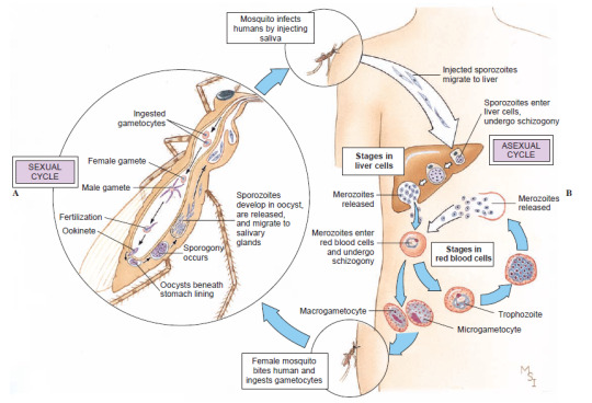
The complex lifecycle of Plasmodium vivax, an agent of malaria. Sporozoites injected by a feeding mosquito travel through the bloodstream to the human liver, where they divide into a mass of merozoites. These take over red blood cells, where they multiply again and again, producing sexual gametes, until some are taken up by another mosquito. In the insect’s gut, the gametes merge to form new sporozoites that colonize the mosquito’s saliva. (Hickman 2008)
3. Unikonta “one flagellum” or Amorphea “shapeless” (in reference to the fact that they have at most one flagellum per cell, and no cell wall, which makes their cells less rigid than Corticata). We, as all animals, are in here, along with fungi and "core” amoebae; see part 5! (That’s right, we are spending the next two parts on plants!)

Summary. Dates (mostly based on Parfrey & al 2011) are tentative, though not as much as in part 1; dashed lines represent unclear lineages. Colored symbols represents event of primary (circle) or secondary (square) endosymbiosis. The yellow circle is the incorporation of Alphaproteobacteria as mitochondria; the teal circles are the incorporation of Cyanobacteria as plastids by Archaeplastida and Paulinella (in Cercozoa); the green squares are incorporations of green algae by Euglenida and Chlorarchniophyta (in Cercozoa), and red squares of red algae by Cryptophyta, Haptophyta, Ochrophyta, and Myzozoa (the latter two might actually be a single event at the root of SAR; see Keeling 2013). Dates are in millions of years.
Sources
Adl & al (2007), “Diversity, Nomenclature, and Taxonomy of Protists” (link)
Adl & al (2019), “Revisions to the Classification, Nomenclature, and Diversity of Eukaryotes” (link)
Brown (2014), Principles of Microbial Diversity, APM Press
Brusca (2016), Invertebrates (3rd edition), Sinauer
Cavalier-Smith (2013), “Early evolution of eukaryote feeding modes, cell structural diversity, and classification of the protozoan phyla Loukozoa, Sulcozoa, and Choanozoa” (link)
Cavalier-Smith (2017), “Kingdom Chromista and its eight phyla: a new synthesis emphasising periplastid protein targeting, cytoskeletal and periplastid evolution, and ancient divergences” (link)
Derelle & al (2015), “Bacterial proteins pinpoint a single eukaryotic root” (link)
He & al (2014), “An Alternative Root for the Eukaryote Tree of Life” (link)
Hickman & al (2008), Integrated Principles of Zoology (14th edition), McGraw-Hill
Keeling (2013), “The Number, Speed, and Impact of Plastid Endosymbioses in Eukaryotic Evolution” (link)
Parfrey & al (2011), “Estimating the timing of early eukaryotic diversification with multigene molecular clocks“ (link)
4 notes
·
View notes
Text
Biotin-Conjugated Dextran
Biotin-Conjugated Dextran: Labeled dextran is a hydrophilic polysaccharide, most commonly used in microscopy studies to monitor cell division, track the movement of living cells, and report the hydrodynamic properties of the cytoplasmic matrix. The labeled dextran is usually introduced into the cells by microinjection.
0 notes
Text
The Love of an Amoeba
Once upon a time, an amoeba was joyfully swimming by itself in a warm pool of water. Suddenly, it felt an intense wave of exhilaration rush through its body. It didn’t need to wonder long where it came from, because a thought inspired by the feeling bloomed inside it; the amoeba loved itself. I adored being by itself and it marveled at how wonderful it was to be an amoeba as amazing as itself.
“When I change my shape,” it thought, “each form I take is perfectly round and squishy. And when I swim and wiggle my pseudopodium, I do so with a transcendent grace that was the envy of any other singe cell organism. By the Carbon, I am so beautiful,” It gushed.
Once again, that feeling of love and happiness burst inside itself, and in that moment it asked the question that every amoeba dreams about, “Do I…” it paused, “Do I want to make another copy of myself? Should I reproduce?”
It stumbled over the last word, because the amoeba knew that this was only something one should do if one truly loved themself, but before it could think any further, it felt a tingle in its elongating nucleus“I’ve never felt this before. Is this what mitotic division feels like?…Oh!” it moaned, “Oh yeah, that feels so good!”
In the glistening water, the Amoeba made sweet, sweet binary fission.“I’m gonna split, I’m gonna split, oh my god I’m gonna split….. ahhh!” It yelled, before it’s nucleus and cytoplasm burst into two.
Suddenly, there was a sister amoeba sitting next to it. “I am even more beautiful now, because there are two of me in this world,” said the two two sister amoebas. The two of them went their separate ways and lived happily ever after, never seeing each other ever again.
1 note
·
View note
Last Seen Blogs

chauhanparkash2005
Bank Routing Number
samesunroastbeef-blog
#beef #japanesebeef #steak #saitama #warabi

kurara-ash
Kurara-Nyan

zombiegorillajesuschrist
ZombieGorillaJesusChrist
yersifanel
Capital de Arcadia