#fundus
Text
Fundus, 23/24
0 notes
Text
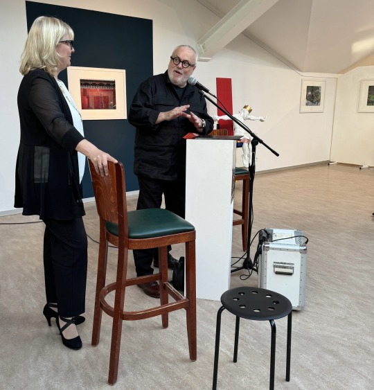
Obwohl Manuela Saß, die Bürgermeisterin der Stadt Werder (Havel), in einem anderen Termin gebunden war, kam sie kurz mit einer Ansprache in die Ausstellung.
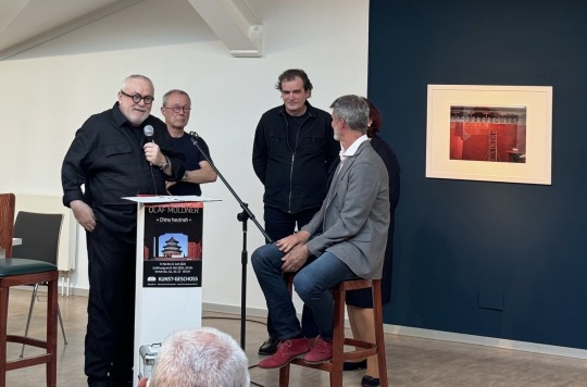
Foto oben: Frank W. Weber stellt die Akteure des Abends vor. Olaf Möldner (sitzend) beantwortete Fragen des Kurators, die allen Besuchern mehr Informationen zur Fotoserie „China hautnah“ gaben.
Am Abend wurden zwei Schenkungen an den Fundus der Stadtgalerie übergeben. Der russische und in Berlin lebende Kunsthandwerker Andrei Zaplatynski (links von Olaf Möldner) und der Leipziger Bildhauer Roland Wetzel (neben dem Kurator) übergaben je ein Werk dem Fundus.
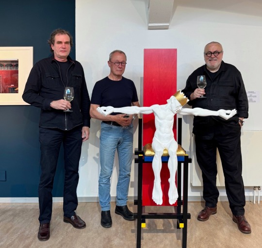
Roland Wetzel (Mitte) hatte 2022 eine Ausstellung in der Stadtgalerie und überließ nun seine Plastik des sitzenden „Nebukadnezar“ dem Fundus. Zwischenzeitlich wurden davon zwei Bronzen gegossen und das Original im Gipsaufbau ging jetzt an die Stadtgalerie. Der Thron des Königs kam aus der Hand von Andrei Zaplatynski. (links) Eine Adaption auf den bekannten Gerrit Rietveld Stuhl von 1916. Der Kunsthandwerker hatte den Stuhl der Stadtgalerie angeboten und geschenkt. Das goldene Sitzkissen des Königs Nebukadnezar passt wie in einem Teamwork abgesprochen exakt auf die Sitzfläche. Der Kurator dazu: „Nebukadnezar, als König umstritten, wird so mit einem modernen Thron in die Jetztzeit transportiert. Dieses neue Gesamtkunstwerk bietet somit neue Interpredationsmöglichkeiten aus der Geschichte auf das Heute. Nebukadnezar hat nun alle Zeit der Welt und darf, wie Pontius Pilatus in „Der Meister und Margarita“ von Michael Bulgakow, über seine Missetaten auf dem Thron sitzend nachdenken.“
Roland Wetzel dazu: „Nebukadnezar verkörpert das Prinzip des Scheiterns als Endpunkt einer Selbstüberschätzung.“
Andrei Zaplatynski redete auf Russisch, was von seiner Frau Elena übersetzt wurde.: „Es ist eine Ehre für meinen Stuhl, jetzt als Thron in der Kunstsammlung der Stadtgalerie zu dienen.“
Obwohl nicht zur Ausstellung „China hautnah“ gehörend wird diese Schenkung an den Fundus der Stadtgalerie während der gesamten Ausstellungszeit bis 23. Juni zu sehen sein.
Die Ausstellung von Olaf Mödner mit 45 ausbelichteten Fotografien und 350 Fotos in einer Bilderschau auf einem Monitor ist eröffnet.
Die Bilderschau läuft als Endlosschleife mit 36 Minuten Dauer während der Ausstellung.
Wie immer Donnerstag, Samstag und Sonntag von 13-18 Uhr, bei freiem Eintritt zu besichtigen.
Am Himmelfahrtstag geöffnet, Pfingssamstag und -sonntag geöffnet. Nicht am Feiertag Pfingstmontag!
Die Fotografien sind als Abzug 40x65 cm für 150€/Stück zu erwerben. Olaf Möldner stiftet diese Einnahmen dem Kindergarten im Hohen Weg für Arbeitsmaterialien.
Fotos: ANDRIOTTA
#kunst geschoss#stadtgalerie#werder havel#fotografie#kunstgalerie#frank w weber#roland wetzel#andrei zaplatynski#olaf moeldner#nebukadnezar#plastik#china#china hautnah#fundus#kunstsammlung
0 notes
Text
Museum: Neue Räume fürs S-Bahn-Museum, Vom Toilettenhäuschen in den Ostbahnhof: Mit einem Weihnachts-Bahn-Souvenirmarkt wird die Eröffnung gefeiert!, aus S-Bahn
06.12.2023
https://sbahn.berlin/was-hast-du-vor/neues-entdecken/artikel/raeume-fuers-s-bahn-museum-im-ostbahnhof/
Die spannende Geschichte des S-Bahnsystems sowie der Stadt-, Kultur- und #Industriegeschichte gilt es, 2024 zu würdigen. Dafür bereiten das Berliner #S-Bahn-Museum und der Deutsche #Bahnkunden-Verband (#DBV) die Eröffnung ihres neuen Ausstellungs- und Projektraumes im Berliner…
View On WordPress
#485#Abschiedstag#Bahnkunden#DBV#Fundus#grüne#Ideenaustausch#Industriegeschichte#Lichtenberg#Nahverkehr#Ostbahnhof#Retrospektive#Schöneweide#Wechselausstellungen#Weihnachts
0 notes
Text
Las villas romanas

View On WordPress
0 notes
Text
Needle Tract Recurrence of Renal Cell Carcinoma after Microwave Ablation by Tella David in Journal of Clinical and Medical Images, Case Reports

Abstract
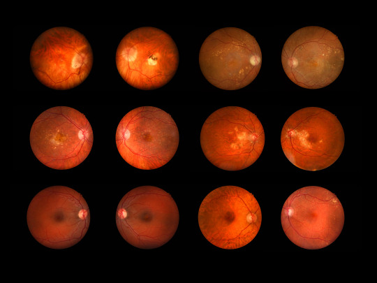
A 21 years-old male was complaining from; decrease in vision, fever, blurring and inability to open the eyes in the sun. The patient attended the Eye hospital in Gaza where Fundus photo and serological tests were carried out in the eye hospital and Islamic University-Gaza laboratories. The patient complains from blurring and retinochoroditis. The ophthalmological examination revealed hypertensive non-granulomatous panuveitis, retinal vasculitis with focus of retinochoroiditis with lesioned central area suggestive of ocular toxoplasmosis. The serological tests proved that IgG was high in the patient serum and recorded 54.50 while IgM was negative. The fundus photo showed a clear lesion. Ocular toxoplasmosis is present among patients attending Eye hospital. The clinical cases could be detected among patients complaining from retinochoroditis. Ocular toxoplasmosis should be considered, and more investigations are needed.
Keywords: Ocular; toxoplasmosis; fundus; eye; Gaza; infection.
Introduction
Thermal ablation is commonly used for management of small renal masses. Tumor seeding due to needle manipulation during renal mass biopsy or thermal ablative treatment is very rare. Seeding upstages a curable, organ-confined disease into a complicated recurrent cancer. This condition is described in few case reports and has not been reported after microwave ablation [3]. We report a patient that underwent transhepatic microwave ablation of a right renal cancer and developed a tumor along the ablation tract.
Case report
A 75-year-old male with complex medical history including heart disease, hypertension, chronic kidney disease, and sleep apnea presented to our clinic with a 4 cm right renal mass discovered incidentally following CT imaging for unrelated abdominal pain (Figure 1). A biopsy of the mass revealed a Fuhrman grade 2 clear cell renal cell carcinoma (RCC). Treatment options were discussed and the patient elected microwave ablation therapy. This procedure was performed by a urology and radiology collaborative team.

Figure 1: Preoperative CT showing right upper pole kidney tumor (thin arrow) and adjacent cyst (thick arrow).
Three PR -15 (NeuWave Medical, Madison, WI) microwave probes were inserted transhepatically into the upper pole renal tumor. Correct positioning of the probes within the mass was confirmed with ultrasound and abdominal CT scan (Figure 2). Immediate post procedure films showed complete ablation of the tumor and the adjacent cyst. Significantly, the entire trans-hepatic tract was ablated during removal of the microwave probes, as is our protocol.

Figure 2: Transhepatically placed microwave probes traversing the posterior aspect of the liver (arrow) with tips located in the right renal mass.
Six months later, the patient underwent a surveillance CT scan which demonstrated no tumor recurrence (Figure 3). MRI imaging at year 2 demonstrated stable post-treatment changes of right upper pole kidney ablation, without evidence of tumor recurrence or metastasis.
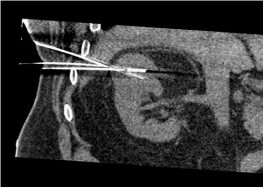
Figure 3: Coronal oblique CT reconstruction showing path of probes through liver edge.

Figure 4: US during ablation showing probe tract through liver edge.
Five years following ablation, the patient presented with gross hematuria and right flank pain. A CT urogram demonstrated a filling defect in the proximal right ureter. Additionally, there was an ill-defined hypo-enhancing lesion at the posterior right hepatic lobe with an adjacent small perihepatic soft tissue nodule, not present on prior studies (Figure 4). The patient underwent right ureteroscopy. Upon entry into the bladder, blood was effluxing from the right ureteral orifice. Ureteroscopy revealed old clot in renal pelvis, but no tumors or active bleeding. Urine cytology was negative for malignancy.
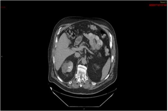
Figure 5: Six month post ablation CT showing non-enhancing right upper pole mass. Liver appears normal.
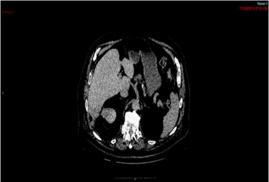
Figure 6: Five year post ablation image showing liver mass (hollow arrow), perihepatic mass (thin arrow), and enhancing upper pole renal mass (arrowhead).
The patient was referred for ultrasound guided percutaneous biopsy of the liver mass. The biopsy revealed metastatic carcinoma, favored to be renal cell carcinoma. Metastatic workup with a chest CT demonstrated scattered (approximately 15) sub-centimeter pulmonary nodules suspicious for metastatic disease. A review of the CT showed 2 cm recurrence of the right upper pole renal mass. The patient has been evaluated by medical oncology and is currently on an active surveillance protocol for his asymptomatic metastatic disease.
Discussion
Percutaneous thermal ablation has been described to be effective in the management of appropriately selected cases of RCC. Few reports of metastasis in the ablation tract after treatment have been published [1, 2]. Metastasis following ablation is suspected to be caused by tumor spillage during cryoablation or during withdrawal of the probe at the end of ablation [3]. Accumulated anecdotal cases note that transhepatic access to the upper pole of the kidney can be performed safely in the rare cases where no other access is possible [4, 5].
Some reports suggest transhepatic radiofrequency ablation can be an advantageous approach for superior or anterior renal masses, especially in patients that are unable to undergo ablation in the prone position [4, 5]. The main disadvantage identified is the violation of the peritoneum, which portends increased risk of seeding. It is estimated that such seeding is a very rare, occurring in <0.01% of cases. These reports discuss tract ablation to mitigate this risk; however, the benefit of this approach is not well defined. To our knowledge, metastasis to the liver following a transhepatic approach has not been previously described after percutaneous microwave ablation of RCC. In the case of our patient, this was an anterior, superior mass. However, this recurrence occurred despite our careful ablation of the liver tract at the end of the procedure.
Conclusions
Seeding metastasis is an unfortunate and rare event, which can occur after percutaneous ablation of a renal mass. We caution all clinicians to avoid transhepatic access for percutaneous ablation of a renal mass, when feasible, to avoid possible hepatic seeding metastasis.
For more details : https://jcmimagescasereports.org/author-guidelines/
#Ocular#toxoplasmosis#fundus#eye#Gaza#infection#hypertensive#panuveitis#retinochoroditis#renal cell carcinoma#hypo-enhancing#Tella David#JCMICR
0 notes
Text

Investing in real estate is a fantastic way to make money, but it can be a bit daunting if you don't have the funds to get started. Luckily, there are a number of ways to get the money you need to invest in real estate. You can look for Funding sources like personal loans or credit cards, or find Real Estate Investment Funds (REIFs). You may also be able to get grants or low-interest loan products from government programs like FHA or USDA. In any case, getting the money you need is essential if you want to start investing in real estate and see big returns. To know more about visit us !
Website:-https://hardmoneyuniversity.com/
Phone:-+1 (800) 622-8209
0 notes
Text
Fundus Examination
People with type 1 and type 2 diabetes are at a heightened risk for eye complications and peripheral neuropathy.
You may have heard that diabetes causes eye problems and may lead to blindness. People with diabetes do have a higher risk of blindness than people without diabetes. But most people who have diabetes have nothing more than minor eye disorders over time.
With regular checkups, you can keep minor problems minor. And, if you do develop a major problem, some treatments often work well if you begin them right away.
1 note
·
View note
Photo

#2011 #fundus #fundstück (hier: Hauptplatz Pfaffenhofen) https://www.instagram.com/p/ChkOEheKnwL/?igshid=NGJjMDIxMWI=
0 notes
Photo

Customer: I'M A LOAN OFFICER AND FUND LOANS; "FUND U" MEANING, I WILL FUND YOUR LOAN.
DMV: YOU, HOSTILE
Verdict: DENIED
#California license plate with text FUNDU#bot#ca-dmv-bot#california#dmv#funny#government#lol#public records
512 notes
·
View notes
Text
das größte verbrechen im tatort saarbrücken ist dass sie leo ständig in weiß und grau stecken, auch wenn ich die gründe natürlich verstehe..... aber kein wunder dass so ein grünes t-shirt gleich alle in aufruhr versetzt. dem mann stehen kräftige farben einfach 100 mal besser. fotobeweise unterm cut
ich mein hallo?? wir erinnern uns:

und das hier?

selbst "langweiliges" dunkelblau oder schwarz


kein weiß oder grau mehr!!!
#danke tess für deinen ausgiebigen fundus an burlakov-pics 👌#vladimir burlakov#spatort#tatort blogging#txt
13 notes
·
View notes
Text
Admittedly, I do like fanfictions and headcanons where Oz is part of the baking club.
Why do I mention this? Because once I thought about it again, I began to wonder, what if Spooky High also had a school festival like in most school manga?
And what if the baking club decided to have a maid and butler cafe for the school festival? But as a cross-dressing variant.

So, whether he liked it or not, Oz had to face his duty.
Vicky picked out the dress for him and stuffed it a little in the chest area.
Amira (despite not being a club member) took care of Oz's hair.
And yes it is Oz's real hair. Since I go along with the theory that Oz can shape his body like he wants, I think it was relatively easy for him to just let his hair grow longer.;)
This is more or less the oldest sketch I have of this idea. The outfit hasn't changed to much in the newer ones. Except for the placement of the bow ribbon.
Maybe let me know if you would like to see more of these?
At least I have some newer sketches lying around in my folder… (Some of them including Amira, Brian and Damien. But mostly just Oz alone.)
#monster prom#monster prom oz#oz yellow#cross dressing#traditional drawing#sketches#my art#do not reupload#Vicky is also a member#She was wearing a butler outfit that day#The costumes come from the theater fundus
15 notes
·
View notes
Note
Hey Abby how’s that reunion going?

Your tail is wagging...?!
"I can still hardly believe she's been alive all this time."
1 note
·
View note
Text
i really miss being an optometrist tech lmao
#i had my own little room and i loved all my machines 💔#the doctor i worked for is gonna open his own practice someday and he wants me as his office manager and head tech#someday i’ll take fundus photos again. until then my beloved Topcon 3D Oct-1 Maestro 2. until then…. ❤️
3 notes
·
View notes
Text
Eye Fundus Camera: Revolutionizing Ophthalmic Imaging
Introduction to Eye Fundus Camera
Eye fundus cameras, also known as retinal cameras, are specialized devices used to capture images of the back of the eye, including the retina, optic disc, macula, and blood vessels. These images, referred to as fundus photographs, play a crucial role in diagnosing and monitoring various eye conditions, such as diabetic retinopathy, glaucoma, macular degeneration, and hypertensive retinopathy.
Importance of Eye Fundus Examination
A comprehensive eye examination typically involves the evaluation of the eye's posterior segment, which includes the retina and optic nerve head. Fundus examination allows ophthalmologists and optometrists to assess the health of these structures, detect abnormalities, and monitor disease progression over time.
Types of Eye Fundus Cameras
Traditional Fundus Cameras
Traditional fundus cameras are stationary devices typically found in ophthalmology clinics and hospitals. They offer high-resolution imaging capabilities and are suitable for detailed retinal examinations.
Handheld Fundus Cameras
Handheld fundus cameras provide flexibility and portability, allowing eye care professionals to capture fundus images outside of traditional clinical settings. These devices are particularly useful for screening programs and telemedicine applications.
Smartphone Fundus Cameras
Smartphone fundus cameras leverage the power of mobile technology to transform smartphones into cost-effective fundus imaging devices. With the use of specialized adapters or attachments, these cameras enable primary care providers and even patients themselves to capture fundus images conveniently.
How Eye Fundus Cameras Work
Fundus cameras utilize various imaging techniques, such as digital photography, scanning laser ophthalmoscopy, and optical coherence tomography, to capture detailed images of the eye's posterior segment. These images are then analyzed by eye care professionals to detect abnormalities and guide treatment decisions.
Benefits of Using Eye Fundus Cameras
Early detection of eye diseases
Monitoring disease progression
Guiding treatment decisions
Facilitating patient education
Enhancing collaboration among healthcare providers
Applications in Ophthalmology
Eye fundus cameras are indispensable tools in ophthalmic practice, with applications including:
Diabetic retinopathy screening
Glaucoma management
Age-related macular degeneration evaluation
Retinopathy of prematurity assessment
Optic nerve head analysis
Challenges and Limitations
Despite their numerous benefits, eye fundus cameras face certain challenges, such as:
Cost of equipment and maintenance
Training requirements for operators
Limited access in underserved areas
Image interpretation variability
Future Trends in Eye Fundus Imaging Technology
Advancements in imaging technology, such as artificial intelligence and telemedicine, are poised to revolutionize eye fundus imaging by improving accuracy, efficiency, and accessibility.
Comparison with Other Imaging Techniques
Compared to other imaging modalities like optical coherence tomography and fluorescein angiography, fundus photography offers a non-invasive and cost-effective approach to visualizing the retina and optic nerve.
Considerations for Purchasing an Eye Fundus Camera
When selecting an eye fundus camera, factors to consider include image quality, ease of use, compatibility with existing systems, technical support, and cost-effectiveness.
Tips for Efficient Use of Eye Fundus Cameras
Ensure proper patient preparation and positioning
Adjust camera settings for optimal image quality
Use appropriate imaging techniques for different eye conditions
Regularly calibrate and maintain equipment
Training and Certification for Eye Fundus Imaging
Eye care professionals should undergo specialized training and certification programs to acquire the necessary skills for operating and interpreting fundus images accurately.
Costs Associated with Eye Fundus Imaging
The initial investment in purchasing an eye fundus camera may vary depending on the type and features of the device. Additional costs include maintenance, accessories, and ongoing training.
Case Studies and Success Stories
Numerous studies have demonstrated the clinical utility and cost-effectiveness of eye fundus cameras in various healthcare settings, highlighting their role in improving patient outcomes and reducing healthcare costs.
Conclusion
Eye fundus cameras have transformed the landscape of ophthalmic imaging, enabling early detection, accurate diagnosis, and personalized management of eye diseases. As technology continues to evolve, these devices will play an increasingly vital role in preserving vision and enhancing patient care.
FAQs (Frequently Asked Questions)
Are eye fundus cameras safe for all patients?
Yes, fundus cameras are non-invasive and safe for most patients, including children and pregnant women.
Can fundus cameras detect all eye diseases?
Fundus cameras can detect a wide range of eye conditions, but they may not capture certain abnormalities that require specialized imaging techniques.
Is dilating the pupils necessary for fundus photography?
Dilating the pupils may be necessary to obtain clear fundus images, especially in cases where a detailed examination is required.
Are smartphone fundus cameras as accurate as traditional devices?
While smartphone fundus cameras offer convenience and accessibility, they may not provide the same level of image quality and diagnostic accuracy as traditional devices in all cases.
How often should fundus examinations be performed?
The frequency of fundus examinations depends on various factors, including age, medical history, and risk factors for eye diseases. It is best to consult with an eye care professional for personalized recommendations.
#eyecare#fundus examination#health#medical care#eye health#eyes#choroida#choroid#medical devices#medical equipment
0 notes
Text
Fluorescein Fundus Camera Market Size, Segments, Growth and Trends by Forecast to 2031
According to a new report published by The Insight Partners, titled, ” Fluorescein Fundus Camera Market Forecast | Share and Size – 2030″. The report provides a detailed analysis of the top investment pockets, top winning strategies, drivers & opportunities, Fluorescein Fundus Camera market size & estimations, competitive landscape, and changing market trends.
The Fluorescein Fundus Camera…
View On WordPress
0 notes
Text
Fundus Examination | Samyak Eye Care Clinic

Fundus Examination
People with type 1 and type 2 diabetes are at a heightened risk for eye complications and peripheral neuropathy.
You may have heard that diabetes causes eye problems and may lead to blindness. People with diabetes do have a higher risk of blindness than people without diabetes. But most people who have diabetes have nothing more than minor eye disorders over time.
With regular checkups, you can keep minor problems minor. And, if you do develop a major problem, some treatments often work well if you begin them right away.
How the eye works
Understanding what happens in eye disorders, helps to understand how the eye works. The eye is covered with a tough outer membrane. The covering in front is clear and curved. This curved area is the cornea, which focuses light while protecting the eye.
After light passes through the cornea, it travels through a space called the anterior chamber (which is filled with a protective fluid called the aqueous humor), through the pupil (which is a hole in the iris, the colored part of the eye), and then through a lens that performs more focusing. Finally, light passes through another fluid-filled chamber in the center of the eye (the vitreous) and strikes the back of the eye, the retina.
The retina records the images focused on it and converts those images into electrical signals, which the brain receives and decodes.
One part of the retina is specialized for seeing fine detail. This tiny area of extra-sharp vision is called the macula. Blood vessels in and behind the retina nourish the macula.
Glaucoma
People with diabetes are more likely to suffer from glaucoma than people without diabetes. The longer someone has had diabetes, the more common glaucoma is. Risk also increases with age.
Glaucoma occurs when pressure builds up in the eye. The pressure pinches the blood vessels that carry blood to the retina and optic nerve. Vision is gradually lost because the retina and nerves are damaged.
There are several treatments for glaucoma. Some use drugs to reduce pressure in the eye, while others involve surgery.
Cataract
Many people without diabetes get cataracts, but people with diabetes are more likely to develop this eye condition. People with diabetes also tend to get cataracts at a younger age and have them progress faster. With cataracts, the eye’s clear lens clouds, blocking sight.
To help deal with mild cataracts, you may need to wear sunglasses more often and use glare-control lenses in your glasses. For cataracts that interfere greatly with vision, doctors usually remove the lens of the eye and replace it with a new artificial lens. In people with diabetes, retinopathy can get worse after the removal of the lens, and glaucoma may start to develop.
Retinopathy
Diabetic retinopathy is a general term for all disorders of the retina caused by diabetes. There are two major types of retinopathy: nonproliferative and proliferative.
Nonproliferative retinopathy
In nonproliferative retinopathy, the most common form of retinopathy is capillaries in the back of the eye balloon and forming pouches. Nonproliferative retinopathy can move through three stages (mild, moderate, and severe), as more and more blood vessels become blocked.
Macular edema
Although retinopathy does not usually cause vision loss at this stage, the capillary walls may lose their ability to control the passage of substances between the blood and the retina. Fluid can leak into the part of the eye where focusing occurs, the macula. When the macula swells with fluid, a condition called macula edema, vision blurs and can be lost entirely. Although nonproliferative retinopathy usually does not require treatment, macular edema must be treated, but fortunately, treatment is usually effective at stopping and sometimes reversing vision loss.
Proliferative retinopathy
In some people, retinopathy progresses after several years to a more serious form called proliferative retinopathy. In this form, the blood vessels are so damaged they close off. In response, new blood vessels start growing in the retina. These new vessels are weak and can leak blood, blocking vision. The new blood vessels can also cause scar tissue to grow. After the scar tissue shrinks, it can distort the retina or pull it out of place, a condition called retinal detachment.
How is retinopathy treated?
Huge strides have been made in the treatment of diabetic retinopathy. Treatments such as scatter photocoagulation, focal photocoagulation, and vitrectomy prevent blindness in most people. The sooner retinopathy is diagnosed, the more likely these treatments will be successful. The best results occur when sight is still normal.
In photocoagulation, the eye care professional makes tiny burns on the retina with a special laser. These burns seal the blood vessels and stop them from growing and leaking.
In scatter photocoagulation (also called panretinal photocoagulation), the eye care professional makes hundreds of burns in a polka-dot pattern on two or more occasions. Scatter photocoagulation reduces the risk of blindness from vitreous hemorrhage or detachment of the retina, but it only works before bleeding or detachment has progressed very far. This treatment is also used for some kinds of glaucoma.
Side effects of scatter photocoagulation are usually minor. They include several days of blurred vision after each treatment and possible loss of side (peripheral) vision.
In focal photocoagulation, the eye cares professional aims the laser precisely at leaking blood vessels in the macula. This procedure does not cure blurry vision caused by macular edema, but it does keep it from getting worse.
When the retina has already detached or a lot of blood has leaked into the eye, photocoagulation is no longer useful. The next option is vitrectomy, which is surgery to remove scar tissue and cloudy fluid from inside the eye. The earlier the operation occurs, the more likely it is to be successful. When the goal of the operation is to remove blood from the eye, it usually works. Reattaching a retina to the eye is much harder and works in only about half the cases.
There are two types of treatment for macular edema: focal laser therapy that slows the leakage of fluid, and medications that can be injected into the eye that slow the growth of new blood vessels and reduce the leakage of fluid into the macula.
A newer retinopathy treatment involves injecting medication directly into the eye. The injection contains a drug that blocks the activity of vascular endothelial growth factor (VEGF). This hormone promotes the growth of new blood vessels and plays a key role in retinopathy by promoting the growth of weak, leaky blood vessels. Anti-VEGF drugs put a stop to problem vessels, improving vision in people with retinopathy. In many cases, these treatments have to be repeated every few months (sometimes every month) to decrease the inflammation in the eye.
There are also some other new treatments with substances that are put into the back of the eye to help it heal. All of these advances in eye care have made a big difference in helping people’s eyes. Prevention is always first, but if damage happens, it can be treated.
Am I at risk for retinopathy?
Several factors influence whether you get retinopathy:
blood sugar control
blood pressure levels
how long you have had diabetes
genes
The longer you’ve had diabetes, the more likely you are to have retinopathy. Almost everyone with type 1 diabetes will eventually have nonproliferative retinopathy. And most people with type 2 diabetes will also get it. But the retinopathy that destroys vision, proliferative retinopathy, is far less common.
People who keep their blood sugar levels closer to normal are less likely to have retinopathy or to have milder forms.
Your retina can be badly damaged before you notice any vision change. Most people with nonproliferative retinopathy have no symptoms. Even with proliferative retinopathy, the more dangerous form, people sometimes have no symptoms until it is too late to treat them. For this reason, you should have your eyes examined regularly by an eye care professional.
Visit the Focus on Diabetes site for more information regarding eye health and diabetes.
high blood pressure and other disorders, because many diseases can have an impact on vision and eye health
1 note
·
View note
Last Seen Blogs

karenfordonte
Caring For Donte

kelvinorange
Kelvin Chan

extraterraangel
i feel fine!

championofapocrypha
Ashes To Ashes, Yams To Yams

shagsvintage
"What a Day For A Day Dream"