#choroid
Text
Eye Fundus Camera: Revolutionizing Ophthalmic Imaging
Introduction to Eye Fundus Camera
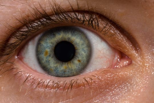
Eye fundus cameras, also known as retinal cameras, are specialized devices used to capture images of the back of the eye, including the retina, optic disc, macula, and blood vessels. These images, referred to as fundus photographs, play a crucial role in diagnosing and monitoring various eye conditions, such as diabetic retinopathy, glaucoma, macular degeneration, and hypertensive retinopathy.
Importance of Eye Fundus Examination
A comprehensive eye examination typically involves the evaluation of the eye's posterior segment, which includes the retina and optic nerve head. Fundus examination allows ophthalmologists and optometrists to assess the health of these structures, detect abnormalities, and monitor disease progression over time.
Types of Eye Fundus Cameras
Traditional Fundus Cameras
Traditional fundus cameras are stationary devices typically found in ophthalmology clinics and hospitals. They offer high-resolution imaging capabilities and are suitable for detailed retinal examinations.
Handheld Fundus Cameras
Handheld fundus cameras provide flexibility and portability, allowing eye care professionals to capture fundus images outside of traditional clinical settings. These devices are particularly useful for screening programs and telemedicine applications.
Smartphone Fundus Cameras
Smartphone fundus cameras leverage the power of mobile technology to transform smartphones into cost-effective fundus imaging devices. With the use of specialized adapters or attachments, these cameras enable primary care providers and even patients themselves to capture fundus images conveniently.
How Eye Fundus Cameras Work
Fundus cameras utilize various imaging techniques, such as digital photography, scanning laser ophthalmoscopy, and optical coherence tomography, to capture detailed images of the eye's posterior segment. These images are then analyzed by eye care professionals to detect abnormalities and guide treatment decisions.
Benefits of Using Eye Fundus Cameras
Early detection of eye diseases
Monitoring disease progression
Guiding treatment decisions
Facilitating patient education
Enhancing collaboration among healthcare providers
Applications in Ophthalmology
Eye fundus cameras are indispensable tools in ophthalmic practice, with applications including:
Diabetic retinopathy screening
Glaucoma management
Age-related macular degeneration evaluation
Retinopathy of prematurity assessment
Optic nerve head analysis
Challenges and Limitations
Despite their numerous benefits, eye fundus cameras face certain challenges, such as:
Cost of equipment and maintenance
Training requirements for operators
Limited access in underserved areas
Image interpretation variability
Future Trends in Eye Fundus Imaging Technology
Advancements in imaging technology, such as artificial intelligence and telemedicine, are poised to revolutionize eye fundus imaging by improving accuracy, efficiency, and accessibility.
Comparison with Other Imaging Techniques
Compared to other imaging modalities like optical coherence tomography and fluorescein angiography, fundus photography offers a non-invasive and cost-effective approach to visualizing the retina and optic nerve.
Considerations for Purchasing an Eye Fundus Camera
When selecting an eye fundus camera, factors to consider include image quality, ease of use, compatibility with existing systems, technical support, and cost-effectiveness.
Tips for Efficient Use of Eye Fundus Cameras
Ensure proper patient preparation and positioning
Adjust camera settings for optimal image quality
Use appropriate imaging techniques for different eye conditions
Regularly calibrate and maintain equipment
Training and Certification for Eye Fundus Imaging
Eye care professionals should undergo specialized training and certification programs to acquire the necessary skills for operating and interpreting fundus images accurately.
Costs Associated with Eye Fundus Imaging
The initial investment in purchasing an eye fundus camera may vary depending on the type and features of the device. Additional costs include maintenance, accessories, and ongoing training.
Case Studies and Success Stories
Numerous studies have demonstrated the clinical utility and cost-effectiveness of eye fundus cameras in various healthcare settings, highlighting their role in improving patient outcomes and reducing healthcare costs.
Conclusion
Eye fundus cameras have transformed the landscape of ophthalmic imaging, enabling early detection, accurate diagnosis, and personalized management of eye diseases. As technology continues to evolve, these devices will play an increasingly vital role in preserving vision and enhancing patient care.
FAQs (Frequently Asked Questions)
Are eye fundus cameras safe for all patients?
Yes, fundus cameras are non-invasive and safe for most patients, including children and pregnant women.
Can fundus cameras detect all eye diseases?
Fundus cameras can detect a wide range of eye conditions, but they may not capture certain abnormalities that require specialized imaging techniques.
Is dilating the pupils necessary for fundus photography?
Dilating the pupils may be necessary to obtain clear fundus images, especially in cases where a detailed examination is required.
Are smartphone fundus cameras as accurate as traditional devices?
While smartphone fundus cameras offer convenience and accessibility, they may not provide the same level of image quality and diagnostic accuracy as traditional devices in all cases.
How often should fundus examinations be performed?
The frequency of fundus examinations depends on various factors, including age, medical history, and risk factors for eye diseases. It is best to consult with an eye care professional for personalized recommendations.
#eyecare#fundus examination#health#medical care#eye health#eyes#choroida#choroid#medical devices#medical equipment
0 notes
Text
Anatomy of Eye | नेत्र रचना शारीर : An overview
Anatomy of Eye | नेत्र रचना शारीर : An overview
Eye, the organ of sight is almost spherical in shape with a diameter of about 2.5 cm. In order to understand the diseases and conditions occurring in one’s eye, one must know the Anatomy of eye.
नेत्र शारीर:-
विद्याद् द्व्यङ्गुलबाहुल्यं स्वाङ्गुष्ठोदरसम्मितम् ।द्व्यङ्गुलं सर्वतः सार्द्धं भिषङ्नयनबुदबुदम् । सुवृत्तं गोस्तनाकारं सर्वभूतगुणोद्भवम् ॥ (सु.उ. 1/10-11)
नयनबुद्बुद (अक्षिगोलक को अपने…
View On WordPress
#Anatomy of Eye#Anatomy of Eye in Ayurveda#Anatomy of Eye in hindi#Anatomy of Eye shalakya Tantra#Anatomy of Eye vaidyanamah#Anatomy of eyes#Anatomy of human eye#Aqueous Humour#Arteries of eye#Chambers of eye#Choroid#Ciliary Body#Conjunctiva#Eye anatomy#Eyeball anatomy#Fovea centralis#Human eye anatomy#Inner coat of eyeball#Iris#Layers of eye#Lens of eye#Lymphatic drainage of eye#Lymphatic system of eye#Macula lutea#Mandala#Middle coat of eyeball#Modern anatomy of Eye#Muscles of eye#Netra dhamni#Netra drushti vichara
0 notes
Text
#Choroidal Neovascularization Market#Choroidal Neovascularization Market Share#Choroidal Neovascularization Market Forecast#Choroidal Neovascularization Market Report#Choroidal Neovascularization Market Growth
0 notes
Text
Dayanita
Gostosa do instagram rebolando muito
Infiel de ecatepec se llama Diana me contacto por medio de xvideos me la cojo cuando el marido sale a trabajar
Ava Devine DP amazing tits and ass
blonde hot babe blowjob
Horny Lesbian Teens
Teen fingering creamy pussy Cristi Ann may be a tiny too cute
Pink Haired Busty Dreamgirl Kiera King Cums Hard on a Big Cock
Sizzling Couple Enjoying all Sex Positions
mirian le gusta la leche
#obligation's#oilway#hobi#choroids#USGA#flouse#semicubit#aristeia#glost#rewards#isochlorophyllin#eschoppe#bullneck#dreamteam#laminations#breachful#lecheries#semisaint#ACTPU#constringency
0 notes
Text
Choroidal Folds Symptoms, Causes, Diagnosis, Treatment
Choroidal Folds Symptoms, Causes, Diagnosis, Treatment
The thick layer of blood vessels and pigmented cells that cover the back of the eyeball, dividing it into two halves, is known as the choroid. To put it another way, it resembles the body of a wine glass because it is one of three parts of the uveal tract. There are three levels to it, and diseases can attack any one of them. It consists of three layers: Bruch’s membrane, capillary layer, and…

View On WordPress
0 notes
Text
Ventricular system
Ventricular system is the extension of the subarachnoid space into the brain, which consist series of interconnecting spaces and channels.
Ventricular system is the extension of the subarachnoid space into the brain, which consist series of interconnecting spaces and channels.
Ventricular system .pdfDownload
Four chambers are filled with cerebrospinal fluid (CSF): the paired lateral ventricles, the unpaired 3rd ventricle, and the unpaired 4th ventricle.
Like and subscribe to support
Connections between the structures occur via…

View On WordPress
#Cerebrospinal fluid#Choroid plexus#Subarachnoid & Circulation of CSF#Typography & Relations
1 note
·
View note

Text
Retinal Fundus Camera: Revolutionizing Eye Health
Imagine a device capable of peering into the depths of your eyes, revealing early signs of potentially sight-threatening diseases. Enter the retinal fundus camera, a groundbreaking tool in ophthalmology that has revolutionized the way eye health is monitored and managed. In this article, we delve into the intricacies of retinal fundus cameras, exploring their history, functionality, applications, and future prospects.
Introduction to Retinal Fundus Camera
At its core, a retinal fundus camera is a specialized imaging device used to capture detailed images of the back of the eye, including the retina, optic disc, macula, and blood vessels. These images, known as fundus photographs, provide invaluable insights into the health of the eye, aiding in the diagnosis and management of various ocular conditions.
History and Evolution of Retinal Fundus Imaging
The roots of retinal fundus imaging can be traced back to the late 19th century when ophthalmologists began experimenting with rudimentary devices to visualize the posterior segment of the eye. Over the decades, advancements in optics, electronics, and imaging technology have led to the development of highly sophisticated fundus cameras capable of capturing high-resolution images with remarkable clarity.
Importance of Retinal Fundus Imaging in Eye Health
Early Detection of Eye Diseases
One of the primary advantages of retinal fundus imaging is its ability to detect subtle changes in the retina that may indicate the presence of various eye diseases, such as diabetic retinopathy, age-related macular degeneration (AMD), and glaucoma, at their earliest stages.
Monitoring Disease Progression
Retinal fundus cameras play a crucial role in monitoring the progression of ocular conditions over time, allowing healthcare providers to assess the effectiveness of treatments and intervene promptly if necessary.
Types of Retinal Fundus Cameras
Traditional Fundus Cameras
Traditional fundus cameras are stationary devices typically found in ophthalmology clinics and hospitals. They consist of complex optical systems capable of capturing high-resolution images of the retina.
Handheld Fundus Cameras
Handheld fundus cameras offer portability and versatility, making them ideal for use in remote or underserved areas where access to traditional imaging equipment may be limited.
Smartphone-Based Fundus Cameras
With the advent of smartphone technology, it is now possible to transform a regular smartphone into a portable fundus camera using specially designed attachments or accessories.
How a Retinal Fundus Camera Works
Fundus imaging involves the capture of digital photographs of the retina, which is the light-sensitive tissue lining the back of the eye. The process typically begins with the dilation of the pupil to provide a clear view of the retina. The camera then emits a flash of light, illuminating the back of the eye, while a specialized lens focuses and captures the resulting image.
Applications of Retinal Fundus Cameras
Diabetic Retinopathy Screening
Diabetic retinopathy is a leading cause of vision loss among individuals with diabetes. Retinal fundus cameras are instrumental in screening for diabetic retinopathy by detecting early signs of retinal damage, such as microaneurysms, hemorrhages, and exudates.
Age-Related Macular Degeneration Diagnosis
Age-related macular degeneration is a progressive eye disease that affects the macula, the central part of the retina responsible for sharp, central vision. Fundus imaging is crucial for diagnosing and monitoring the progression of AMD, enabling timely intervention to preserve vision.
Glaucoma Detection
Glaucoma is a group of eye conditions characterized by damage to the optic nerve, often resulting in irreversible vision loss. Retinal fundus cameras help in identifying optic nerve abnormalities and monitoring changes in the optic disc that may indicate the presence of glaucoma.
Retinopathy of Prematurity Screening
Retinopathy of prematurity is a potentially blinding condition that affects premature infants. Fundus cameras are used to screen premature babies for signs of retinopathy, allowing for early intervention to prevent vision loss.
Advantages of Retinal Fundus Imaging
Non-Invasive Procedure
Retinal fundus imaging is a non-invasive procedure that does not require direct contact with the eye, making it safe and comfortable for patients of all ages.
High-Quality Images
Modern fundus cameras are capable of capturing high-resolution images with exceptional detail, allowing for accurate diagnosis and treatment planning.
Early Disease Detection
By detecting subtle changes in the retina at an early stage, retinal fundus imaging enables healthcare providers to initiate timely interventions and prevent vision loss.
Challenges and Limitations
Despite its numerous advantages, retinal fundus imaging is not without its challenges and limitations.
Accessibility Issues
In many parts of the world, access to retinal fundus cameras may be limited, particularly in rural or underserved areas where healthcare infrastructure is lacking.
Cost
The initial cost of acquiring and maintaining retinal fundus imaging equipment can be prohibitive for smaller clinics or healthcare facilities with limited budgets.
Operator Skill Requirement
Interpreting fundus images requires specialized training and expertise, highlighting the importance of ensuring that healthcare providers receive adequate education and training in fundus photography.
Future Trends in Retinal Fundus Imaging
Artificial Intelligence Integration
Advancements in artificial intelligence are poised to revolutionize retinal fundus imaging, enabling automated analysis of fundus photographs for the early detection of eye diseases.
Miniaturization and Portability
The development of compact and portable fundus cameras is opening up new possibilities for remote screening and telemedicine, particularly in resource-limited settings.
Telemedicine Applications
Retinal fundus imaging has the potential to transform the delivery of eye care by facilitating remote consultations and telemedicine services, allowing patients to access specialized care from the comfort of their homes.
Conclusion
Retinal fundus cameras represent a paradigm shift in the field of ophthalmology, offering a non-invasive and effective means of assessing and monitoring eye health. As technology continues to advance, the role of retinal fundus imaging in preventing vision loss and preserving sight will only continue to grow.
FAQs
Are retinal fundus cameras safe?
Yes, retinal fundus imaging is a non-invasive procedure that poses minimal risk to patients.
Can retinal fundus cameras detect all eye diseases?
While retinal fundus cameras are highly effective for detecting many eye diseases, they may not be able to identify certain conditions that require specialized testing or imaging techniques.
How often should someone undergo retinal fundus imaging?
The frequency of retinal fundus imaging depends on various factors, including age, medical history, and risk factors for eye diseases. It is best to consult with an eye care professional to determine an appropriate screening schedule.
Are retinal fundus cameras only used by ophthalmologists?
While retinal fundus cameras are commonly used by ophthalmologists, they may also be utilized by optometrists and other eye care professionals trained in fundus photography.
What should I expect during a retinal fundus imaging procedure?
During a retinal fundus imaging procedure, your eyes may be dilated with eye drops to provide a clear view of the retina. You will then be asked to focus on a target while the camera captures images of the back of your eye.
#eyecare#eyes#health#medical care#medical devices#eye health#choroida#choroid#fundus examination#medical equipment
0 notes
Text
What if Antari eyes had a tapetum lucidum layer?
Eyes reflect light, but some eyes have the tapetum lucidum, an iridescent cellular or noncellular layer of the choroid, which is responsible for the metallic reflex seen at night in the eyes of many mammals and for their impressive night vision. It is not present in the human eye. And although this structure improves night vision, its presence decreases visual acuity during daylight.
And now I'm just sitting here, imagining a multitude of scenarios, if this were to be true.
The nights spent by Kell and Rhy in the castle, freaking the servants out, because Kell's eyes glow.
Lila robbing people at night, and they just think they came face to face with some demon.
The Danes having to order Holland to stop prowling around at night, because it creeps them out.
Nasi waking up next to Kosika, just to find this pair of glowing eyes staring at her.
I find this idea truly hilarious.
#adsom#ve schwab#lila bard#fragile threads of power#the fragile threads of power#holland vosijk#kell maresh#kosika#rhy maresh
38 notes
·
View notes
Text
Handy blog on eyeshine, important to understand when squatching at night…
Eyeshine in animals is produced by a special membrane, called the tapetum lucidum (tapestry of light), a reflective surface that is located directly behind the retina. When the small rays of light found in the night, like starlight or moonlight, enter the eye, they bounce off the membrane, giving the eye a second chance to use the light. For animals that have this membrane, it is like having a built in flashlight that lights a path from the inside out.
The tapetum lucidum, coupled with big eyes and lots of light-sensing rod cells, allow nocturnal mammals to see well in dark or dim conditions. But eyeshine isn’t limited to mammals. Once, while at the edge of a pond listening to the midnight chorus of frogs, my flashlight caught the glimmering, emerald-green eyes of a huge bullfrog. And in my obsession over eyeshine, I am eagerly looking forward to the summer, when I will be searching the forest floor for the ruby red glow of a wolf spider’s eyes. I only wish that my eyes would glow, a fierce sapphire blue in the darkest of night, but although humans have many interesting adaptations, good night vision is not one of them. Our abundance of cones and lack of rods mean we see more colors than most other animals, but we can’t see in the dark. And we don’t have a tapetum lucidum – when our eyes appear red in photographs, it’s a reflection of the camera’s flash off the red blood cells of the choroid, which is a vascular layer behind the retina.
Eyeshine color varies by species, from the amber glow of a bobcat to the red glint of a black bear. The different colors are produced by the mineral content and the structure of the tapetum lucidum, as well as varying pigments in the retina. There does seem to be some overlap of colors, like bobcat and raccoon having yellow/amber eyeshine.
So is it at all possible to identify an animal by eyeshine color alone? According to ecologist and long-time tracker Dr. Rick van de Poll, eyeshine is somewhat variable so that even within the same species the color can look a bit different. Factors that influence individual eyeshine color, according to van de Poll, include the age and individual chemistry of the animal, as well as seasonal variation and the angle and intensity of the light hitting the eye. But this doesn’t deter van de Poll from using eyeshine as a clue to identifying mammals. “It’s part of the information” he said, “but you have to also be paying attention to the animal’s behavior, the shape and placement of the eyes, and how the animal moves away from the light, or if it even moves away from the light at all.”
#cryptozoology#cryptids#bigfoot#bigfootmountain#bigfootsighting#bigfootisreal#bigfootsightings#ufo#lochnessmonster
35 notes
·
View notes
Text
Hey Tumblr! So, i used to share science photos all the time, and I definitely miss doing it. I figured my tumblr could be another social space for this, so here's a photo of a embryonic day 15.5 mouse brain! 🧠🐭
(Not the best focus, but if you look towards the brain stem [bottom], you can see the 4th ventricle choroid plexus!)

#science#black women#women in stem#stem#stemblr#stem academia#phd life#phdblr#phd student#mouse#brain#big brain
23 notes
·
View notes
Text
There is a huge lobby for normalization of SARS Cov 2. Entire industries depend on the public’s return to normal consumer and working behaviors. As such, the rationalizations and reassurances to the public that SARS Cov 2 is a normal seasonal Coronavirus are relentless. These are constructed like homilies and catch-phrases, such as “we must learn to live with it,” and, “it’s endemic,” with the implication of its endemicity referring to the abandonment of efforts which acknowledge its existence, such as testing.

It is a complete misconception that introduction of a virus to the immune system makes subsequent infections like a common cold, and that virulence is due to novelty. If nerves, organs, and immune systems could speak, they would tell a tale of exceptional inflammation, aging, and death, which we must turn to science to hear. Professor Fuhrer would be taken aback to find there are efforts to examine specific mechanisms which tell another tale than his own.
Here, I will give you, the reader, clear enumerations where SARS Cov 2 is unlike a common cold.
SARS Cov 2 triggers a unique, long-lived inflammatory overreaction unseen in Sepsis and influenza. https://genomemedicine.biomedcentral.com/articles/10.1186/s13073-023-01227-x
It caused cells of the immune system to react in a way to create further inflammation and activation of the immune system for an extended amount of time. For technical facets of this, please see the paper.
SARS Cov 2 sends T cells into the brain while lethal influenza does not.

SARS Cov 2 directly causes autoimmunity by reprogramming a special type of T cell called the T regulatory cell, which has never been observed before. https://www.frontiersin.org/articles/10.3389/fimmu.2020.589380/full
The human genetic line has not propagated any sarbecovirus elements therefore never has faced Sarbecovirus infection to the extent to evolutionarily adapt, except in the unlikely theoretical possibility of extremely negative selection (meaning infected humans did not create progeny.)
There are more exceptional facets but these are simple and digestible. There is also more to write about but I must make a confession. The status quo has morphed in such a way as to browbeat scientists into disavowing a harsh reality in order to acquiesce to corporate and business interests. As we see the average life expectancy decline, we have been left intellectually out in the cold. The truth tellers have been assaulted and crushed, and the individuals that comprise the public, in denial, will put off the realization of a below 70s life expectancy until each one approaches retirement in piecemeal, just as all the grains of sand in an hourglass do not fall at once.
23 notes
·
View notes
Text
Progressive Retinal Atrophy in cat.
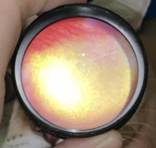
The cat was presented with gradual blindness over several months. After trying several hospitals with unsatisfactory results, they finally reached me.
I used the indirect ophthalmoscope technique with a 20D aspheric lens (no I did not buy from Volk.) and a focal light source (Incandescent ophthalmology pen light) to obtain the view at the back of the eyeball, where the retina is located. Producing an image taken as shown above.
This image clearly shows the hyper-reflection of the fundus, indicative of retinal degeneration. It also shows the blood vessels had become very faint. I was unable to get a good image of the optic disk, it is just slightly out of frame at the top right (where you can see the faint blood vessel leading to) (also image the lens is showing is upside down.)
(I could make another post about how to take a fundic photograph without a fundus camera, it's difficult and produces a somewhat poor quality image as shown above, but it's better than nothing.)
For comparison, here is the normal feline fundus
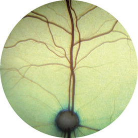
image credit : https://veteriankey.com/retina-choroid-sclera/
note the lower reflection of the tapetum (yellow-green background). The light doesn't try to blind you, because the layer of retina was in between you and the tapetum. If the retina degenerates, it become more transparent and the light reflecting shines right back at your face.
note2 blood vessels in the fundus should look thick as shown in this example, not near-non-existence like my patient above.
Unfortunately, there is no cure for Progressive Retinal Atrophy. There is no clear cause either, as we've ruled out the possible toxicity. This disease will often spring upon us without any reason. Genetics is one of the possible explanation, but I cannot prove it, as the cat was of a mixed breed.
The cat was already well adjusted to the owner's household. I gave some recommendations for blind cat environmental care, and assured them the condition was not painful, and there is no additional danger other that the cat will continue to be more blind as time goes by.
The owner was happy with the visit.
It seems, some cases are more about healing the owner's anxiety than the animal's illness. I was not able to produce any better cure than any of the previous hospital's visit, but knowing what's going on, what to expect and what to do goes a long way for a pet parent who loves their furball family member with all their hearts.
27 notes
·
View notes
Note
[ that art you rbed has me thinking. did you talk once about a hc that there's something slightly Off about john's eyes or did i dream that. because if so: BARKING WITH DELIGHT. ]
YES I ABSOLUTELY DID!! basically i headcanon that, since the demon blood received from nergal 35 years ago has been able to integrate so thoroughly with constantine's system that his blood is now classified as a unique demon-human hybrid (blows a kiss to city of demons), it's also created a few other odd physical distinctions as well!
this includes the development of a tapetum lucidum, an additional reflective layer in the choroid of the eye. it's what cats have that makes their eyes reflect light in the dark, and it lets constantine see better in the dark. he DEFINITELY doesn't know that he has it, he just knows that he's got superb vision for a 70 year old guy & that people get freaked out when he lurks in the shadows, and he has refused to investigate any further than that.
so yeah, his eyes glow in the dark! i picture it more like this (jumpscare warning for large staring cat) than the ringed heterochromia that legend & icon ratblazer draws him with, but the effect is very much the same. / @nightmarecountry
#( ooc. ) OUT OF CIGS.#thank you SO MUCH for remembering this asghjdk this is one of my FAVORITE headcanons!!!!#i like when he is Kind Of A Freak!!! it's fun!!!#the demon constantine on my multi has the same thing but it's bright-ass red bc he's got bright-ass red eyes#i love reflective eyes on creachers it is one of my favorite tropes. let those bitches have a lil cryptid to em! as a treat!#nightmarecountry#( headcanons. ) I'M JUST LIKE THE BASTARDS I'VE HATED ALL ME LIFE.#( answered. ) THIS IS JOHN CONSTANTINE. FUCK OFF.
8 notes
·
View notes
Text
the songs already beautiful especially with his family actually singing the choroid but the imagery gets me too i love the for sale sign doubling as him carrying the cross bc being able to keep land within black families thru generations is a weight that’s often burdened on the youngest or most successful within the family in black communities, him retaining land as a symbol of finally being “free” is so very important to us and reminds me of reading the house of eve and raisin in the sun growing up and going ah ok so this fear and burden is always gonna be there
8 notes
·
View notes
Last Seen Blogs

jessicawozny
Jessica Wozny

efreeiii
Erek Freeman Photography

supermace
I don't know karate, but I do know crazy.

pitchiou
e.k

pinkdodes
safe from the world though the world will try