
shalinijainsamyak
Shalini Jain
Samyak
Eye Care Clinic is one of the Best Eye Hospitals in Ghaziabad. We have the
best eye doctor for eye care treatment and surgeries. Do visit us.
5 posts
Don't wanna be here? Send us removal request.
Last Seen Blogs

nelnel62
neruko wa sodatu.

pureromanccewithkayla
PureRomance

cruz-de-la
CRUZ

homeofficesetups
Home Office Setups

csenge04-blog
Elkeseredett😭
Text
Fundus Examination
People with type 1 and type 2 diabetes are at a heightened risk for eye complications and peripheral neuropathy.
You may have heard that diabetes causes eye problems and may lead to blindness. People with diabetes do have a higher risk of blindness than people without diabetes. But most people who have diabetes have nothing more than minor eye disorders over time.
With regular checkups, you can keep minor problems minor. And, if you do develop a major problem, some treatments often work well if you begin them right away.
1 note
·
View note
Text
Fundus Examination | Samyak Eye Care Clinic
Fundus Examination
People with type 1 and type 2 diabetes are at a heightened risk for eye complications and peripheral neuropathy.
You may have heard that diabetes causes eye problems and may lead to blindness. People with diabetes do have a higher risk of blindness than people without diabetes. But most people who have diabetes have nothing more than minor eye disorders over time.
With regular checkups, you can keep minor problems minor. And, if you do develop a major problem, some treatments often work well if you begin them right away.
How the eye works
Understanding what happens in eye disorders, helps to understand how the eye works. The eye is covered with a tough outer membrane. The covering in front is clear and curved. This curved area is the cornea, which focuses light while protecting the eye.
After light passes through the cornea, it travels through a space called the anterior chamber (which is filled with a protective fluid called the aqueous humor), through the pupil (which is a hole in the iris, the colored part of the eye), and then through a lens that performs more focusing. Finally, light passes through another fluid-filled chamber in the center of the eye (the vitreous) and strikes the back of the eye, the retina.
The retina records the images focused on it and converts those images into electrical signals, which the brain receives and decodes.
One part of the retina is specialized for seeing fine detail. This tiny area of extra-sharp vision is called the macula. Blood vessels in and behind the retina nourish the macula.
Glaucoma
People with diabetes are more likely to suffer from glaucoma than people without diabetes. The longer someone has had diabetes, the more common glaucoma is. Risk also increases with age.
Glaucoma occurs when pressure builds up in the eye. The pressure pinches the blood vessels that carry blood to the retina and optic nerve. Vision is gradually lost because the retina and nerves are damaged.
There are several treatments for glaucoma. Some use drugs to reduce pressure in the eye, while others involve surgery.
Cataract
Many people without diabetes get cataracts, but people with diabetes are more likely to develop this eye condition. People with diabetes also tend to get cataracts at a younger age and have them progress faster. With cataracts, the eye’s clear lens clouds, blocking sight.
To help deal with mild cataracts, you may need to wear sunglasses more often and use glare-control lenses in your glasses. For cataracts that interfere greatly with vision, doctors usually remove the lens of the eye and replace it with a new artificial lens. In people with diabetes, retinopathy can get worse after the removal of the lens, and glaucoma may start to develop.
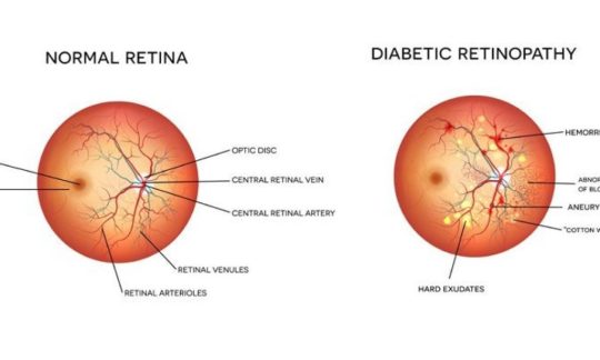
Retinopathy
Diabetic retinopathy is a general term for all disorders of the retina caused by diabetes. There are two major types of retinopathy: nonproliferative and proliferative.
Nonproliferative retinopathy
In nonproliferative retinopathy, the most common form of retinopathy is capillaries in the back of the eye balloon and forming pouches. Nonproliferative retinopathy can move through three stages (mild, moderate, and severe), as more and more blood vessels become blocked.
Macular edema
Although retinopathy does not usually cause vision loss at this stage, the capillary walls may lose their ability to control the passage of substances between the blood and the retina. Fluid can leak into the part of the eye where focusing occurs, the macula. When the macula swells with fluid, a condition called macula edema, vision blurs and can be lost entirely. Although nonproliferative retinopathy usually does not require treatment, macular edema must be treated, but fortunately, treatment is usually effective at stopping and sometimes reversing vision loss.
Proliferative retinopathy
In some people, retinopathy progresses after several years to a more serious form called proliferative retinopathy. In this form, the blood vessels are so damaged they close off. In response, new blood vessels start growing in the retina. These new vessels are weak and can leak blood, blocking vision. The new blood vessels can also cause scar tissue to grow. After the scar tissue shrinks, it can distort the retina or pull it out of place, a condition called retinal detachment.
How is retinopathy treated?
Huge strides have been made in the treatment of diabetic retinopathy. Treatments such as scatter photocoagulation, focal photocoagulation, and vitrectomy prevent blindness in most people. The sooner retinopathy is diagnosed, the more likely these treatments will be successful. The best results occur when sight is still normal.
In photocoagulation, the eye care professional makes tiny burns on the retina with a special laser. These burns seal the blood vessels and stop them from growing and leaking.
In scatter photocoagulation (also called panretinal photocoagulation), the eye care professional makes hundreds of burns in a polka-dot pattern on two or more occasions. Scatter photocoagulation reduces the risk of blindness from vitreous hemorrhage or detachment of the retina, but it only works before bleeding or detachment has progressed very far. This treatment is also used for some kinds of glaucoma.
Side effects of scatter photocoagulation are usually minor. They include several days of blurred vision after each treatment and possible loss of side (peripheral) vision.
In focal photocoagulation, the eye cares professional aims the laser precisely at leaking blood vessels in the macula. This procedure does not cure blurry vision caused by macular edema, but it does keep it from getting worse.
When the retina has already detached or a lot of blood has leaked into the eye, photocoagulation is no longer useful. The next option is vitrectomy, which is surgery to remove scar tissue and cloudy fluid from inside the eye. The earlier the operation occurs, the more likely it is to be successful. When the goal of the operation is to remove blood from the eye, it usually works. Reattaching a retina to the eye is much harder and works in only about half the cases.
There are two types of treatment for macular edema: focal laser therapy that slows the leakage of fluid, and medications that can be injected into the eye that slow the growth of new blood vessels and reduce the leakage of fluid into the macula.
A newer retinopathy treatment involves injecting medication directly into the eye. The injection contains a drug that blocks the activity of vascular endothelial growth factor (VEGF). This hormone promotes the growth of new blood vessels and plays a key role in retinopathy by promoting the growth of weak, leaky blood vessels. Anti-VEGF drugs put a stop to problem vessels, improving vision in people with retinopathy. In many cases, these treatments have to be repeated every few months (sometimes every month) to decrease the inflammation in the eye.
There are also some other new treatments with substances that are put into the back of the eye to help it heal. All of these advances in eye care have made a big difference in helping people’s eyes. Prevention is always first, but if damage happens, it can be treated.
Am I at risk for retinopathy?
Several factors influence whether you get retinopathy:
blood sugar control
blood pressure levels
how long you have had diabetes
genes
The longer you’ve had diabetes, the more likely you are to have retinopathy. Almost everyone with type 1 diabetes will eventually have nonproliferative retinopathy. And most people with type 2 diabetes will also get it. But the retinopathy that destroys vision, proliferative retinopathy, is far less common.
People who keep their blood sugar levels closer to normal are less likely to have retinopathy or to have milder forms.
Your retina can be badly damaged before you notice any vision change. Most people with nonproliferative retinopathy have no symptoms. Even with proliferative retinopathy, the more dangerous form, people sometimes have no symptoms until it is too late to treat them. For this reason, you should have your eyes examined regularly by an eye care professional.
Visit the Focus on Diabetes site for more information regarding eye health and diabetes.
high blood pressure and other disorders, because many diseases can have an impact on vision and eye health
1 note
·
View note
Text
Samyak Eye Care Clinic is one of the Best Eye Hospitals in Ghaziabad. We have the best eye doctor for eye care treatment and surgeries. Do visit us.
#best eye specialist in vaishali ghaziabad#best eye hospital in ghaziabad#vision therapy exercises in ghaziabad
1 note
·
View note
Text
#bostonsight scleral lenses#scleral contact lenses in ghaziabad#scleral lenses in vaishali ghaziabad
1 note
·
View note
Text
What are Scleral Lenses and How do they work their magic?

There are a lot of people suffering from various eye conditions, but many aren’t good candidates for using contact lenses or have difficulties wearing contact lenses. The large diameter gas permeable (GP) lenses known as scleral lenses are an effective alternative for such patients as they can provide the same benefits that conventional GP lenses can give.
Vision problems caused by Keratoconus and other cornea irregularities can be corrected by the smooth optical surface of the scleral lens. Scleral lenses are filled with isotonic fluid before fixing, so that the space between the cornea and the back surface of the lenses acts as a tear reservoir. This tear reservoir continually provides moisture and oxygen to prevent eye injuries like corneal abrasions.
Why Scleral Lenses are a Better Option?
The key advantages of scleral lenses are that it provides sharper vision, greater durability, easier handling and less risk of complications compared to standard lenses. With BostonSight Scleral lenses it is easy to custom design a lens to provide the best possible vision, eye health and comfort for each patient. Scleral lenses vault over the entire corneal surface and rest on the sclera of the eyes while the conventional GP lenses cover only a portion of the cornea. Scleral lenses are more stable on the eyes because of its larger size so there is less chances of getting accidentally dislodged from the eye. This also makes scleral lenses more comfortable especially for sensitive eyes or irregularly shaped corneas. In addition to correcting your vision, scleral lenses also help improve the health of your eyes and to reduce the need for surgical intervention for those with severe ocular surface diseases.
Types of Scleral Lenses
Scleral lenses come in three different categories, differentiated by their size running from 14.5mm to 24mm and the primary contact it has with the front surface of the eye. The larger size allows it to cover the entire cornea, extending to the sclera. During the contact lens exam, your eye specialist will decide which size of contact lens to use depending on the complexity of the vision problem.
Corneo-scleral and semi–scleral lenses
Much larger than GP lenses, Corneo-scleral and semi-scleral lenses are designed to rest near the junction between the cornea and the sclera. Patients who require contacts after laser eye surgery or corneal refractive surgery can benefit from this type of scleral lens to correct irregular astigmatism.
Mini scleral lenses
Mini scleral lenses are large diameter rigid lenses designed to bind over the entire corneal surface and rest on the anterior sclera. Patients whose corneal shape is distorted by eye conditions, scarring or after a corneal graft procedure can get their vision rehabilitated by using this type of scleral lens.
Full scleral lenses
The largest of all scleral lenses, full scleral lenses provide the most amount of clearance between the back surface of the lean and the cornea. Patients who suffer from complex eye conditions like advanced keratoconus, severe dry eyes or severe ocular disease can benefit from using the full scleral lens.
With lens materials that are highly breathable rigid gas permeable, all types of scleral lenses keep the eyes healthy and comfortable by supplying plenty of oxygen to the front surface even though the entire cornea is covered.
Who is a Good Candidate?
Scleral lenses in Vaishali Ghaziabad are a good choice for anyone looking to get the best possible vision with contact lenses. People with problems like corneal irregularities caused by either keratoconus or surgical procedure, ocular surface disease, severe refractive errors, and hard-to-fit eyes can benefit more by wearing scleral lenses. Scleral lenses can also benefit those with dry eye disease as it keeps the eyes moisturized and provides a more comfortable contact lens experience. This is possible because the ample space between the back surface of the lens and the cornea acts as a tear reservoir. Your ophthalmologist in Ghaziabad will conduct a contact lens exam to tell you which type of scleral lenses will be best for you. Scleral lenses are often fitted using special automated measuring tools and imaging devices.
Cost of Scleral Lenses
The cost associated with the fitting or replacing scleral contact lenses in Ghaziabad will vary from patient to patient depending on the nature of the lens. As scleral lenses need to be custom-made to the exact specifications prescribed by your eye specialist, their price can even go higher than that of Ortho K lenses in Vaishali Ghaziabad. You can ask your eye specialist for specific cost advice for scleral contact lenses in Vaishali Ghaziabad that meet individual needs. Some of the expenses associated with scleral lenses can be covered under your vision insurance policy.
Dr Shalini Jain’s Samyak Eye Care Clinic is a trusted name for ensuring patients the highest quality results with scleral lenses. Book an appointment with our eye specialist to know if you are a good candidate for scleral lenses and discuss how it can help improve your vision.
#scleral contact lenses in ghaziabad#scleral lenses in vaishali ghaziabad#bostonsight scleral lenses
4 notes
·
View notes