#Optic Thalamus
Text
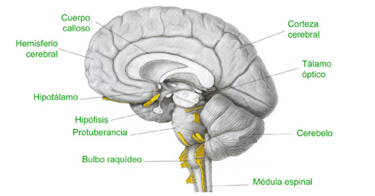
#Cerebro#Brain#Cuerpo Calloso#Hard Body#Hemisferio Cerebral#Cerebral Hemisphere#Hipotálamo#Hypothalamus#Hipófisis#Hypophysis#Protuberancia#Protuberance#Bulbo Raquídeo#Medulla Oblongata#Corteza Cerebral#Cerebral Cortex#Tálamo Óptico#Optic Thalamus#Cerebelo#Cerebellum#Médula espinal#Spinal Cord
0 notes
Text
Anatomy of the Nervous System
Structure of the Nervous System
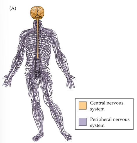
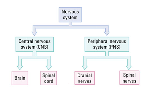
The brain is inside the skull; the spinal cord is inside the vertebral column (spine).
Somatic nervous system:
controls skeletal muscles
conscious control
Autonomic nervous system:
balances internal organs
unconscious control
Functions of the Nervous System
Control of behaviors:
Sensory function: get and integrate information about the world and create a sensory reality.
Motor function: control movements.
Plasticity: adapt to the world by changing its physical or chemical properties; functions to adapt to environmental change or to compensate for injury.
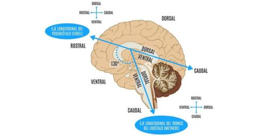
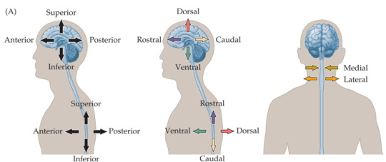
Navigating Your Nervous System
front/back = anterior/posterior
up/down = superior/inferior
belly/back = ventral/dorsal
beak/tail = rostral/caudal
along the middle = medial
left or right side = lateral
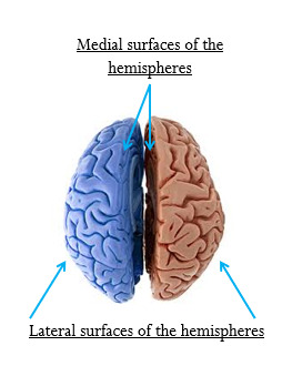
How can we visualize brain structures?
A. Dead Brain
Postmortem examination:
Brain is stored in fixative solution
Sliced with a knife to see brain structures
B. Live Brain
Optically sliced:
Computer tomography (CT)
Magnetic resonance imaging (MRI, structural or functional)
Positron emission tomography (PET)
Functional MRI and PET help to assess activity of brain regions
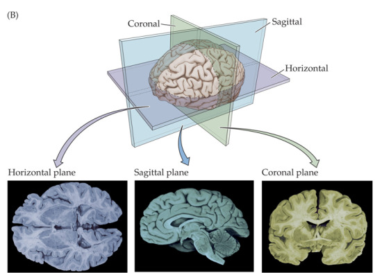
Rat Brains vs. Human Brains
Rats are commonly used as research models. Brain structures have not changed very much during mammalian evolution, so the basic brain plan is similar for rats and humans.
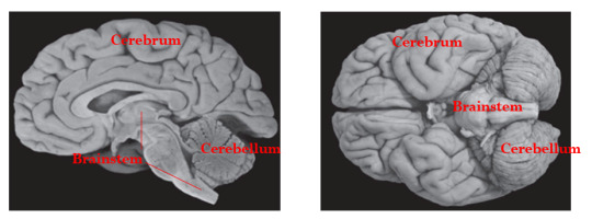
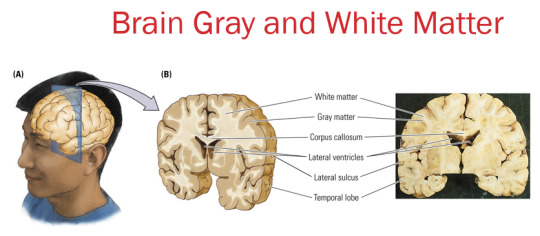
Brain Divisions (based on surface features)
A. Cerebrum: consists of two virtually identical hemispheres (left and right)
B. Cerebellum (”little brain”)
C. Brain stem
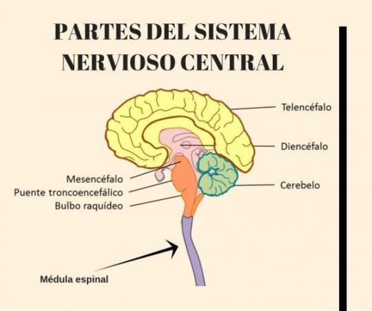
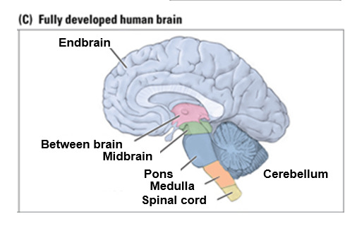
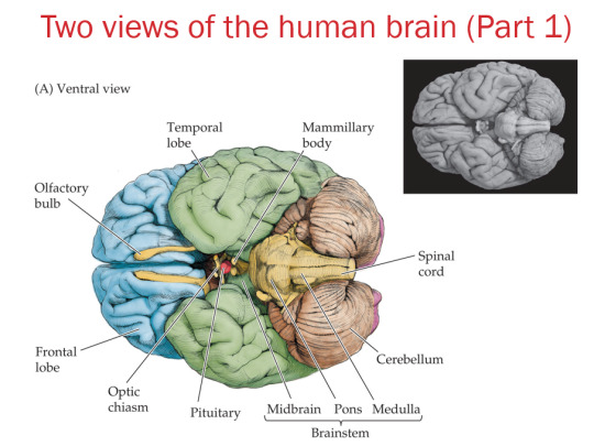
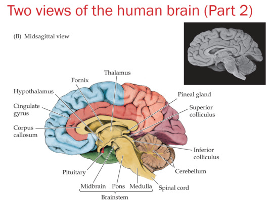
Brain divisions (based on development and evolution)
Forebrain
A. End brain
Cortex
Corpus callosum
Limbic system
Basal ganglia
Olfactory bulb
B. Between brain
Thalamus
Hypothalamus
Midbrain
Tectum
Tegmentum
Hindbrain
Pons
Cerebellum
Medulla
2 notes
·
View notes
Text
Can ""signals that are supposed to go to the human brain" (e.g. electrical signals from photoreceptors)" be interpreted by AI? Can "what the thalamus is supposed to relay to the human brain" be interpreted by AI? Is such able to be done via BCI?
I agree with this answer that someone else wrote on Quora:
If you mean, can the signals from millions of individual retina cells that travel down the optic nerve fibers to the brain’s visual cortex be “interpreted” by AI as the triggering image? Yes, of course it could.
The hard part: How to intercept each of the many millions of individual optic-nerve fibers, so that the data can be extracted for AI processing. The AI interpretation is the easy part - but no near-term brain-computer interface (BCI) is anywhere close to being that intrusive.
The thalamus is another matter - far more complex to interpret - but the same problem with instrumentation arises. One must accurately detect millions (billions) of nerve signals while incurring no damage to the nerves themselves. Moreover, unlike the optic nerve, regions such as the thalamus are highly interconnected, and the brain’s “natural” interpretation involves not only the signals being passed, but the trillions of interconnect possibilities as they distribute signals to other collections of neurons. Merely taking a “snapshot” of a billion signals passing a given point would be insufficient for useful analysis and interpretation.

0 notes
Text
How the Human Eye and Brain Perceive Colors

One unique human capacity is the ability to see the world in color. This is enabled by specialized cells known as cones and rods that cover the retina, at the back of the eye. They react to various wavelengths in ways that are transmitted through the optic nerve, via electrical signals, to the brain’s thalamus.
The thalamus processes the signals as color, reprocessing and repackaging some into information that it distributes to other regions of the brain. The visual cortex receives such information, with some cells reacting to the shapes of what is seen and others to the color. These converge as a fully processed image, for example of a basketball that is orange.
The image is then sent to other areas of the brain for further processing, including the prefrontal cortex. Situated behind the forehead, the prefrontal cortex makes sense of the entire range of information received and directs action. For example, if the basketball is traveling toward the head at a high speed, the natural reaction is to shield the face or duck.
Abnormal color vision becomes more prevalent as one ages, with at least half of people over 70 experiencing color recognition issues, particularly when perceiving blue or yellow colors. Lighter shades of blue may be mistaken for purple, while yellow may be mistaken for yellow-green or green. Among the potential causes are decreased pupil size, changes in vision pathway acuity, and a yellowing of the lens within the eye.
0 notes
Text
Module 2 - Perceptions
Perception is a central theme of cognitive psychology. Nothing can be explained or determined without stating that there will always be differences in perception or errors in perception.
Perception is the intake of sensory information around us that informs our view and understanding of the world around us.
Path Through the Brain
Visual perceptions follow a specific path from the eye into the brain: the image appears upside down and mirrored in the back of the retina. That info then travels through the rods and cones collecting activity and sending it to the optic nerve which travels into the brain through the thalamus and into the Primary Visual Cortex (V1). The image gets more detailed as it travels from the simple cells (showing just lines), to the complex cells (direction and orientation of the lines), and finally to the hypercomplex cells (movement and length of the lines).
There are two processing patterns:
Bottom-up processing - driven by sensory info from the physical world
Top-down processing - seeking and extracting from sensory info driven by our knowledge, beliefs, and expectations.
The brain notices patterns in our perceptions and those patterns build a mental version of the world. People make assumptions and miss key information all the time and this often leads to errors in our patterns and perceptions.
Problems in Perception
There are two problems of perception:
Too much input: A person cannot possibly look around a notice every detail about everything they see, feel, or hear all at once.
Not enough input: Everything we perceive is incomplete, so the brain has to fill in the blanks based on our understanding of the world.
These errors are impossible to correct at the moment due to their nature, and they often have strong consequences.
Cognitive Biases
Cognitive biases are systematic inaccuracies in the way we think and they're often caused by the two problems of perception.
For example, authority bias is the assumption that someone who claims or appears to be an authority figure will always be correct. This bias comes from not having enough input. We assume that they know what they're talking about despite not knowing for sure if they have the expertise. We assume because of their appearance or title rather than their knowledge.
An example of a cognitive bias that comes from having too much input is confirmation bias. There is so much input about any given thing that not all of it can get through, so people are more likely to remember or recognize information that supports a belief that already exists.
Inverse Problem
Another problem with perception is the inverse problem, which states that A) It is possible to derive a specific perception from a given stimulus, but B) it is impossible to derive a specific stimulus from a given perception.
This problem means that we can perceive things without realizing what is causing that perception (For example, I have an intense hatred of Ram trucks and I have no idea why).
0 notes
Text
12 days of Cranial Nerves: Optic nerve
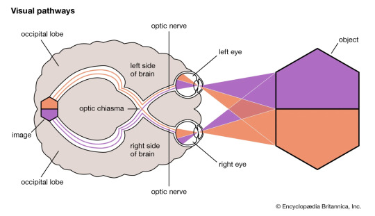
Optic nerve governs nasal visual (sees lateral view) and temporal visual (sees above the nose) fields. It originates in the retina and half of the axons (parts of nerve cells) cross in the optic chiasm. Half of the nerve fibres (axons) from your left eye continue to the left side of your brain. Half of your right eye’s nerve fibres connect to the right side of your brain. The remaining nerve fibres join together. Your brain receives signals from both eyes at the same time to create a cohesive visual image (binocular vision).
Fun fact - to take the brain out of the skull, optic nerve needs to be cut! From the optic chiasm information is relayed to thalamus via optic tract and further to the occipital lobe via optic radiations.
What if it isn’t working properly?
You might experience distorted vision in forms of colour blindness, night blindness, peripheral vision loss, partial or complete vision loss, eye pain, headaches, nausea and tinnitus (ringing in the ears)
What conditions might affect the optic nerve?
Many. Some of them you are born with, some of them can occur spontaneously throughout life targeting only vision or other body systems as well.
Glaucoma: Fluid buildup in the front part of your eye that puts pressure on the optic nerve and further damages it. Leading cause of blindness in adults over 60 years old.
Anterior ischemic optic neuropathy: Loss of blood flow to the optic nerve that causes sudden vision loss.
Optic atrophy: Lack of blood to the optic nerve resulting in its shrinkage. Might be inherited or be a result of trauma, stroke, infection, brain tumour or hydrocephalus.
Optic nerve drusen: Protein and calcium deposits (drusen) builds up on the optic nerve.
Optic nerve gliomas: Tumors (growths, usually benign) on the optic nerve. Often affect people who have an inherited condition called neurofibromatosis 1 (NF1).
Optic nerve meningiomas: Slow-growing tumors (rare and benign) that can lead to severe vision loss.
Optic neuritis: Inflammation of optic nerve caused by ifections and autoimmune diseases like multiple sclerosis.
Papilledema: Swelling of the optic nerve caused by pressure around your brain from a traumatic brain injury, brain tumors, meningitis,
Neuromyelitis optica, or Devic’s disease: Occurs when the immune system mistakenly attacks the optic nerves and spinal cord.
0 notes
Text
E-PORTFOLIO
IN PHYSIOLOGICAL &
BIOLOGICAL PSYCHOLOGY
(PY48)
MRS. ABIGAIL INTERNO (COURSE PROFESSOR) LAIRA ANDREI S. OSIAS (STUDENT)
MIDTERM ASSIGNMENT #2
PHYSIOLOGICAL & BIOLOGICAL PSYCHOLOGY
NAME: OSIAS, LAIRA ANDREI S.
SUBJ CODE: PY48
UNIT 3 TOPIC:
Sensory Physiology
Endocrine Glands
Muscles
SPECIFIC GUIDE QUESTIONS:
A. Sensory Physiology
Discuss the following:
undefined
Sensory receptors are primarily classified as chemoreceptors, thermoreceptors, mechanoreceptors, or photoreceptors.
Broadly, sensory receptors respond to one of four primary stimuli:
Chemicals (chemoreceptors)
Temperature (thermoreceptors)
Pressure (mechanoreceptors)
Light (photoreceptors)
Cutaneous Sensations
Touch, pressure, and temperature receptors are found on the surface of the skin. The connections between receptors and cutaneous sensations are not well understood. Touch sensitive Meissner corpuscles and deep pressure sensitive Pacinian corpuscles Ruffini ends communicate warmth, while Krause's bulbs communicate cold. Information is sent from the receptors to nerve fibers in the spinal cord, which then travel to the brainstem. They are then sent to a cortical area in the parietal lobe. Skin senses are also subjected to sensory adaptation. A hot tub, for example, can be unbearably hot at first, but after a while, one can sit in it without discomfort.
Pain. The majority of pain receptors in the skin are free nerve endings. Information is transmitted by two types of pathways to the brain by way of the thalamus.
The fast (myelinated) route recognizes localized pain and transmits it to the cortex quickly.
The unmyelinated slow route carries less localized, longer acting pain information (such as that concerning chronic aches).
Taste & Smell
Taste and smell are two distinct sensations with independent receptor organs, but they are inextricably linked. Taste buds, which are made up of unique sensory cells, detect tastants, which are substances found in foods. These cells convey messages to specific parts of the brain when activated, making us aware of our taste sense. Similarly, odorants, or airborne odor molecules, are picked up by specific cells in the nose. Odorants trigger a neuronal response by activating receptor proteins present on hairlike cilia at the ends of sensory cells. Taste and smell messages eventually converge, allowing us to detect food flavors. This strong association is most evident in our perception of food flavors. Food "tastes" differently when the sense of smell is impeded, as anyone who has had a head cold will attest. Actually, the flavor of the food, or the combination of taste and smell, is what is being altered. This is because just the taste of the meal is detected, not the scents. Taste is concerned with discriminating between compounds that have a sweet, salty, sour, bitter, or umami flavor (umami means "savory" in Japanese). Taste and smell interactions, on the other hand, improve our perceptions of the meals we eat. Specialized sensory neurons in a small patch of mucus membrane along the roof of the nose detect airborne odor molecules called odorants. These sensory cells' axons flow through perforations in the overlying bone and enter two extended olfactory bulbs on the underside of the frontal lobe.
Vestibular Apparatus and Equilibrium
The vestibular system is the inner ear's sensory equipment that aids in maintaining postural balance. The vestibular system's input is also crucial for coordinating the position of the head and the movement of the eyes.
The Ears and Hearing
The ear is the organ of hearing and balance. The parts of the ear include:
External or outer ear, consisting of:
undefined
Tympanic membrane (eardrum). The tympanic membrane divides the external ear from the middle ear.
Middle ear (tympanic cavity), consisting of:
undefined
Inner ear, consisting of:
undefined
The ability to see the world around you is determined by your vision. Several components within your eye and brain work together to give you vision. These components are:
Lens
Retina
Optic nerve
Many different parts of your eye and brain work together to assist you in seeing. The following are the primary elements of your vision:
Cornea: The front layer of your eye is called the cornea. The cornea is a dome-shaped structure that bends light entering your eye.
The pupil is the black dot in the middle of your eye that serves as a light gateway. In dark light, it extends, and in brilliant light, it contracts. The iris is in charge of it.
Iris: Your eye color is usually attributed to this area. The iris is a muscle in your eye that regulates the size of your pupil and the amount of light that enters it.
The lens is located behind the iris and pupil. Like a camera, it works with your cornea to concentrate the light that enters your eye. The lens sharpens the image in front of you, allowing you to see all of the details clearly.
The retina is a layer of tissue located in the back of the eye that converts the light that enters your eye into electrical signals. These signals are transmitted to the brain, which recognizes them as images.
Optic nerve: This aspect of your vision serves as a link between the retina and the brain. The electrical signals created in the retina are transmitted to the brain via the optic nerve. The brain then generates visuals.
Tears: Tears are supposed to keep your eyes moist and help you focus effectively, despite the fact that they are most usually associated with sobbing. They also aid in the prevention of eye discomfort and infection.
Endocrine Glands
Discuss the following:
undefined
The endocrine system is made up of hormone-secreting endocrine glands. Despite the fact that there are eight primary endocrine glands spread throughout the body, they are nevertheless considered one system since they have comparable functions, influence mechanisms, and interrelationships.
Non-endocrine portions of certain glands serve purposes other than hormone release. The pancreas, for example, includes both an exocrine and an endocrine part that secretes digestion enzymes and hormones. Hormones are secreted by the ovaries and testes, which also create eggs and sperm. Although several organs, such as the stomach, intestines, and heart, create hormones, this is not their major role.
Mechanisms of Hormone Action
Hormones activate target cells by diffusing through the target cell's plasma membrane (lipid-soluble hormones) to bind a receptor protein in the cell's cytoplasm, or by binding a particular receptor protein in the target cell's cell membrane (water-soluble proteins).
Pituitary Gland
The pituitary gland is a small pea-sized gland that plays a major role in regulating vital body functions and general wellbeing. It is referred to as the body's 'master gland' because it controls the activity of most other hormone-secreting glands.
Adrenal Glands
Adrenal glands are small, triangular-shaped glands that sit on top of both kidneys. Hormones produced by the adrenal glands serve to regulate your metabolism, immunological system, blood pressure, stress response, and other vital activities.
Thyroid and Parathyroid Glands
Iodine from meals is used by the thyroid gland to produce two thyroid hormones that control how the body uses energy. The parathyroid glands are a group of four small glands that sit behind the thyroid gland. The parathyroid glands make a hormone (parathyroid hormone) that helps regulate calcium levels in the blood.
Pancreas and Other Endocrine Glands
Glands are organs in the body that generate and release chemicals. The pancreas has two primary functions: exocrine and endocrine. Exocrine function: produces chemicals (enzymes) that aid digesting. Endocrine function: releases hormones that regulate the quantity of sugar in your blood.
Paracrine & Autocrine Regulation
(Autocrine glands generate hormones that operate on their own glandular cells, such as prostaglandins; paracrine glands create hormones that are released into the extracellular matrix and diffuse to neighboring cells, such as islets of Langerhans - somatostatin.) Diffusible chemicals bind to receptors on the same cell from which they were released in autocrine signaling. Insulin was the first transmitter to be implicated in the autocrine regulation of -cell function.
Muscles
Discuss the following:
Skeletal Muscles
Skeletal muscles make up 30 to 40% of your total body weight. They're the muscles that attach to your bones and allow you to move and operate in a variety of ways. Skeletal muscles are voluntary, which means you may choose when and how they perform.
Mechanisms of Contraction
Abstract. When the thin actin and thick myosin filaments slip past each other, muscle contraction occurs. Cross-bridges that stretch from myosin filaments and cyclically engage with actin filaments when ATP is hydrolyzed are thought to be the driving force behind this activity.
Contractions of Skeletal Muscles
The neuromuscular junction, which is the synapse between a motoneuron and a muscle fiber, is where skeletal muscle contraction begins. The presynaptic membrane's voltage-gated calcium (Ca2+) channels open when action potentials are sent to the motoneuron and then depolarized.
Energy Requirements of Skeletal Muscles
The breakdown of ATP provides the energy required for muscle contraction, but the amount of ATP in muscle cells is only enough to fuel a brief contraction.
Neural Control of Skeletal Muscles
Concentric, eccentric, and isometric contractions, muscle fiber recruitment, and muscle tone are all controlled by neural control. The role of motor units in nervous system control of skeletal muscles is critical.
Cardiac & Smooth Muscles
Cardiac muscle cells are found in the heart's walls, appear striped (striated), and are controlled involuntarily. Except for the heart, smooth muscle fibers are found in the walls of hollow visceral organs (such as the liver, pancreas, and intestines), are spindle-shaped, and are controlled involuntarily.
0 notes
Photo
New Without tags
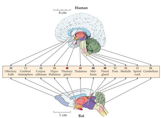
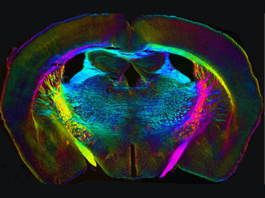
Allowing scientists to snoop on life in dark places, microscopy continues to adapt to new challenges. One problem is that lasers aimed into delicate tissues like the brain usually need something to aim at – perhaps molecules of chemical dyes that label or ‘tag’ important structures. Yet labels can interfere with microscopic life, or hide subtle details behind their glare. Here, researchers have used a technique called QLIPP to take a detailed look inside an adult mouse’s brain without labelling. Instead, they measure what happens to laser light as it hits or passes through the tissues – changes in orientation reveal shapes and textures (highlighted in different colour) while delays in the light’s waves, their retardance, reveal different densities (highlighted by brightness). The team used use deep-learning to pinpoint areas like the thalamus (middle, blue) and optic tract (bottom yellow and purple) – in the future they may spot subtle changes in health and disease.
Written by John Ankers
Image from work by Syuan-Ming Guo and colleagues
Chan Zuckerberg Biohub, San Francisco, CA, USA
Image originally published with a Creative Commons Attribution 4.0 International (CC BY 4.0)
Published in eLife, July 2020
You can also follow BPoD on Instagram, Twitter and Facebook
6 notes
·
View notes
Text
The Endocrine physiology overview

Part 1
Endocrine glands = Glands that secretes hormones directly into the blood stream= No canal.
Hormones = chemical substances that act on certain cells and guide processes such as: metabolism, growth etc.
The endocrine system in our body has :
Hypothalamus & the pituitary gland
Pineal gland
Thyroid gland
Parathyroid glands
Pancreas
Adreal glands
And the gonads (testes and ovaries)
The hypothalamus:
Nuclei [ supraoptic & paraventricular]
The ceo of the whole system! It senses the information from the body and then reacts by secreting CEO hormones!
The romantic relationship between the hypo and the pituitary:
Between the hypo and the anterior lobe of the pituitary ->
The hypothalamic-hypophyseal portal system (which is a system of blood vessels)
Between the hypo and the posterior lobe of the pituitary ->
The pituitary stalk (which is made by the axons coming from the nuclei in the hypo)
The hypothalamus hormones:
Hormones that affect the cells in the anterior lobe of the pituitary to trigger the secretion of certain hormones :
•- Stimulatory:
TRH = Thyrotropin Releasing Hormone
CRH = corticotropin Releasing Hormone
GNRH = Gonadotropin Releasing Hormone
GHRH= Growth hormone Releasing Hormone.
•- Inhibitory:
GHIH = Growth Hormone Inhibiting Hormone (Somatostatin)
Prolactin Inhibiting Hormone (Dopamine)
Hormones that are secreted by the hypo and stored in the posterior lobe of the pituitary (in Herring bodies) :
ADH (Vasopressin)
Oxytocin
The pituitary gland:
The co-ceo of the endocrine system. Lies in the sella turcica near the hypothalamuc underneath the optic chiasm.
Has two lobes: Anterior and posterior.
The posterior works as a storage for the hypo's two hormones (ADH & oxytocin) that secretes them when needed.
The anterior is made of glandular tissue = is able to produce hormones.
The Pituitary hormones:
ACTH = Adrenocorticotrophic hormone
TSH = Thyroid-stimulating hormone
LH = Luteinising hormone
FSH = Follicle-stimulating hormone.
PRL = Prolactin
GH = Growth hormone
MSH = Melanocyte- stimulating hormone.
Now how do these hormones work?
Now it is a great chance to learn 3 things:
Why the hypothalamus is the ceo?
What is the romantic relationship between it and the pituitary?
And the Endocrine /axes/
So, basically we can recap that like this:
TRH-> TSH-> Thyroid gland -> T4 & T3
CRH-> ACTH-> Adrenal cortex -> Cortisol & androgen
GNRH-> FSH/LH -> Gonads -> sex hormones
GHRH-> GH-> Liver-> IGF-1 / IGF-BP3
Example explained:
TRH stimulates the pituitary to secrete TSH in response to certain conditions and TSH works on certain cells in the thyroid gland (the follicular cells) to activate the producing of T3 & T4 .
What about ADH and oxytocin?
⬇ Blood volume -> ADH -> Distal nephron -> water balance.
Oxytocin = the hormone of motherhood and cuddle!
Motherhood:
- Uterus during delivery = stimulates uterine contractions.
- Breast = During breastfeeding
Cuddle:
Such as social interactions (snuggles~ ) and orgasms!!
The Thyroid Gland:
Butterfly-shaped gland located in the anterior neck just below the laryngeal prominence (adam's apple) . It has left and right lobe wrapping around the trachea and connected in the middle by an isthmus.
Thyroid hormones:
The thyroid gland has many follicles which walls are lined by follicular cells (thyrocytes) that produce:
T4: Triiodothyronine
T3: Thyroxine
Both affect metabolism.
Parafollicular cells (C-cells) produce: Calcitonin
-> calcium and phosphate -> bone metabolism.
Parathyroid glands:
Tiny, round structures found embedded in the posterior surface of the thyroid gland. Most people have 4 parathyroid glands.
Hormones:
PTH = parathyroid hormone -> calcium regulation.
Pineal gland:
Endocrine structure of the diencephalon of the brain . Inferior and posterior to the thalamus. Made of pinealocytes.
Hormones:
Melatonin -> circadian rhythm.
Pancreas:
Located in the abdomen behind the stomach. Has endocrine and a digestive exocrine function.
The endocrine part is called 'Langerhans islets' constitutes about 1-2% of the pancreas volume.
There are five types of cells in these islets:
Beta-> insulin -> ⬇Blood sugar
Alpha -> Glucagon -> ⬆ Blood sugar
PP cells -> pancreatic polypeptide
Delta -> somatostatin -> inhibits the secretion of other pancreatic hormones.
Epsilon -> ghrelin -> 'the hunger hormone'
The Adrenal Glands:
Two small glands located on top of each kidney
Has two parts :
Cortex:
3 Zones:
Zona glomerulosa -> Aldosterone -> ⬆ Blood pressure
Zona Fasciculata -> Cortisol -> Stress & diabetogenic
Zona reticularis -> Androgens.
Medulla:
Epinephrine & norepinephrine -> flight or fight response.
The Gonads:
Ovaries:
Located along the lateral wall of the uterus under the external iliac artery and in front of the internal iliac artery. The region that has the ovaries is called 'the ovarian fossa'
Hormones:
Estrogen : Estradiol, Estrone & Estriol.
Progesterone
Testes :
Oval-shaped organs, contained in the scrotum that hangs outside the body in front of the pelvic region.
Hormones :
Testosterone.
#studyblr#study motivation#medical student#2020 quarantine challenge#medical school#medicine#education#student#notes#biology#endocrinewithmarie#medicinewithmarie#physiology#pathology#the endocrine system#science#endocrine notes#study inspo#human biology#pituitary#medblr#med school#biomedicine#thyroid#human body#usmle#usmle endocrine#med
86 notes
·
View notes
Text
Sensation and Perception
For us to sense the world around us, our sensory organs receive stimuli. The messages those organs produce go through a process known as transduction. This means that the signals are transformed into impulses that travel through the thalamus to the appropriate cortices. Our bodies are constantly sending signals to the brain, but if it were to register every sensation, it would be overwhelming. Imagine feeling, seeing, hearing, touching, etc.. everything. This doesn’t happen due to the combination of sensory adaptation (the decrease in responsiveness to stimuli due to constant stimulation) and sensory habituation (our perception of sensation is partially due to how focused we are on them.) Now that I mention it, you likely will start to feel the socks on your feet, even though you hadn’t noticed that before, as you weren’t focused on the sensation. This connects to the cocktail-party phenomenon which is the idea that as your focus changes, you ignore certain stimuli, for example, at a party, if you’re focused on your friend talking, and someone yells your name across the room, your focus may involuntarily shift to whoever is yelling at you, causing you not to hear what your friend is saying.
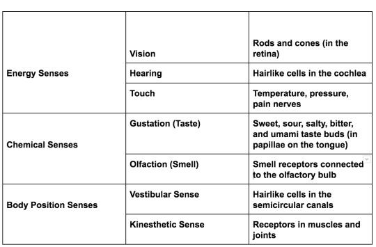
ENERGY SENSES
Vision
Step 1: Gathering Light: Light reflects off of surfaces and hits our eyes. The colour we perceive that surface to depend on a few things; one, the light intensity which is to say, how much energy is in the light that hits the eye. The second is the light wavelength, which determines the hue that we see. (Or if we even see it at all- wavelengths longer than visible light such as infrared waves, microwaves and radio waves or shorter than visible light such as UV waves and X-rays aren’t registered by the eye) A mix of all the visible hues of light produces white. When an object appears a certain colour, it has absorbed all other colours and reflected the colour we see it as. A green plant, for example, would not be able to survive in the green light, as it would reflect the green, causing it to starve.
Step 2: Within the Eye: Reflected light enters the eye through the cornea which is a clear protective covering over the eye. The light travels through a hole known as the pupil. The iris (what make your eyes a certain colour) is a collection of muscles which work to constrict the pupil in extremely light situations, and expand the pupil in dark places- this is known as accommodation. The curved lens focuses the light like a camera focuses on an image. The image is flipped upside down as it travels through the lens. This now focused, inverted image project itself on the retina which works as a screen. On this screen are specialised neurons which are activated by light.
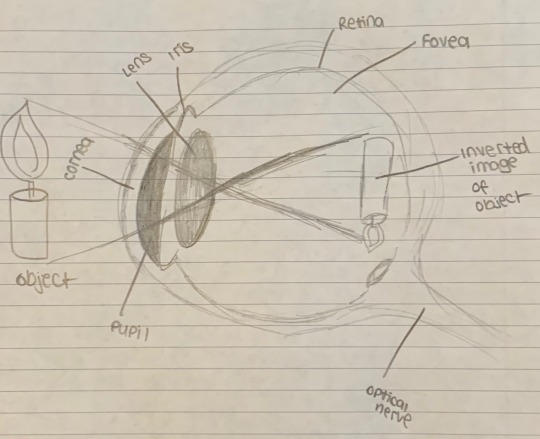
Step 3: Transduction: The light needs to be translated into neural impulses to be registered by the brain. There are two kinds of neurons which do that job. Cones, which are activated by colour and rods, which respond to black and white. Rods outnumber cones, and are especially useful in dark situations when colour cannot be registered at all; this is why in the dark, it is nearly impossible to figure out what colour an object is. There just isn’t enough coloured light being reflected for the cones to be activated properly. Cones are concentrated in the centre of the retina. At the very centre of the retina is the fovea; where the highest concentration of cones exists. Peripheral vision depends on cones, while your centred of vision depends on cones (in light, of course). Once enough rods and cones fire, they activate the next layer of bipolar cells; ganglion cells, whose axons make up the optic nerve, sending impulses to the lateral geniculate nucleus (LGN) of the thalamus. Messages are relayed to the visual cortex in the occipital lobes. The optic nerve has no rods or cones and is known as your blind spot (different to the driving blind spot). There are two parts to the optic nerve. Impulses from the right side of both retinas go to the right hemisphere, while impulses from the left side of both retinas go to the left hemisphere; it is that separation of the optic nerve that allows this to take place. The spot where the optic nerves cross each other is the optic chiasm.
Here’s a fun experiment to show where the blind spot is. (Here is the original link for anyone curious: https://visionaryeyecare.wordpress.com/2008/08/04/eye-test-find-your-blind-spot-in-each-eye/- it has a few more interesting experiments to try as well)

Sit with your nose between the cross and black circle. Cover your left eye, and look at the cross with your right eye. Move towards the computer while still using your right eye. Eventually, the black circle will disappear and reappear, showing you your blind spot. Do the same with your left eye, except now focus on the black circle. Weird right?? (Science rules!)
Step 4: In the Brain: There is still a lot of debate as to how our visual cortex actually works to register vision. Some say the visual cortex is where sensation ends and perception begins. Some say interpretation begins at the retina. Some say it begins at the thalamus. Scientists David Hubel and Torsten Wiesel were responsible for finding that groups of neurons in the visual cortex respond to different types of visual images. There are feature detectors for vertical lines, curves, motion, and so on.
Theories of Colour Vision
A lot is still unknown about how we see colour. The oldest theory is the Young-Helmholtz Trichromatic theory which hypothesised that we have 3 different typed of cones which detect, red, blue and green. They are activated in different combinations to produce the spectrum of colour we see. There are issues with this theory; namely afterimages and colour blindness. That’s where the opponent-process theory of colour vision comes in. (Not to be confused with the opponent-process theory of emotion. I’ll get to that in a later post). This theory states that sensory receptors are arranged in pairs: red/green, yellow/blue, and black/white. When one sensor is stimulated, its pair is inhibited. This explains after images. Try this illusion out: Stare at this image for 30 seconds, and then look at a white surface. If the flag doesn’t immediately show up try blinking a few times.

What’s happening is that you’ve exhausted your cones, and so the opponents will fire. This is why dichromatic colour blindness also tends to be consistent as to which two colours are affected such as red-green colour blindness, or yellow-blue colour blindness
Hearing
While both vision and hearing are considered energy senses, they are very different stimuli. Sound waves are vibrations in the air, instead of electromagnetic waves used to produce images. The vibrations are captured by our ears and transduced into neural impulses which allow the brain to register and interpret the sound. Sound waves have amplitude and frequency. Amplitude is the height of the wave and determines how loud the sound will be. Frequency is the length of the waves and determines the pitch of the sound. High-frequency waves are densely packed together, while low-frequency waves are spaced apart.
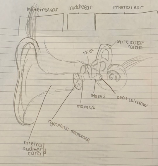
The process of hearing works much like a Rube Goldberg machine, where one thing moving activates the next part of the sequence. The pinna (outer ear) collects sound, which travels down the auditory canal. The sound reaches the tympanic membrane (the eardrum) which vibrates as sound hits it. Attached to the tympanic membrane are the ossicles; the smallest bones in your body. The tympanic membrane is connected to the hammer/malleus, which is connected to the anvil/incus, which is connected to the stirrup/stapes, which all transmit the vibration to the oval window, a similar membrane to the eardrum. The windows membrane is connected to the cochlea, a snail-like structure filled with perilymph, and endolymph. The vibration of the oval window causes these fluids to move. At the base of the cochlea is the basilar membrane, lined with hair cells connected to the Organ of Corti, which are neurons activated by the moving hair cells. The Organ of Corti fires neural impulses and those impulses travel to the brain via the auditory nerve.
Pitch Theories
Place Theory: Place theory states that hair cells in the cochlea respond to different frequencies based on their location. Some bend to high pitches, and others to low pitches.
Frequency Theory: Research has shown that while place theory is accurate for high pitches, but not for low tones, who are sensed by the rate at which the cells fire. We sense pitch because hair cells fire at different rates (frequencies) in the cochlea.
Deafness
For each step in the hearing process, there are just as many ways those steps can go wrong. Conduction deafness refers to a problem conducting sound to the cochlea (so any point in the ear canal, to the eardrum, ossicles, or oval window). Sensorineural deafness occurs when the hair cells in the cochlea are damaged. Often this is caused by overstimulation, causing a high pitched ringing. Prolonged exposure to that much stimulation can severely damage the hair cells which do not regenerate.
Touch
There is one more type of energy sense and it is just as different as the other two. Our skin contains millions of different types of receptors. Some (nociceptors) respond to pain, some (thermoreceptors) respond to temperature. Some (Messiner’s Corpuscles) respond to plain old touch. Some (Pacinian Corpuscles) respond to pressure, all of which can be found all over the skin. The relationship between these receptors is not well understood. What we do know is that the brain interprets the amount of indentation (temperature change) and the intensity of touch. Where nerve endings fire also determines where the touch is coming from. Certain parts of our body have more concentrated amounts of receptors.
For example, in my physiology class, we did an interesting experiment where a friend took a paper clip and unfolded it until it had 2 ends. The friend would poke me on different parts of my skin with either 1 or 2 of the ends and see if I could guess how many ends I was being poked with. On the fingertip, I guessed with 100% accuracy, while on my shoulder, I was guessing the whole time.
Gate-control theory explains how pain is experienced. Some pain messages are more important than others. When a priority message is sent, the “gate” swings open for it and swings shut for lower priority messages. This is why scratching an itch gets rid of the itch. The scratching is a higher priority message, and so the itch is temporarily ignored in order to prioritise the itching. That’s also why you shouldn’t scratch an itch- you’re not doing yourself any good. Endorphins also work to shut this gate, working like morphine to shut off any pain.
CHEMICAL SENSES
Gustation/Taste
Fun fact before I start: I remember that taste is gestation because of Gusteau from Ratatouille who was named after the French word for taste. When you eat food, the tastebuds are not responding to energy, but rather the chemicals on the tongue. These tastebuds are located on papillae, which are bumps on the tongue. There are 5 different kinds of tastebuds scattered throughout the tongue which respond to Sweet, Salty, Bitter, and Umami (savoury). The more densely packed the taste buds, the more intense the taste is. The tongue is not unique to taste; you’ll know this if you’ve ever tried to eat a jalapeno because spicy food stimulates nociceptors, which is why the experience is agonising for some people. Weaker nociceptors increase a person’s spice tolerance, which is why certain cultures do better eating spicy foods; they’ve adapted to the pain.
Smell/Olfaction
Olfaction is created by chemicals entering the nose and being absorbed by receptors on the mucous membrane. A lot is not known about these receptor cells- some researchers estimate there are as many as 100 different types. These cells are linked to the olfactory bulb which gathers messages from olfactory receptor cells and relays them to the brain. The nerve fibres from the olfactory bulb connect to the brain differently than all the other senses; instead of going to the thalamus, the message is sent to the amygdala and then the hippocampus. This could be why smells can trigger memories much more strongly than other senses can.
BODY POSITION SENSES
Let's get something out of the way: you do not have 5 senses. How many you have depends on who you ask, but a prominent one is the sense of position.
Vestibular Sense
Your vestibular sense helps you determine where your body is in space. The semicircular canals in the inner ear are responsible for this. The canals are tubes partially filled endolymph. When the head moves, the fluid moves too. The hair cells activate, sending signals to the brain telling it that the head has moved. Dizziness is caused when the brain receives several conflicting signals in a short period of time, caused by rapid movement, or when you’re in a boat, and the eyes register a static view, while the inner ear is registering all kinds of movement.
Kinesthetic Sense
While vestibular sense tracks the overall orientation of the body, kinesthetic sense tracks the position and orientation of specific body parts. Receptors in muscles and joints send information to the brain, helping the brain track the body.
PERCEPTION
Perception is the process of understanding and interpreting sensations. Psychophysics is a branch of psychology which studies the interaction between sensations and how we experience them.
Thresholds
Senses have limits. The absolute threshold is the smallest amount of stimulus we can detect. There are several videos online helping people track the absolute threshold of pitch that they can detect by playing higher and higher frequencies. The proper definition of the absolute threshold is the minimal amount of stimulus we can detect 50% of the time as things such as other sensory inputs can make studying the absolute threshold extremely difficult. Stimuli below the absolute threshold are referred to as subliminal.
Another important threshold is the difference threshold. This is also referred to as the just-noticeable difference and is rather self-explanatory. It is the smallest amount of change in a stimulus needed for us to detect a change. This threshold is determined by Weber’s Law, discovered by psychophysicist Ernst Weber (it is sometimes called the Weber-Fechner law.) The change needed is proportional to the original intensity of the stimulus. The more intense a stimulus, the more it will need to change before we notice a difference. Each sense varies according to a constant (which differs between the senses). The constant for hearing is 5%- when listening to a 50-decibel tone, it would need to reach 52.5 decibels to notice a difference.
Perceptual Theories
Signal Detection Theory: This theory investigates the effects of distractions and interference we normally experience in our lives. This area of research revolves around attempting to predict what we will perceive. It takes into account how motivated we are to perceive specific stimuli and our expectation. These are known as response criteria or receiver operating characteristics. A false positive is when we think we are perceiving a non-present stimulus. While a false negative is the failure to perceive a present stimulus.
Top-Down Processing: When we use top-down processing, we perceive by filling in gaps what we sense. For example, when reading this sentence: I _m t_king p_ych_ _ gy, you can pretty easily fill in the blanks based on the context of what you read before, and the letters I did give you. In other words, top-down processing uses background knowledge to fill in gaps. Our schemata are mental representations of how we think the world should be. They can create a perceptual set or a predisposition to experience something a specific way. https://www.youtube.com/watch?v=Bafuyg98no4- Watch this video about backmasking (”hidden” messages in songs when played backwards.) Try looking away when the song starts playing and then reading the supposed message. I know personally, when I did this, I couldn’t understand the hidden message until I read what I was supposed to be hearing, and then I could hear it clearly. Several parents became worried about this phenomenon, likely because they had schemata of the music their kids were listening to as dangerous.
Bottom-Up Processing: This is also known as feature analysis, where only the features of the object are used to build a perception. This is an automatic process, however, is much slower than top-down processing. As a result, it is less prone to error.
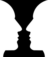
Principles of Visual Perception
There are numerous rules for visual perception. One of the first decisions the brain makes is the figure-ground relationship. This is self-explanatory; what is the figure and what is the background? A famous illusion connected to this principle is the vase face illusion. Do you see the background as black or white? Do you see the figure as the vase or the face?
Gestalt Rules
Gestalt psychologists at the beginning of the twentieth century noted several principles which explain how we perceive groups of objects, and theorised that we perceive images as groups rather than individuals. (Hence the name “Gestalt”) Here is a great diagram from https://www.verywellmind.com/gestalt-laws-of-perceptual-organization-2795835 illustrating those principles.
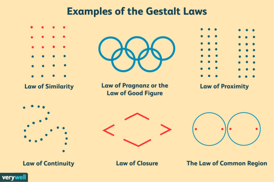
Proximity: Objects that are close together are more likely to be perceived as a part of the same group
Similarity: Objects that look similar to each other are likely to be perceived as part of one group
Continuity: Objects creating a continuous form are likely to be perceived as part of the same group
Closure: Similar to the sentence connected to top-down processing. Objects making a recognisable image are likely to be viewed as part of the same group
Law of Pragnanz: You are more likely to perceive and interpret complex images as the simplest form possible
Law of Common Region: Objects grouped together within the same region of space tend to be viewed as their own group.
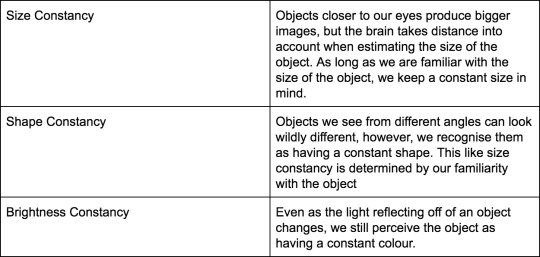
Constancy
Our ability to maintain a constant perception of an object despite a constantly changing world is known as constancy. Here are a few types.
Perceived Motion
Our brains can detect how fast images move across the retinas and use our own motion to determine the speed the object is moving. This causes some interesting phenomena where the brain thinks a still object is moving. For example, you may be familiar with the stroboscopic effect. This is what causes perceived motion in flipbooks. There is also the phi phenomenon. Movie Marquees use this phenomenon by turning on and off lights sequentially, creating the appearance of one moving light.
Depth Cues
Depth perception is what allows us to perceive the world in 3-dimensions rather than 2. Eleanor Gibson performed an interesting experiment known as the visual cliff experiment, which found that crawling toddlers can perceive depth. In this experiment, the infant is placed onto one side of a glass table, creating the appearance of a cliff. The infant would not crawl onto the glass. There are two categories of cues that we use; monocular (needing one eye) and binocular (needing two)
Monocular Cues
Artists tend to use monocular cues to create the illusion of depth in their art. A wonderful artist on deviant art created a really helpful diagram, illustrating each of these ideas. https://www.deviantart.com/akenon/art/Monocular-Cues-34065283
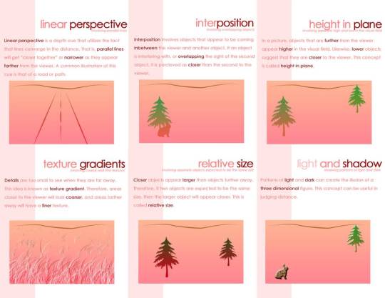
Linear Perspective- In order to draw a faraway house, artists tend to draw two lines which converge at a focal point, taking advantage of the linear perspective cue.
Relative Size Cue- When drawing two similarly sized cats, one far away and one nearby, an artist will draw the faraway cat as significantly smaller, creating the illusion of distance.
Interposition Cue- If the artist draws a cat that is partially obscured by a tree, we will view the tree as closer than the cat.
Texture Gradient- Another way artists create the illusion of distance is by making close up objects more detailed than those faraway
Shadowing- By using shading in an image, an artist can imply a light source, further implying depth in the image.
Binocular Cues
Binocular Disparity/Retinal Disparity- Each eye sees an object at a slightly different angle. The farther away an object is, the more similarly the eyes will perceive it, the closer it is, the more retinal disparity there will be. Notice in this diagram how the trees don’t seem to move, but the finger moves drastically?
Convergence- As an object comes closer to our face, the eyes must move towards each other to keep it in focus. The more the eyes converge, the closet the object is.
How Culture Affects Perception
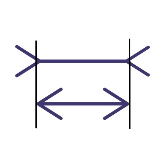
One of the more interesting (in my opinion) parts of psychology is exploring how culture affects us. Some of the perceptual rules that for years psychologists believed to be innate are actually learnt. Cultures which don’t use monocular depth cues in their art don’t see depth in pictures using these cues. It also affects how optical illusions affect our brains. The famous Muller-Lyer illusion often does not affect cultures that don’t use right angles and corners in architecture.

#AP Psych#AP Psychology#psychology studyblr#psych studyblr#human anatomy studyblr#physics#psychophysics#physiology studyblr#sensation#perception#eye anatomy#ear anatomy#anatomy#human anatomy#biology#biology studyblr#studyblr#bio#human bio#human biology#physio#human physio#human physiology
28 notes
·
View notes
Text
Interesting Papers for Week 44, 2020
Hippocampal Network Reorganization Underlies the Formation of a Temporal Association Memory. Ahmed, M. S., Priestley, J. B., Castro, A., Stefanini, F., Solis Canales, A. S., Balough, E. M., … Losonczy, A. (2020). Neuron, 107(2), 283-291.e6.
Information Closure Theory of Consciousness. Chang, A. Y. C., Biehl, M., Yu, Y., & Kanai, R. (2020). Frontiers in Psychology, 11, 1504.
Experience-Dependent Reorganization Drives Development of a Binocularly Unified Cortical Representation of Orientation. Chang, J. T., Whitney, D., & Fitzpatrick, D. (2020). Neuron, 107(2), 338-350.e5.
Consistent patterns of distractor effects during decision making. Chau, B. K., Law, C.-K., Lopez-Persem, A., Klein-Flügge, M. C., & Rushworth, M. F. (2020). eLife, 9, e53850.
Top-Down Control of Inhibitory Granule Cells in the Main Olfactory Bulb Reshapes Neural Dynamics Giving Rise to a Diversity of Computations. Chen, Z., & Padmanabhan, K. (2020). Frontiers in Computational Neuroscience, 14, 59.
Neural dynamics of perceptual inference and its reversal during imagery. Dijkstra, N., Ambrogioni, L., Vidaurre, D., & van Gerven, M. (2020). eLife, 9, e53588.
Learning-related population dynamics in the auditory thalamus. Gilad, A., Maor, I., & Mizrahi, A. (2020). eLife, 9, e56307.
Cortical Observation by Synchronous Multifocal Optical Sampling Reveals Widespread Population Encoding of Actions. Kauvar, I. V., Machado, T. A., Yuen, E., Kochalka, J., Choi, M., Allen, W. E., … Deisseroth, K. (2020). Neuron, 107(2), 351-367.e19.
Mechanistic determinants of effector-independent motor memory encoding. Kumar, A., Panthi, G., Divakar, R., & Mutha, P. K. (2020). Proceedings of the National Academy of Sciences of the United States of America, 117(29), 17338–17347.
Phylogenetic variation in cortical layer II immature neuron reservoir of mammals. La Rosa, C., Cavallo, F., Pecora, A., Chincarini, M., Ala, U., Faulkes, C. G., … Bonfanti, L. (2020). eLife, 9, e55456.
Inhibition of impulsive action by projection-defined prefrontal pyramidal neurons. Li, B., Nguyen, T. P., Ma, C., & Dan, Y. (2020). Proceedings of the National Academy of Sciences of the United States of America, 117(29), 17278–17287.
A Model of Memory Linking Time to Space. Löffler, H., & Gupta, D. S. (2020). Frontiers in Computational Neuroscience, 14, 60.
Measuring habit formation through goal-directed response switching. Luque, D., Molinero, S., Watson, P., López, F. J., & Le Pelley, M. E. (2020). Journal of Experimental Psychology: General, 149(8), 1449–1459.
Development of Binaural Sensitivity: Eye Gaze as a Measure of Real-time Processing. Peng, Z. E., Kan, A., & Litovsky, R. Y. (2020). Frontiers in Systems Neuroscience, 14, 39.
Vision perceptually restores auditory spectral dynamics in speech. Plass, J., Brang, D., Suzuki, S., & Grabowecky, M. (2020). Proceedings of the National Academy of Sciences of the United States of America, 117(29), 16920–16927.
A taxonomy of seizure dynamotypes. Saggio, M. L., Crisp, D., Scott, J. M., Karoly, P., Kuhlmann, L., Nakatani, M., … Stacey, W. C. (2020). eLife, 9, e55632.
Unveiling Stimulation Secrets of Electrical Excitation of Neural Tissue Using a Circuit Probability Theory. Wang, H., Wang, J., Thow, X. Y., Lee, S., Peh, W. Y. X., Ng, K. A., … Lee, C. (2020). Frontiers in Computational Neuroscience, 14, 50.
Two Forms of Knowledge Representations in the Human Brain. Wang, X., Men, W., Gao, J., Caramazza, A., & Bi, Y. (2020). Neuron, 107(2), 383-393.e5.
Universal inference. Wasserman, L., Ramdas, A., & Balakrishnan, S. (2020). Proceedings of the National Academy of Sciences of the United States of America, 117(29), 16880–16890.
Base rate neglect and neural computations for subjective weight in decision under uncertainty. Yang, Y.-Y., & Wu, S.-W. (2020). Proceedings of the National Academy of Sciences of the United States of America, 117(29), 16908–16919.
#science#Neuroscience#computational neuroscience#Brain science#research#neurobiology#psychophysics#scientific publications#cognition#cognitive science
3 notes
·
View notes
Photo
Visual perception is the potential of the mind to discern situations around us through the light that enters the eye. According to research done at Massachusetts Institute of Technology (MIT) in 2017, the mind does visual perception in a timespan of 13 milliseconds, and the ability to see colour is just one of the outcomes of it.
Light travels in different wavelengths, and each colour possesses its respective wavelength. As wavelengths of light enter the eye, photoreceptors sense them and respond by forming electrochemical signals to be sent to the brain. The signals travel through the optic nerve in the back of the eye, followed by the thalamus, to reach the cerebral cortex, a thin layer of the brain. Through this, the brain interprets wavelengths of light as colour to ensure we distinguish features of the environment better.
1 note
·
View note
Text
nomu is my kismesis
homestucks only
anyway
i fucking hate the nomu because as a biology/neuroscience student, i’m just like, what the ever-loving shit is happening there. why are the eyes THERE. how the fuck are the optic nerves supposed to get to the optic chiasm and into the superior colliculus and thalamus? what the FUCK are they doing buried in the goddamn like
what the fuck is that spot
the fucking frontal lobes???????? UJIKO YOU STUPID OLD NUTSACK ASS BITCH
those brains make no sense. they make NO sense. there is no somatosensory cortex or motor cortex and i am having a fucking seizure just looking at them. the anatomy. the audacity. the hubris to come up with a walking sasquatch hunk with no discernible brain anatomy. i’m so mad i need to sit down.
nomu brains look like a goddamn fucking vagina. i’m gonna ram a strap on through those brains just to teach them a lesson not to screw with me.
39 notes
·
View notes
Text
TAFAKKUR: Part 18
Your Nervous System: Part 1
Finally, I have come to say goodbye to you. As you probably know, there is a saying “Leave the best till last.” I am the greatest of all the organs and systems that have described themselves to you so far. I am an integrative system that forms a chain between every organ in your body. Just as your veins are spread out to carry nutrients and oxygen to every part of your body, I also embrace your entire system like a network, without leaving the tiniest space; I am informed of everything that goes on inside your body. Even if a tiny insect settles on your arm, you sense it immediately. I make you aware of a tiny drop of sweat on your body. I induce pain in suitable measures to inform you of any illnesses in your inner organs. In fact, I not only inform you, I also warn you to seek help.
However, it is hard for me to describe myself. When you hear the words “nervous system,” what comes to mind is a cluster of cells called neurons. But this is a great mass of cells, and we should always remember that we are referring to the most complex matter in all of creation. The very important main nervous systems, which are very close to one another, are the huge masses positioned beneath the skull, the extensions of my system and secondary nervous system; this latter is spread out through various regions of the body. It would take up too much of your time to describe each region and branch of my system individually to you each month, but in this way I could prove what a perfect and incredible duty each of them performs within your body. However, I will try to explain the subject briefly to avoid boring you. Nevertheless, please forgive me if I ramble on too much; we are describing the most excellent organ created by God so it is inevitable that there will be some complicated matters that need clarification.
Instead of allowing each of my sections to describe themselves to you individually, I will speak on their behalf as the “brain.” It may be easier for you to understand the system if we divide it into two. One of them is me and the nervous system which I lead; we can briefly describe this as the thalamus, hypothalamus, cerebellum, the medulla, and the spine. The other part is the peripheral nervous system, which emerges from the central nervous system and is distributed, rather like fiber optic telephone cables, throughout the entire body. In addition to me, the brain, and my two large cerebral hemispheres, there is another smaller section, which is known as the brain stem. The brain and its cerebral hemispheres and the sections of the brain stem which are protected beneath the skull (the cerebellum, medulla, thalamus, and hypothalamus) are very important. The spinal cord, which is also a part of the central nervous system, however is not in the skull, but positioned within the vertebrae that constitute the spine. Due to its connection with the central nervous system, any damage to the spinal cord can endanger life.
Although damage to regions of the body where the nerves are distributed from the central nervous system may cause paralysis, or functional disorder for the specific organ, such an incident is not life threatening.
If you recall, when the heart and circulation system described themselves they boasted – indeed the veins also seemed to brag a bit when they stated that they measured 75,000 miles (long enough to go around the world almost three times). But the nerves are approximately 477,000 miles, long enough to stretch from the earth to the moon, and back again… the nerves which are distributed throughout the various parts of your body measure 250,000 miles, and the total length of the central nervous system is 228,000 miles. Almost 200,000 signals pass through just one cell at a time, which means that every moment thousands of signals pass through millions of my cells all throughout your body, and flow from the central nervous system to the whole body and then back to the central nervous system. There are about 30 billion cells in my system. 10 billion of these cells are in the cortex, 10 billion in the cerebellum, and the remainder forms the structure of the nerves and other sections. As a comparison, a fly’s brain contains 100 thousand cells, and a rat’s brain has 10 million cells. The total number of connections and contact points (synapses) that my 30 billion cells use to send and receive signals is 100 trillion. The number of combinations that these connections can establish to send signals to one another is greater than the number of atoms in the universe. At the beginning of a thinking process, the number of cells activated is between 10 and 100 million, and according to the depth and intensity of the activity, these figures can increase to astounding numbers. Every second 4 billion signals are exchanged between the left and right hemispheres. When you were an embryo, just a few weeks old, I consisted of 92% water. When you were first born, the ratio of water was 90%. And when you were fully developed, the water ratio remains at 77%.Can you imagine, a heap of mass consisting of 77% water, the remainder made up of various element. Our Lord, the bearer of eternal power places me in you, in the head of the most honorable creation, and with me you form civilizations; you invent and discover. And even more important, with my mediation you have the ability to contemplate and reach your Creator. What we are learning about here is how with me you are able to recognize the wisdom of the entire universe. The electric signals of the various sense organs, such as the eyes, ears, nose, tongue and skin, all of which have previously described themselves, are transmitted by the receptive cells on various wavelengths; these are then conveyed to you in the form of sight, noise, smell and taste. In fact I am inducing you to write these words at this very moment. The evaluation of everything you do passes through me, but you are not even aware of it. When you walk, eat, talk, speak or sleep, the information I receive from every part of your body is reviewed and responded to in a suitable manner. Could a single nucleus of a single one of my cells possibly position itself alone?
#allah#god#muhammad#prophet#sunnah#hadith#islam#muslim#muslimah#hijba#help#quran#ayah#dua#salah#pray#prayer#revert#convert#reminder#religion#welcome to islam#how to convert to islam#new muslim#new revert#new convert#revert help#convert help#islam help#muslim help
1 note
·
View note
Photo

Near Death Experiences
The term “near-death experience” (NDE) is well-known throughout America, but the phenomenon is not restricted solely to the Western world. Most cultures have an equivalent experience; even children have related NDEs.
An NDE might involve walking toward a bright light at the end of a tunnel, meeting gods, speaking with relatives who are long-dead, out-of-body experiences (OBEs) or feeling bathed in light.
Almost unanimously a significant life experience, conversations about NDEs are often accompanied by discussions of the afterlife and the mind surviving the mortal body.
These kinds of esoteric tales would normally be banished to the realms of pseudoscience and parapsychology, but their pervasive nature – an estimated 3 percent of Americans report having experienced an NDE – has sparked a smattering of genuine scientific research and a wealth of conjecture.
What do NDEs consist of?
One Dutch study, published in The Lancet, set out to investigate the regularity of NDEs and tried to tease apart causal factors.
The investigators reported that 50 percent of individuals who experienced an NDE mentioned an awareness of being dead, 56 percent regarded it a positive experience, 24 percent reported an OBE, 31 percent described traveling through a tunnel and 32 percent spoke of interacting with deceased people.
The study also showed that, of the patients they interviewed, although all were clinically dead at one point, only a small percentage (18 percent) experienced, or remembered, the NDE. The likelihood of having an NDE was not related to the level of cerebral anoxia (lack of oxygen to the brain), the amount of preceding fear or the type of medication they were taking.
According to the paper, NDEs were more often experienced by patients under 60, and women more commonly described deeper experiences. Conversely, those with memory deficits following resuscitation were less likely to report NDEs, which is to be expected.
There is obviously something driving these experiences, but the factors that impact them are still very much up for debate.
What is behind NDEs?
A phenomenon so widely experienced cannot be dismissed as just another old wives’ tale, there has to be something biological at work to explain its prevalence.
Some observers claim that NDEs display a rift in current neuroscientific theory, and that the experience shows another, more esoteric facet to our existence.
Can neuroscience unpick the mysteries of the NDE?
Many believe we should split the mind from the functions of the brain, once and for all.
However, this type of thinking is not necessary to explain NDEs; rather than claiming paranormal origins, the field of cognitive neuroscience has attacked the problem as it would any other: as an output of the brain.
Out-of-body experiences
OBEs are commonly a part of NDEs and sometimes include autoscopy – seeing one’s body from above. Although this seems like an otherworldly event, neuroscientists know that OBEs also happen in settings other than the near-deathbed.
For instance, during an attack of sleep paralysis, which affects up to 40 percent of people at some point in their lives, OBEs are common. Sleep paralysis occurs when an individual is still essentially in REM sleep, but their brain awakens partially.
During REM sleep, the brain effectively paralyzes the body to prevent it from acting out dreams. The brain, still believing that the person is asleep, keeps this lock on the body, subjecting the individual to a terrifying, literal, waking nightmare. The experience often involves a sensation of floating from one’s body and viewing the room from the ceiling’s perspective.
Other researchers have demonstrated that by stimulating the right temporoparietal junction (TPJ), they could induce OBEs artificially. The TPJ is a section of the brain that collates information from the thalamus (regulator of consciousness, sleep, and alertness), limbic system (involved in emotion, behavior, motivation, and long-term memory), and the senses.
Meeting the dead
Meeting and greeting the dead is another commonly reported aspect of NDEs and can be partially explained away by expectations. Cultures are often filled to the brim with tales of heaven or some other type of afterlife where long-dead relatives eagerly await us.
Added to this, people with Alzheimer’s and Parkinson’s are known to have vivid hallucinations of ghost-like entities; some report seeing dead relatives in their homes. These types of apparitions have been linked to pallidotomy lesions – a kind of neurosurgery used in some Parkinson’s patients.
Ghostly apparitions are not necessarily rooted in another dimension.
The experiences are considered to be due to dysfunction in the pathways of dopamine, a neurotransmitter involved in the brain’s reward pathways that is known to cause hallucinations.
In truth, it is surprising that we do not hallucinate more than we already do. Our brains weave our senses into the experience of perception in such a way that we forget what a difficult and amazing job they do.
Any cracks in the perceptual sphere are seamlessly back-filled by the brain; as a quick example, we all have a blind spot where the optic nerve meets the retina. In this section of our visual field, we can see nothing at all, but we never notice because our brain simply fills in the blanks.
But on occasion, if under duress or when receiving confusing inputs, rather than penciling in a chair, a patch of wallpaper, or a door, it fills the void with a goblin or ghoul.
In macular degeneration, the center of the visual field gradually fails; patients report the hallucination of ghosts relatively frequently. This might be due to the brain attempting to make sense of the neural “noise” being generated from the faulty or partial messages it is receiving.
The tunnel of light
Possibly the most well-known facet of an NDE is the feeling of being drawn into a long tunnel with a bright light at the end. Some researchers believe that this phenomenon can be explained by retinal ischemia (lack of oxygen to the retina).
The theory goes that, as the retina is starved of oxygen, peripheral vision slowly decays and only the center of the visual field can be seen. Tunnel vision is a symptom of both extreme fear and oxygen loss (hypoxia), both of which are often present during the process of dying.
No doubt, NDEs are a complex phenomenon with a myriad of mechanisms behind them. From a lack of oxygen affecting the visual system to a brain struggling to make sense of strange emotions; from the drug-like triggering of reward pathways and a host of cultural expectations. Being close to death (or believing that you are) is a unique physiological and psychological experience. It is little wonder that it produces such a confusion of sights and sounds.
The precise nature of each NDE will not be unraveled for many years. After all, catching them in action, at one of the most critical points of an individual’s life is no easy task, and the ethics of experimental interventions could prove tricky.
10 notes
·
View notes
Photo

Peace is divine, but men have misused its name.
The Earth is one constituent of the universe, but because men see it underneath Heaven, they regard Heaven as its antagonist; for division is the first function of Nature. But just as the two eyes see two images, which are made one in the optic thalamus, so should man forget antagonism and create unity in himself.
Peace is the reconciliation of Heaven and Earth. But to say this to the worldly is a waste of breath, for the worldly man does not know the meaning of either his Heaven or his Earth. His body came from earth, and his Spirit from Heaven desiring to know earth, but the body has denied the Spirit which gives it life. So man looks for God in the starry sky, and makes the intellectual mistake of dividing the Spirit of Heaven from the Spirit of Earth.
Image-Ptah
Isha Schwaller De Lubicz-The Opening of the Way; A Practical Guide to the Wisdom Teachings of Ancient Egypt
493 notes
·
View notes
Last Seen Blogs

serenefreakgeek
multi.fandom.blog

inkonhair
Untitled

shigellasonnei
sonnei

iammdmasudgazi
Md Masud Gazi

ducksarescary
Pro Lurker