#hypodermis
Text
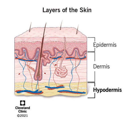
Subcutaneous Layer
The first layer below the cutaneous membrane is called the subcutaneous layer. It is also known as the hypodermis and superficial fascia. Although this layer helps stabilize the integumentary system and shares some characteristics, it is not technically a part of the integumentary system.
https://my.clevelandclinic.org/health/body/21902-hypodermis-subcutaneous-tissue
Within the subcutaneous…
View On WordPress
#animals#biology#blog#collagen#college#cutaneous membrane#dermis#elastic#epidermis#estrogen#facts#follow#hypodermic needle#hypodermis#Integumentary system#layers of the skin#nature#photography#science#skin#subcutaneous#subscribe#testosterone
3 notes
·
View notes
Text
Laser therapy and application of Aloe vera for wound treatment after mastopexy complications: A case report by Emerson Barbosa da Silva in Journal of Clinical and Medical Images, Case Reports

Abstract
Mammoplasty is a surgery aimed at reducing breasts, being an invasive procedure and with the possibility of post-surgical complications. In the case presented, the patient had healing difficulties which progressed to an intense inflammatory process that evolved to necrosis of the areolas, laser therapy sessions were applied using lasers with red LED and blue LED in order to treat the inflammatory process, stimulate regeneration of tissue and collagen synthesis, treat the patient's pain, hydrate the tissues, fight infections and skin diseases and whiten the area. It was associated with the application of Aloe vera in natura to treat wounds and heal the local. It was observed that after treatment there was a significant improvement in areola necrosis, healing of the injured part and improvement in collagen production. Making an evaluation during the 24 sessions, we were able to assess that in synergy between the Red Laser, Infrared, Blue LED and the use of the active ingredient Aloe vera corroborate the evolution of tissue healing.
Keywords: Aloe vera; laser therapy; mastopexy; surgical complications.
Introduction
Mastopexy or reduction mammoplasty is a surgery performed to reduce the breasts are usually more invasive because it involves larger incisions, tissue, skin, fat and repositioning of the areolas. These patients have back and neck pain due to the weight of the breasts, in some cases, the clinical picture even presents curvature of the back, some patients seek this type of surgery because they are unhappy with their aesthetic appearance [1]. Necrosis occurs when cells in a particular region of the body fail to receive enough oxygen. There are risks that are managed by the patient and risks inherent to the surgery technique itself. There are more common factors after surgery, which are headaches and in the surgery itself, bleeding at the site, keloids, infection, necrosis and thrombosis. It is not common to happen necrosis in the breast after mammoplasty, the probability is around 1%, factors that interfere with healing are smoking and diabetes [2, 3]. In performing this type of surgery, it is necessary to raise the areola, due to a structure called the Areolocapillary Complex - CAP, it can suffer some injury, when this tissue does not receive oxygen causing necrosis [3, 4].
The doctor responsible for the surgery cannot observe if there is any type of injury in the NAC, this usually occurs in breasts with ptosis because the greater the distance that the areola will travel during the surgery, the greater the chances of any injury to the NAC. Several authors sought to describe studies that showed assessments regarding complications in breast reduction surgery. The most common complications found in the literature were described, related to blood perfusion of the nipple-areola complex (NAC), operative site infection, dehiscence, asymmetries and changes in sensitivity secondary to the surgical procedure. The type of injury that occurred was scarring below the right areola in the stitches and tissue necrosis in the left areola, the probable cause may have been because of the NAC injury. Each author in the literature as well as those present on the table describes the safety of each flap, with its particularities, but without comparing the different techniques with each other1. The treatment performed was Red, Infrared, Blue Led laser therapy and the Aloe and vera plant [4].
The laser performs an amplification of light by stimulated emission of electromagnetic radiation that emits coherent and collimated light that can have different powers, for therapeutic use, we use low power laser that increase lymphocytes and phagocytosis, increasing fibroblasts and intensifying reabsorption of fibrin and due to the characteristics Biostimulators accelerating tissue repair, due to mitotic activation of epithelial cells, produces collagen and decreases the synthesis of inflammatory meters.
Treatment Red and infrared laser therapy (light amplification by stimulated emission of radiation) Red Light (660nm): Red light treats inflammatory processes, stimulates tissue regeneration and collagen synthesis, improves vascularization and angiogenesis, increases ATP production. It acts on the epidermis, dermis, hypodermis, muscle fascia, muscle tissue, tendinous ligament [5]. Infrared Light (808nm): Infrared light has the analgesic function (pain treatment) as the main point, acts in lymphatic drainage and edema, has an anti-inflammatory effect and increases the absorption of products by 40 %. It acts on the epidermis, dermis, hypodermis, muscle fascia, muscle, ligament, tendon, nervous and bone tissue [6]. Blue Light (470nm): Blue light has bactericidal and fungicidal action, promotes tissue hydration, fights infections and skin diseases and has a whitening effect. It acts mainly on the epidermis and on open lesions in the dermis [5, 6]. The healing caused by the blue LED is a mechanism of molecular events divided into three phases: inflammatory, proliferative and remodeling. It is in the remodeling phase that the recovery of tissue structure occurs through maturation of elements and changes in the extracellular matrix, where the deposit of proteoglycans and collagen occurs.
Case Presentation
Patient 27 years old, female, denies smoking and alcohol consumption, denies diabetes mellitus, underwent mastopexy surgery to reduce and improve the sagging of the breasts, after performing the surgery below the right areola, she presented difficulty in healing in the stitches and in the left areola necrosis and difficulty healing the stitches. Patient underwent breast reduction surgery, after surgery the breasts present exudate thickening of the skin in the right areola in the stitches it began to show difficulty in healing, in the left areola that was not healing, there was a darkening of the areola and after two weeks the tissue it was already completely necrotic. The plastic surgeon who performed the surgery indicated the use of Kollagenase (collagenase) Cristália®, intended for the treatment of skin lesions when debridement is indicated in wounds, ulcers and necrotic lesions. Without evolution, the professional indicated Lasertherapy to the patient. The methodologies applied during the first 20 sessions were performed 3 sessions per week, red and infrared laser therapy doses of 8 J/cm2 and blue LED using the Laser Therapy EC - DMC® equipment. 24 laser therapy sessions were carried out, in all of them both blue and red led were used. The first 20 sessions were held 3 times a week, after which 4 sessions were held, one per week. The patient applied Aloe vera in natura 3 times a day on the lesion, before application, asepsis of the lesion was recommended.
The patient underwent anchor mastopexy with the desire to improve the aesthetic aspect of the breast, in addition to improving physical aspects such as pain in the spine, after a week of the surgical procedure the patient noticed that the left breast took longer to heal when compared to on the right. The lesion turned red and began to produce fluid, the shape of the areola was changing due to the intense inflammatory process. After a few days, a blackened stain began to appear, which characterized a necrosis process that soon extended, the incision became swollen and without the possibility of healing as seen in (Figure 1).
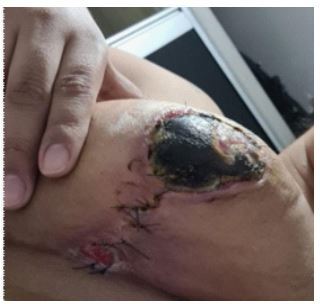
Figure 1: Left breast with extensive necrotic ulceration in the areola.
In the first consultation, skin debridement was performed, which aims to remove the largest amount of dead tissue, in order to reduce the process of tissue necrosis and reduce the possibility of infection by opportunistic pathogens, which would complicate the situation. The lesion was now cleaner (Figure 2) and it was possible to carry out an effective treatment plan.
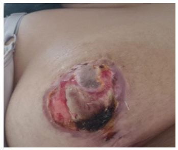
Figure 2: Left breast after debridement and cleaning of the lesion.
The application of Infrared and Blue LED was started, three times a week until completing the twenty sessions, during the treatment the patient performed the topical application of Aloe vera in natura to assist in the healing process in the skin and enhance the treatment. The lesions became redder, since the treatment increases tissue perfusion, improving nutrient delivery and access to defense cells that help in the healing process (Figure 3).
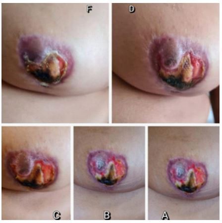
Figure 3, A-F: Evolution of the lesion between the first 5 sessions.
After the tenth session, the tissue improvement in relation to necrosis was already noticed and until the twentieth session, areas of necrosis in the breast were no longer seen, which showed the efficiency of the treatment (Figure 4).
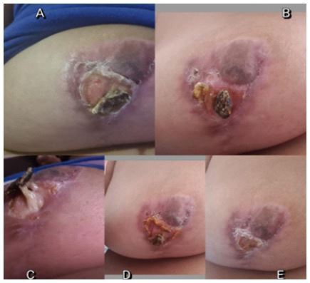
Figure 4, A-E: Aspect of the lesion from the tenth and twentieth sessions.
After the twenty-fourth session, the tissue has already been fully recovered and fibrosis at the site where the lesion was, showing the tissue completely healed, without bleeding and without production of fluids from the inflammatory process as seen in (Figures 5 and 6), leaving the optimistic patient and with improved self-esteem and non-psychological trauma suffered after the surgical complication.
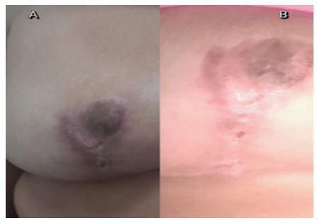
Figure 5, A-B: Result of the twenty-fourth session.

Figure 6: Aspect in the breast at the end of treatment.
Aloe vera, popularly known as aloe, is a medicinal plant that brings benefits to skin health. The hydrating action of the plant has already been proven in several studies. However, there is still not enough scientific evidence to confirm its healing effect [7]. Pyrocatechol, cinnamic acid, ascorbic acid and p-coumaric acid are some of the substances involved in the bactericidal (destroying bacteria) and bacteriostatic (preventing the proliferation of bacteria) effect of Aloe vera. The plant has antimicrobial action and fights some types of fungi, viruses and bacteria [7, 8, 9].
The use of Aloe vera can aid in healing and re-epithelization (repair of skin tissue) in a short period, if properly indicated, in case of burns [10]. Infrared density therapy has an anti-inflammatory effect, through vasodilation, it also has a beneficial effect on nerve cells, decreasing sensitivity and blocking the pain transmitted by these cells to the brain [9]. The laser penetrates deep into tissue and accelerates cell reproduction and growth. In this way, it increases the energy available to the cell so that it can absorb nutrients more quickly and get rid of waste products. As a result of laser exposure, damaged cells are repaired more quickly [10, 11].
Blue light laser therapy stimulates the development of fibroblasts in the damaged tissue. Fibroblasts are the building blocks of collagen, which is the essential protein needed to replace old tissue to repair tissue damage. Thus, the technique is effective in improving the aesthetic appearance of surgical scars and in the treatment of open wounds and burns, reducing the formation of fibrous tissue and keloids [12]. Thus, we noticed that the therapy associated with laser brings several benefits, mainly by accelerating the healing process of the skin, leading to the best appearance of the tissue and reducing the lesion, being in this situation the best treatment available for the conditions and access to the technologies that the patient can enjoy [13].
Conclusion
Although mammoplasty is a surgery aimed at improving not only the quality of life of the patient, also helps in self-esteem. Is important stress the necessary care during these procedures so that no negative side effect occurs, as in the case presented? When the patient goes through this situation, she has a worsening in her health and self-esteem, because there may be cases where recovery is not possible, affecting that person's life permanently. Fortunately, with the technologies we currently have, it is possible to perform treatments for help with the problem. It was observed that the synergistic treatments that were performed corroborate the improvement in the recovery of breast tissue.
Acknowledgments
The Sara Magna Clinic – Dermatologist and Naturopath, the Coordination of the Biomedicine and Pharmacy Course at the Centro Universitário Ítalo Brasileiro and the Centro Universitário FMABC.
For more details : https://jcmimagescasereports.org/author-guidelines/
#Mammoplasty#Aloe vera#laser therapy#mastopexy#surgical complications#Necrosis occurs#hypodermis#FMABC#Emerson Barbosa da Silva#JCMICR
0 notes
Text
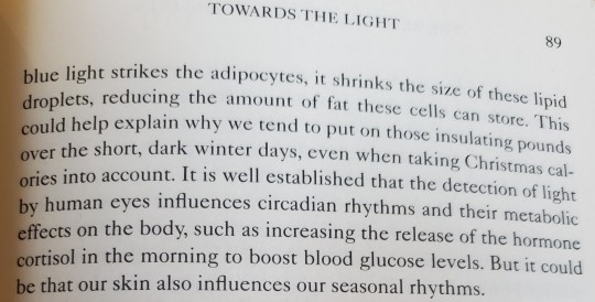
#seasonal weight gain#the remarkable life of skin#monty lyman#an intimate journey across our largest organ#UVA#UVB#UVC#blue rays#weight#seasonal#weight fluctuations#light to lose weight#UVA dive through layers into fat cells#adipocytes#hypodermis#winter weight gain
1 note
·
View note
Text
Wanna rewire my entire personality to one one that my fp adores tbh I know exactly what they desire and I can tweak it anytime and they probably wouldn't realize I really really really just want to be by your side but.... I don't want to be different. It's taken me so long to even accept this and connect all these fragments into the same person I've been the same person all along but I still struggle to believe that
#i kinda have a thing for reading people#its difficult online or even on the phone. way easier in person#even though i cant make direct eye contact with you for more than a second#that would be too embarrassing for me#something about me's changed and you haven't liked it for years now#wow just the thought of that makes me want to attempt to slit my arm down to the muscle in one swipe#i will Not do that i cannot go to hospital again im not strong enough#i was just there for food poisoning they would be like what the fuck man#did the toxins eat your brain!!!#considering what im saying rn perhaps they did#i dont think ive ever even cut to muscle#dear god imagine the pain you get with a bruise and thats protected by your skin#and your epidermis dermis and hypodermis to muscle all while there's blood vessels in there and nerve endings good god
4 notes
·
View notes
Note
So interesting ^^ most definitely not something I would’ve come up with. Please write more biology things cause they’re fun lol. I also liked that one time you mentioned bombers having tadpole tails when they’re young

hey, gladly! i do think that they have tadpole-esque tails when very young as a result of evolutionary atavism - they're not structurally developed to the extent that the rest of the individual's body is, even at that young and quite literally tender an age, and are either absorbed back into the body for nutrients or shrink until fallen off during the formation of the protective epidermis
which probably requires some explanation in itself, because i've been thinking that the various colored parts we see on jetters bombermen are different layers of skin development on their body, with the three primary layers being hypodermal (visible only on the face, where much of it is adapted to become spongy and semiporous in order to imbibe food and drink), dermal (seen on the limbs and antenna, often either white or a similar color to the hypodermis), and epidermal (frequently brightly-colored, covers the torso and resembles a jumpsuit)
the hypodermal layer is by far the most sensitive and malleable, allowing for emotive facial expression but a liability to have exposed on parts of the body less necessary for social communication, while the dermal layer is an in-between mainly just doing the everyday work of keeping an individual safe from infection and minor, mundane physical harm. the epidermal layer is specially adapted to withstand heavy damage like could be sustained through bombs - typically explosive or incendiary - and is much less sensitive than the layers underneath, with enough effectively dead tissue at the surface (think like human nails or hair) that it can be used to attach things like pins or brooches
#fractions thoughts#while all three are smoother than human skin the epidermis in particular has a texture somewhere between rubber and the material of a tarp#touch on the hypodermis (i.e. face) is understandably and kinda obviously an intimate gesture in their society as the equivalent is in ours
5 notes
·
View notes
Text
The amount of times the ROTTMNT fandom talks about Donnie and subcutaneous trackers has unironically helped me memorize shit for my anatomy class, so thanks!
6 notes
·
View notes
Text
I love having a well of infromation that makes people say "why/how do you know that?"
#my mysterious persona#and its mostly political and insane shit which makes them more confused#like how do i say that i think the one scene in act 2 of bg3 (with the nurses) has weird gore#like i know it wouldnt look like thst#where is the dermis and hypodermis#but if i say that i get weird questions
1 note
·
View note
Note
A general question how do they look naked? (No sexual purpose I’m just curious about what’s the difference between us and them) are they half devil??
Hello anon, thanks for your question! :)
The reply has gotten a little more lengthy, sorry for that. I hope it answers all your questions.
First of all, I do believe that thanks to the "Blood of Asmodeus" curse, tieflings are to some degree blood-related to devils, though they are not half-devils. That blood, however, does affect their looks.
I will definitely be talking more about tiefling skin in general in the future, from the nice ridges on their tails to the skin around their horns, and the bumps along their chests and backs, but those are already visible in the game. Have a quick look for example at the back of Poem, my tiefling character:
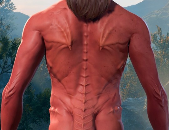
The way those ridges work I believe, is to provide additional protection, almost like a layer of body armor created by a tiefling's own body, shielding more vulnerable places like the spine, but they also exist on areas of skin like the elbow and knee that are already thicker in humans, as the skin there has to withstand mechanical stretching from those joints having quite a bit of mobility and being used all the time.
Due to fewer oil glands in this area, the skin there gets drier and flakier, and retains less water. I believe the thicker skin there also serves protective purposes, but not exactly the same way as the ridges on the spine and ribs. They are probably mostly there to ease friction, but also protect the sensitive nerves there. (Yes, the funny bone is actually a nerve called the Ulnar nerve, and tieflings probably have no issues hitting it, because it is much better protected.)
Then, of course, there are genitals, which were kept fairly standard for tieflings in BG3.
As much as I personally like to imagine their genitals to be ribbed or nubby, I don't think it would make much sense. The bumps and ridges on their skin are for protection, and while genitals are definitely a sensitive area, they should probably not have this particular type of protection.
My reasoning here would be how these ridges work vs. what you would typically want.
I've quickly doodled something to explain this a little better, please excuse the quality here and my terrible handwriting haha
Please note that I drew up this little diagram as a more general sort of overview and the yellow subcutaneous tissue has its color from the fat that is typically part of this layer, but is missing in clitoris, penis, eyelids, and other similar areas of skin. That's why these areas of skin feel different, by the way!
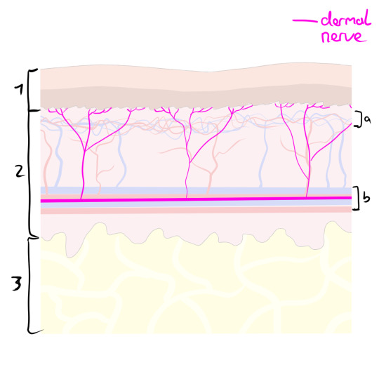

Epidermis (Outermost layer of skin)
Dermis (Middle layer of skin containing connective tissue, the superficial arteriovenous plexus [a] with all those fine little blood vessels, and also the deep arteriovenous plexus [b], so the thicker blood vessels, but also the dermal nerve fibres that I marked in pink here.)
Hypodermis/Subcutaneous tissue (Layer right below the skin, anchoring the skin to the connective tissue of the body)
As you can see, the nerve does not reach all the way into the epidermis (which is good, you absolutely would not want that). But that also means adding a protective layer to it, as I have done in tiefling-red on the right, you create an even thicker barrier before reaching the nerve, making the skin a lot less sensitive.
Obviously, you do not want a tiefling's genitals to be insensitive like this. You want them to enjoy having sex after all.
Additionally, according to this article, as well as the player handbook, the Asmodeus curse has made tiefling genetics very dominant, meaning that tieflings will always produce tiefling offspring, serving "a purpose for the Hells in that it strengthens their presence in the mortal realms".
This leads me to speculate whether tieflings might have higher libido than humans and if, perhaps, they are more potent.
Additionally, due to the way their pelvis is shaped thanks to their tails (I will get more into this in the tail essay), there is a chance that - compared to humans - giving birth is slightly less painful for them. While probably far from an enjoyable process, they could potentially see some benefits from their sacrum being turned outward.
As far as shapes and sizes go, it seems logical for them to be at least compatible with other humanoids to ensure that they can produce offspring with one another. Tiefling vaginas would probably be similar to those of humans, not much deeper or wider, since too many differences would make impregnation harder for any non-tieflings.
Tiefling penises probably also wouldn't be much longer or thicker or strangely shaped, as that could do significant damage to a human or elven body, which would once again not lead to creating more tiefling offspring for Asmodeus to extend his reign.
But it is possible for them to have differences! I think that's mostly where people's headcanons can come in. Anything that doesn't make it biologically more difficult to conceive a child is fair game!
Penises with more tapered tips? Possible.
Larger or more sensitive clitoral glans? Hell yeah!
Want your tiefling to have an additional g-spot that Asmodeus put in there to be cheeky? Go for it!
--
I hope that did answer your question well enough, but please feel free to follow up if there is anything else you would like to know.
62 notes
·
View notes
Text
Dermis
Layers Of The Dermis
The dermis layer is located inferior to the epidermis layer in the cutaneous layer. This layer is made of connective tissue and can be divided into two layers:
Papillary LayerReticular Layer
https://www.researchgate.net/figure/Adult-human-skin-is-a-layered-organ-consisting-of-an-epidermis-and-a-dermis-The_fig1_233976352
The papillary layer of the dermis is the most…

View On WordPress
#aging#anatomy#animals#biology#blog#collagen#college#cutaneous#cutaneous membrane#epidermis#facts#fibers#follow#human anatomy#hypodermis#nature#Nerves#neurons#papillary layer#photography#plexus#reticular layer#science#skin#stretch marks#subscribe#tension lines
3 notes
·
View notes
Text
Humans are Weird, “Skin, and the sun”
Thought you guys might have missed some of the more humans are weird related Dr. krill writes a medical report stuff.
Consider this a PSA from Krill to anyone who happens to possess skin
https://www.patreon.com/empyreaniris?fan_landing=true
https://starr-fall-knight-rise.tumblr.com/post/182501791735/master-post
https://docs.google.com/document/d/1jzEIdDAB4omdO2JcQVMObfrhLJ5kX4ONmSsLypM1ks0/edit?usp=sharing.\
-
The Intergalactic Journal of Xenomedical Biology
Author: Dr. Krill
ID# 374622
Status: Revision Phase Not Cleared for Publication
Issues - Informal language
I think by this point my contribution to the scientific community is enough to openly acknowledge that I am the universe’s number one leading nonhuman expert in human biology. I have been told it is presumptive to claim that I probably know more than the humans do, so I have been forced to put in “nonhuman expert” against my will.
Human skin is an object of some interest in the xenobiological community. It should hardly be so interesting, considering that every species in the galaxy has skin to some degree or another, but human skin has some interesting qualities I wish to discuss here.
Human skin is the soft outer layer most visible on the human body, skin tones on a human can range from almost white to almost black with brown and cream comprising the midtones, though despite the color variations the underlying structure remains the same. Now human skin is known, commonly, to have three layers, in its most simple model, as there are other models discussing the layers within the layers, but for the purpose of this paper, I will be discussing the Epidermis, the dermis and the hypodermis.
The skin in general, but especially the epidermis acts as protective barrier, specifically to keep out germs and bacteria from infecting the inner structures of the body. Now, while it is possible for a human to get a skin infection, the sheer fact that they don’t spend their entire lives infected with all the nasty things that they insist on touching is beyond me. I mean humans put their own fingers in their mouths for architect’s sake, and as you know from my previous paper, “A comprehensive analysis on the human mouth.” The fact that they don’t simply dissolve from flesh eating bacteria is a miracle in itself.
Furthermore, the outer layer creatures new skin cells at a rapid rate. Do not be surprised, when upon acquiring a human, your living space suddenly becomes more dusty. You will be interested to learn that this is the fault of your human companion who cannot help but shed the entirety of their outer dermal layer every thirty days, so if you have a human, and your ship is dusty, simply know that most of that is dead human skin just floating around, absolutely disgusting. Humans can’t go anywhere without leaving little pieces of themselves behind.
Earth is a planet with an open atmosphere and dynamic cloud cover, which allows sunlight, and thus radiation down through its atmosphere and onto the ground. You would think after a thousand years of evolution human skin would do a bang up job of protecting the human in question from radiation burn.
And in fact it does a bang up job. In the same way your granny makes a banging noise as she falls down the stairs. Human skin is absolutely shit at resisting stellar radiation. Now don’t you dare ask a human about this, because you are bound to hear a crock of bullshit come out of their mouths like, “Oh I don’t burn, I tan!” or “My skin is too dark to burn, so I’m good.”
All bullshit.
Human skin does indeed contain melanin, which is the structure that allows for skin color and tanning. On different parts of earth, different levels of light are allowed through the atmosphere, and over the years this has caused a varying degree of melanin to be distributed throughout the human population. Humans with ancestry that hails from the north, where effects of sunlight are lessened will have naturally pale skin, while humans with ancestry more towards the equator will have darker skin, which is an adaptive response to varying degrees of sunlight.
However, dark skin still does a shit job at protecting from the sun. Yeah sure pale skin is shittier, but shitty and shitteir are pedantic when it comes to the possibility of skin cancer. Research suggests that darker skin humans have a natural SPF (sun protection factor) of 13, while fair skinned people might have half of that.
This may seem like a boon, however, human experts recommend an SPF of at least 30 which is TWICE the amount of natural SPF provided by higher melanin content. But try telling that to a human! You can scream at the top of your lungs all day and the bastards will just insist that they will get a tan instead. Its this diabolical human belief that they are somehow IMMUNE TO THE SUN!!!!? So please, if you must, grab your human and marinade them in sunscreen, no matter how much melanin they happen to possess.
Because of the erroneous belief that darker skin is effective at blocking out UV radiation, Darker skinned humans may be more likely to die of skin cancer than light skinned humans. Light skinned humans are more aware of the dangers, due to their natural proclivity for burning instead of tanning, and the visibility of dermal abnormalities like moles, or freckles. In many cases cancer on darker skinned humans is caught later because of the erroneous belief by both civilians AND medical professionals in some cases, that darker skinned people are not susceptible to skin cancer.
So I don’t care if your human insists they either don’t like or don’t need sunscreen. TO BAD! They are WRONG! And they deserve to be SMACKED. Even a SINGLE sunburn can increase a human’s chance of getting cancer by FIFTY PERCENT. A SINGLE sunburn. Never in my life have I ever met a human who didn’t have at least TWO OR MORE sunburns.
The “I don’t get burned I just tan.” humans are FULL OF SHIT, “Tanning comes as a direct result of SUN DAMAGE. I don’t care how pretty it looks, it is NOT good for you. I had a light skinned human once who fell asleep in the sun and the skin BLISTERED. That is a SECOND degree burn from RADIATION.
Humans can get SUN POISONING.
MMMmmmmm
The worst part about all this is I can’t even lock the humans under a rock somewhere because humans NEED sunlight in order to get their vitamin D, and without Vitamin D, they could be tired, sore, weak, and may fall into heavy depression. In fact some humans become depressed in seasonal cycles where the sun may not provide them with ENOUGH light.
So apparently humans are INCAPABLE of being happy without getting to stand in the CANCER RAY
I have been stuck with these monstrosities for almost a decade now a decade! And I thought maybe at some point in my life humans would stop making me angry, but it just keeps getting worse!
455 notes
·
View notes

Note
Salt pookie do u have any advice on how to draw gore
Look at it/J
Learn organ placements and what they look like from diagrams, and learn the skeletal structure. If you want to draw skinning then you may need to learn about muscle.
As for cuts, PLEASE JUST LEARN THE LAYERS OF THE SKIN!!! I AM TIRED OF JUST SEEING RED WHEN NEW GORE ARTISTS DRAW DEEP CUTS!!! THE HYPODERMIS IS NOT RED ITS ORANGE!!! AND IT LOOKS LIKE BEANS!!!!
For the layer above the hypodermis, the dermis, you can just use a grayish white and add some red to it, unless you want the scar to already be bleeding instead of fresh. Anything above that can be a red line.
For blood itself, it’s a thick liquid that stains everything. So please when drawing it, the “head” of the drop should be thicker than the trail itself. Also, play around in multiply to make it actually look like blood and not a saturated red mess, it’s going to need several layers, all in different shades of red
17 notes
·
View notes

Text
Mod 3: Gymnosperms
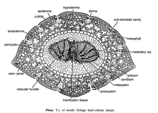
Pinus Needle T.S.
It is circular in outline in P. monophylla, semicircular in P. sylvestris and triangular in P. longifolia, P. roxburghii, etc.
Outermost layer is epidermis, which consists of thick-walled cells. It is covered by a very strong cuticle.
Many sunken stomata are present on the epidermis.
Each stomata opens internally into a substomatal cavity and externally into a respiratory cavity or vestibule.
Below the epidermis are present a few layers of thick-walled sclerenchymatous hypodermis. It is well developed at ridges
In between the hypodermis and endodermis is present the mesophyll tissue.
Cells of the mesophyll are polygonal and filled with chloroplasts. Many peg-like infoldings of cellulose also arise from the inner side of the wall of mesophyll cells.
Few resin canals are present in the mesophyll, adjoining the hypodermis. Their number is variable but generally they are two in number.
Endodermis is single-layered with barrel-shaped cells and clear casparian strips.
Pericycle is multilayered and consists of mainly parenchymatous cells and some sclerenchymatous cells forming T-shaped girder, which separates two vascular bundles. Transfusion tissue consists of tracheidial cells.
Two conjoint and collateral vascular bundles are present in the center. These are closed but cambium may also be present in the sections passing through the base of the needle.
Xylem lies towards the angular side and the phloem towards the convex side of the needle.
Idk where to find xeric nature info
Williamsoniaceae
Occurrence of Williamsonia
Williamsonia belongs to family Williamsoniaceae of Bennettiales.
It has been reported from Upper Triassic period but was more abundant in Jurassic.
This was earlier discovered under the name Zamia gigas by Willamson in 1870 but has now been named as Williamsonia.
Professor Birbal Sanhi (1932) described W. sewardiana from Rajmahal Hills of Bihar (India).
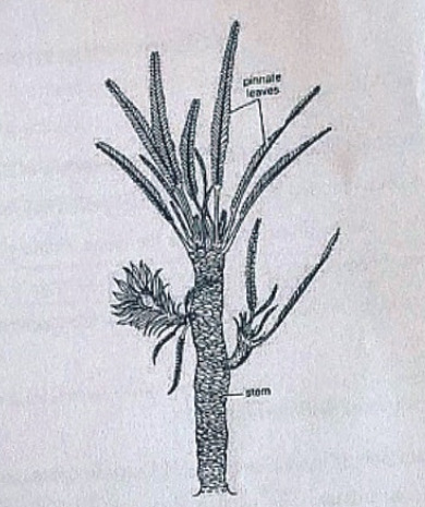
External Features of Williamsonia
Williamsonia resembled Cycas in appearance, but its best-knows species is W. sewardiana. The plant had an upright, branched, and stout stem covered by persistent leaf bases.
A terminal crown of pinnately compound leaves was present. For the stem genus Bucklandia, Sharma (1991) opined that features of leaf bases such as their shape, size and arrangement pattern are of taxonomic significance.
He observed that leaves in Williamsoniaceae show syndetocheilic stomata with rachis possessing collateral endarch vascular bundles.
Reproduction in Williamsonia
The fructifications of Williamsonia were large and attained a diameter of about 12 cm.
They were borne on a peduncle.
Many spirally arranged bracts were present around the base of the floral axis.
In W. gigas the cones were present among the crown of leaf bases while in W. sewardinia they were present on the short lateral branches.
Williamsonia plants were unisexual.
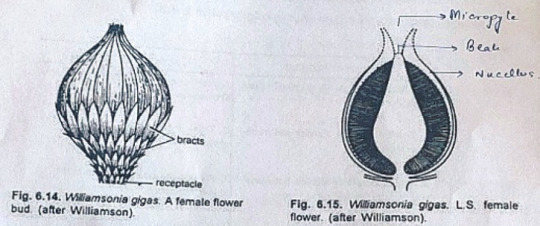
Female Flower
The female 'cones' of W. gigas and W. sewardiana have been investigated in detail. Instead of 'strobili' or 'cones', Sporne (1965) proposed to use the term 'flower'.
The conical receptacle was surrounded by many perianth-like bracts. The ovules were stalked.
The apex of the receptacle was naked and sterile. The nucellus was surrounded by a single vascularize integument, which was fused with the nucellus. The nucellus had a well-marked beak and a pollen chamber. In young ovules the micropylar canal was long and narrow.
In mature ovules, the canal widened because of the formation of nucellar plug and disappearance of interlocking cells. In the apical part of the endosperm, Sharma (1979) observed 2 or more archegonia.
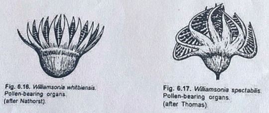
Male Flower
Male flowers consisted of a whorl of microsporophyll's which were united to form a more or less cuplike structure. In majority of the investigates species the sporophylls were un-branched but in some species they were also pinnately branched.
Sitholey and Bose discovered W. santalensis from Upper Gondwana, and observed that microsporophyll's in the species were bifid.
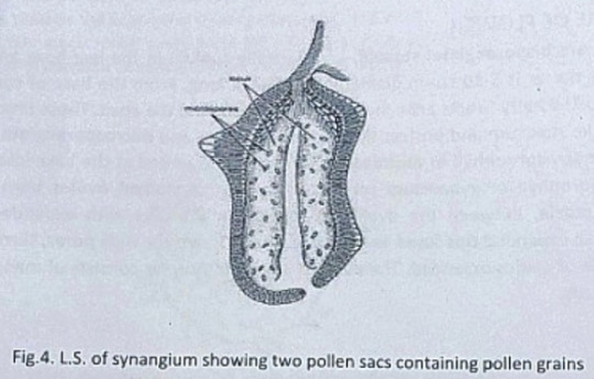
One of the branches of microsporophyll was fertile while the other was sterile. The fertile part has finger-like structures called synangia. Each synangium had two rows of chambers enclosing microsporangia.
The fertile branch of the bifid sporophyll possessed many purse-like capsules, in each of which there were present many monocolpate pollen grains.
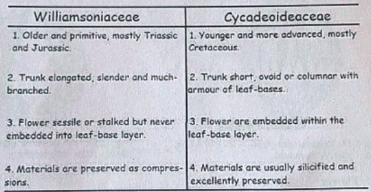
Cycadeoideaceae
Classification:
Division - Cycadeoidophyta
Order - Cycadeoideales
Family - Cycadeoideaceae
Genus - Cycadeoidea
Introduction:
Cycadeoidea is the only genus of family Cycadeoidaceae, represented by thirty species. They are entirely extinct and resemble cycads in the outward stumpy appearance of trunk and an apical crown of pinnate compound leaves. This fossil group of plants flourished during the Triassic to Cretaceous periods of the Mesozoic era. They are reported from various places in the world, in India the Cycadeoidales are found in Rajmahal Hills in Bihar. The petrified trunks of C. entrusca are the oldest fossil ever collected by man.
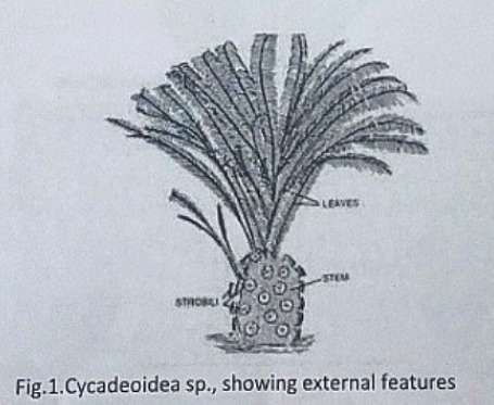
External Features:
The genus Cycadeoidea had a short, branched, or unbranched spherical, conical, or irregular trunk. The diameter of the trunk is 50cm and the highest rarely reached a meter except in C. jenneyana, it attended the height of several meters. These trunks are covered by rhomboidal leaf bases having multicellular hairs in between. Crown of 10ft long pinnate compound leaves are present at the top.
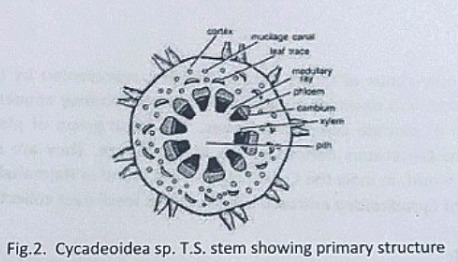
Anatomy of Stem:
The transverse section of the stem shows roughly a circular outline. The epidermis is not very distinct due to the presence of heavy armor of leaf bases. The cortex is parenchymatous and traversed by mucilage canals and numerous leaf traces. The primary vascular structure consists of a ring of endarch, collateral, conjoint, and open vascular bundles encircling the pith. Pith is wide and parenchymatous. A ray-like extension passes between the vascular bundles that make their appearance discrete.
There is a cambium ring with a thin zone of secondary wood. The secondary wood encircles the primary xylem and consists of tracheids with scalariform and bordered pits. The secondary medullary rays traverse the secondary xylem and secondary phloem.
The C-shaped leaf traces arise singly from the primary vascular strand and entering the cortex divided into several masarch strands and enters straight into the leaf.
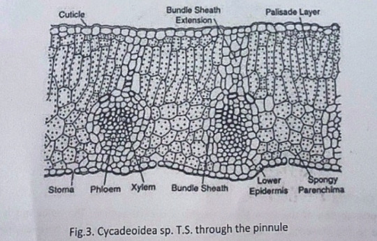
Anatomy of Leaf:
The pinnules show xerophilous structure. The upper and lower epidermis is heavily cutinized and thick walled. The mesophyll cells are distinguished into palisade and spongy parenchyma. The vascular bundles are mesarch and surrounded by bundle sheath.
Reproduction:
The reproductive structure is represented by flowers. In most of the species, the flowers are bisexual and arise in the axil of each leaf.
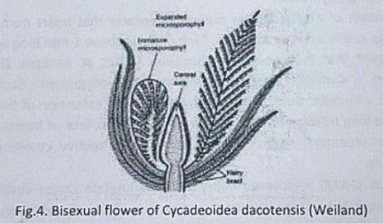
Structure of Flower:
The flowers are bisporangiate, stalked, and partially sunken in the leaf base armor. Rach such mature flower is 5-10cm in diameter and 10cm long. From the base of such flowers about 100 to 150 hairy bracts arise in close spiral little below the apex. These bracts formed a perianth like structure and protect the megasporangiate and microsporangiate parts of a flower. The microsporophyll or androecium forms a whorl united at the base into a sheath. The megasporophyll or gynaecium consists of numerous stalked ovules born around a central receptacle. Between the ovules, interseminal scales with expanded tips are present. These expanded tips fused to form a continuous surface with pores, through which the micropyle of ovules extended. The vascular supply of flowers consists of many branches from leaf traces.
Microsporophyll or Androecium:
The microsporophyll is 10-12cm long, consists of a central rachis bearing numerous pinnae. The pinnae bear two rows of bean-shaped shortly stalked pollen capsules or synangia. These pollen capsules are born on the trabeculae within the fertile region of microsporophyll. A line of dehiscence is also visible at the base of each microsporophyll. This suggest that the entire microsporophyll might have been shed as a unit. The pollen capsule or synangia measures about 3.5x2.5mm and its wall is several layers thick, the outer layer made up of palisade like cells, and the inner layer is made up of thin-walled cells followed by a tapetum. The tapetum was not demarcated. A ring of microsporangia arranged around the periphery of each synangium. The microsporangia dehisce longitudinally and release the microspores into the synangial cavity. At maturity, the synangia liberate these microspores outside by an apical opening that splits into two valves. The liberated microspores or pollens are oval, measures up to 68µ that represents the male gametophytes. Pollen grains of Cycadeoidea are multicellular.
Megasporophyll or Gynoecium
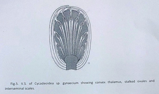
The gynoecium consists of a spherical or conical receptacle that bears numerous stalked orthotropous ovules and interseminal scales. Each ovule is about 1mm long and consists of the single integument that fused with the nucellus except at its apex.
According to Lignier, in C. morieri, nucellus is free from the integument. Each ovule has a pollen chamber and a nucellar beak. This nucellar beak is the extension of the integument. The ovules also have long micropyle, extended from the flat surface of interseminal scales. The fused tips of interseminal scales form an external protective covering or pericarp surrounding the seeds.
Crepet and Delevoryas discovered many of bisporangiate cones from the Cretaceous of black hills. They studied the structure of these ovules in detail. These ovules are urn-shaped and resemble with the ovules of C. wellsii. According to them the micropyle of these ovules are funnel-shaped due to the constriction below the flaring. The inner wall of the micropyle is lined with large cells, considered to be epidermal cells. The integument has three distinct layers. The outer fleshy layer of radially elongated cells, the middle stony layer made up of thick-walled cells, and the inner layer is fleshy.
The young nucellus is made up of thin-walled cells. The cells at the micropylar end are much elongated (80µ long) in comparison to the cells of the chalazal end. The cell at the nucellar tip is pointed up tp whereas cells on either side are bend outward to give the nucellus a distinct shape.
Crepet and Delevoryas reported a linear tetrad or row of three cells in the center of the nucellus.
The seeds are somewhat elongated or oval and possessed two cotyledons.
#exam season#send help#biology#notes#science#botany#gymnosperms#pines#pine trees#pine needles#plants#plant science#plant biology#nature#long post
8 notes
·
View notes
Last Seen Blogs

chocobocodin
dio apologist

wallyelledge419-blog
Yellowish Page Advertising and marketing Tactics That Work

tabris3000
Tubris


artemeis
sit sine spina