#centrioles
Text
Centriole Formation
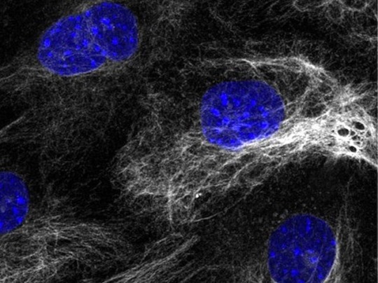
Protein called SAS-6 role in the maintenance of the architecture of centrioles – specialised structures involved in organising cell contents during division – as they form early in embryonic development in cells that generate mouse embryonic stem cells
Read the published article here
Image from work by Marta Grzonka and Hisham Bazzi
Department of Cell Biology of the Skin and Department of Dermatology and Venereology, Medical Faculty, University of Cologne, Cologne, Germany
Image originally published with a Creative Commons Attribution 4.0 International (CC BY 4.0)
Published in eLife, February 2024
You can also follow BPoD on Instagram, Twitter and Facebook
#science#biomedicine#immunofluorescence#cells#stem cells#cell division#centrioles#embryo development#developmental biology
12 notes
·
View notes
Text
Hot young stud with a hard cock sucks a tranny then fucks her tigh ass
Blonde milf dildo ass Krissy Lynn in The Sinful Stepmother
Gostosas com cenas maravilhosas
ESPOSA DE QUATRO PARA O AMIGO DO CORNO
Zoey Holloway hot milf love black dick in her
Ashlee stuffs her pussy until it explodes with a climax
Amateur lesbian babes are playing with their toys all day long
Asian Girl Handcuffed for Backshots
BurningAngel Dirty Waitress Anna Bell Peaks Gets Banged Hard By An Old Perv
Wet Japanese Pussy Of Wife Chiaki Sugai Is Hungry For Some Rough Sex
#misanthropi#女装#outprays#suber#eloped#swallow-tailed#Lenoxdale#variegated#Connarus#sauceline#frontend#enarbor#uneye#lightened#centriole#Eleutherios#undawning#sore-won#Teteak#nonsystem
0 notes
Text
Where do the microtubules of the spindle originate during mitosis in animal cells a centromere B centrosome C Centriole?
Where do the microtubules of the spindle originate during mitosis in animal cells a centromere B centrosome C Centriole?
Where do microtubules originate in animal cells?
Where do the microtubules of the spindle Fibres originate?
Where do the microtubules of the spindle apparatus originate during mitosis in both plant and animal cells?
Where are microtubules in mitosis?
Where do microtubules grow from in animal cells?
Where do the microtubules originate from?
Where do microtubules originate during mitosis in animal cells?
Where are microtubules found?
What structure do the spindle fibers originate from?
What phase does spindle microtubules form?
Where do the microtubules in the spindle apparatus connect?
Where do the microtubules of the spindle originate during mitosis in both plant and animal cells a centromere B centrosome C Centriole D chromatid e kinetochore?
Where do microtubules originate in plant and animal cells?
Where do microtubules of the spindle apparatus originate during mitosis?
Are microtubules in both plant and animal cells?
Where can microtubules be found?
Where do microtubules attach during mitosis and meiosis?
What is microtubules in mitosis?
Are microtubules in meiosis?
Where do microtubules grow from?
How do microtubules grow?
Where do the microtubules of the spindle originate grow from during mitosis in animal cells?
Where does the microtubules of plant cell came from and how is it important during cell division?
What pathway produces microtubule proteins in cells?
#Where do the microtubules of the spindle originate during mitosis in animal cells a centromere B centrosome C Centriole#Bookvea
0 notes
Text
lab courses will have you look at the grungiest slide in the universe with no stain on it and ask you to identify the centrioles. like sir that is not happening but i can identify all the floaters in my eye for you instead
120 notes
·
View notes
Text
i've officialy lost it.
#i have a biology test tomorrow.#i've been preparing for days. i'm sick of it.#but lysosomes absolutely fuck#it's really late so that explains whatever this is anfjfjskdnfj#niko rambles
36 notes
·
View notes
Text
Organelles
Nucleus
-- located near the center of the cell
-- contains the genetic material of the cell (DNA) and nucleoli
-- site of ribosome and messenger RNA synthesis
Nucleolus
-- located in the nucleus
-- site of ribosomal subunit assembly
Ribosomes
-- located in the cytoplasm
-- site of protein synthesis
Rough Endoplasmic Reticulum
-- located in the cytoplasm
-- has many ribosomes attached
-- site of protein synthesis
Smooth Endoplasmic Reticulum
-- located in the cytoplasm
-- site of lipid synthesis
-- participates in detoxification
Golgi Apparatus
-- located in the cytoplasm
-- modifies protein structure
-- packages proteins in secretory vesicles
Secretory Vesicle
-- located in the cytoplasm
-- contains materials produced in the cell
-- formed by the Golgi apparatus
-- secreted by exocytosis
Lysosome
-- located in the cytoplasm
-- contains enzymes that digest material taken into the cell
Mitochondria
-- located in the cytoplasm
-- site of aerobic respiration
-- major site of ATP synthesis
Microtubule
-- located in the cytoplasm
-- supports cytoplasm
-- assists in cell division
-- forms components of cilia and flagella
Centrioles
-- located in the cytoplasm
-- facilitate the movement of chromosomes during cell division
Cilia
-- located on the cell surface with many on each cell
-- move substances over the surface of certain cells
Flagella
-- located on sperm cells
-- one per cell
-- propels sperm cell
#medblr#studyblr#notes#my notes#anatomy and physiology#anatomy#physiology#biology#bio#bio notes#biology notes#anatomy notes#physiology notes#cells#cells notes#microbiology#microbiology notes#studyblr notes
62 notes
·
View notes
Text
kinetochores are so weird like why is there a whole other part of the centriole microtubles attach to why cant they just be like "ya centrioles are were the bullshit happens" like what?
#auburn's rambles <3#i should not be up this late studying for bio#sillies and goofies amiright#THIS IS A RHETORICAL QUESTION BTW DONT ACTUALLY ANSWER IT IM TIRED
20 notes
·
View notes
Text


Lezley Irene Saar is an African American artist whose artwork is responsive to race, gender, female identity, and her ancestral history. Her works are primarily mixed media, 3-dimensional, and oil & acrylic on paper and canvas.
Her mother Betye Saar (née Brown), is an African-American assemblage artist. Via Wikipedia
#bornonthisday Lezley Saar
Centrioles Later, 2014
Let's talk of space sadness, 2014
Detail “Not born under a rhyming planet”
Mitochondrial Muses, 2014
All from the Monad Series
Let's read it together again: Betye Saar, Her Daughters, and the House That Never Stopped Making Art
www.harpersbazaar.com/culture/features/a36182098/betye-saar-daughters-in-conversation/
The pioneering artist and her three daughters on family, creativity, and why being able to see beauty, even in difficult times, is the true mother of invention. (2021)
#TracyeSaarCavanaugh #AlisonSaar #BetyeSaar #LezleySaar #artherstory #artbywomen #womensart #palianshow #art #womenartists #femaleartist #artist #daughters
3 notes
·
View notes
Text
New Beginnings
The cell was preparing for mitosis; the process by which it would divide and replicate itself in order to create two identical daughter cells. Prophase, the first stage of mitosis, was already underway. The cell's nucleus, containing its genetic material in the form of threadlike chromosomes, was beginning to break down. Proteins called histones, which normally helped to package the DNA into an orderly structure, were now starting to unwind and coil themselves around the chromosomes.
This process of coiling was crucial; if the DNA didn't condense sufficiently, it would make cell division difficult and potentially result in unequal distribution of genetic material between the two daughter cells. At the same time, the sister chromatids - the two identical strands of DNA that made up each chromosome - were beginning to separate from each other, aided by a specialized structure called the centrosome. The centrosome contained a pair of structures called centrioles, which helped to organize microtubules that would later be used to pull the sister chromatids apart during a process called anaphase.
The cell's cytoskeleton was also beginning to undergo changes. Microtubules, long protein fibers that formed the cell's skeleton, were starting to appear and begin to connect with the centrosome. Soon, they would stretch out across the cell, forming a spindle-shaped structure that would play a critical role in separating the chromosomes during mitosis.
2 notes
·
View notes
Text
Mitosis Help- College Biology (AP)
Introduction
What do your intestines, the yeast in bread dough, and a developing frog all have in common? Among other things, they all have cells that carry out mitosis, dividing to produce more cells that are genetically identical to themselves.
Why do these very different organisms and tissues all need mitosis? Intestinal cells have to be replaced as they wear out; yeast cells need to reproduce to keep their population growing; and a tadpole must make new cells as it grows bigger and more complex.
What is mitosis?
Mitosis is a type of cell division in which one cell (the mother) divides to produce two new cells (the daughters) that are genetically identical to itself. In the context of the cell cycle, mitosis is the part of the division process in which the DNA of the cell's nucleus is split into two equal sets of chromosomes.
The great majority of the cell divisions that happen in your body involve mitosis. During development and growth, mitosis populates an organism’s body with cells, and throughout an organism’s life, it replaces old, worn-out cells with new ones. For single-celled eukaryotes like yeast, mitotic divisions are actually a form of reproduction, adding new individuals to the population.
In all of these cases, the “goal” of mitosis is to make sure that each daughter cell gets a perfect, full set of chromosomes. Cells with too few or too many chromosomes usually don’t function well: they may not survive, or they may even cause cancer. So, when cells undergo mitosis, they don’t just divide their DNA at random and toss it into piles for the two daughter cells. Instead, they split up their duplicated chromosomes in a carefully organized series of steps.
Phases of mitosis
Mitosis consists of four basic phases: prophase, metaphase, anaphase, and telophase. Some textbooks list five, breaking prophase into an early phase (called prophase) and a late phase (called prometaphase). These phases occur in strict sequential order, and cytokinesis - the process of dividing the cell contents to make two new cells - starts in anaphase or telophase.
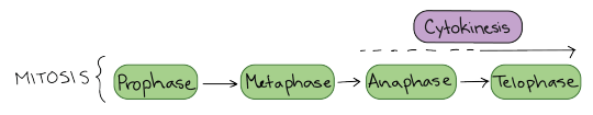
Stages of mitosis: prophase, metaphase, anaphase, telophase. Cytokinesis typically overlaps with anaphase and/or telophase.
You can remember the order of the phases with the famous mnemonic: [Please] Pee on the MAT. But don’t get too hung up on names – what’s most important to understand is what’s happening at each stage, and why it’s important for the division of the chromosomes.
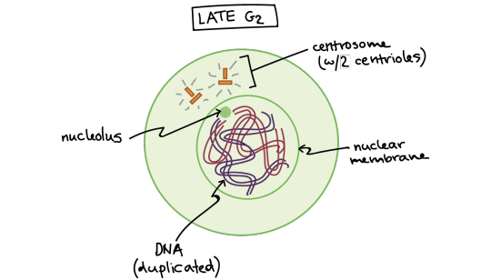
Late G2 phase. The cell has two centrosomes, each with two centrioles, and the DNA has been copied. At this stage, the DNA is surrounded by an intact nuclear membrane, and the nucleolus is present in the nucleus.
Let’s start by looking at a cell right before it begins mitosis. This cell is in interphase (late G\[_2\] phase) and has already copied its DNA, so the chromosomes in the nucleus each consist of two connected copies, called sister chromatids. You can’t see the chromosomes very clearly at this point, because they are still in their long, stringy, decondensed form.
This animal cell has also made a copy of its centrosome, an organelle that will play a key role in orchestrating mitosis, so there are two centrosomes. (Plant cells generally don’t have centrosomes with centrioles, but have a different type of microtubule organizing center that plays a similar role.)

Early prophase. The mitotic spindle starts to form, the chromosomes start to condense, and the nucleolus disappears.
In early prophase, the cell starts to break down some structures and build others up, setting the stage for division of the chromosomes.
The chromosomes start to condense (making them easier to pull apart later on).
The mitotic spindle begins to form. The spindle is a structure made of microtubules, strong fibers that are part of the cell’s “skeleton.” Its job is to organize the chromosomes and move them around during mitosis. The spindle grows between the centrosomes as they move apart.
The nucleolus (or nucleoli, plural), a part of the nucleus where ribosomes are made, disappears. This is a sign that the nucleus is getting ready to break down.
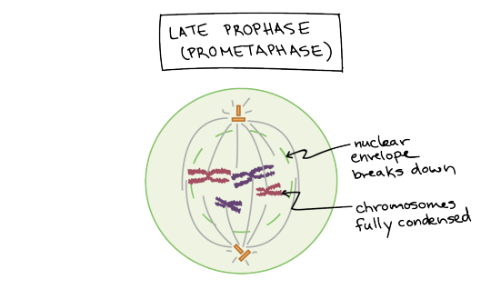
Late prophase (prometaphase). The nuclear envelope breaks down and the chromosomes are fully condensed.
In late prophase (sometimes also called prometaphase), the mitotic spindle begins to capture and organize the chromosomes.
The chromosomes become even more condensed, so they are very compact.
The nuclear envelope breaks down, releasing the chromosomes.
The mitotic spindle grows more, and some of the microtubules start to “capture” chromosomes.

Anatomy of the mitotic spindle. Diagram indicating kinetochore microtubules (bound to kinetochores) and the aster. The aster is an array of microtubules that radiates out from the centrosome towards the cell edge. Diagram also indicates the centromere region of a chromosome, the narrow "waist" where the two sister chromatids are most tightly connected, and the kinetochore, a pad of proteins found at the centromere.
Microtubules can bind to chromosomes at the kinetochore, a patch of protein found on the centromere of each sister chromatid. (Centromeres are the regions of DNA where the sister chromatids are most tightly connected.)
Microtubules that bind a chromosome are called kinetochore microtubules. Microtubules that don’t bind to kinetochores can grab on to microtubules from the opposite pole, stabilizing the spindle. More microtubules extend from each centrosome towards the edge of the cell, forming a structure called the aster.
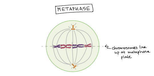
Metaphase. Chromosomes line up at the metaphase plate, under tension from the mitotic spindle. The two sister chromatids of each chromosome are captured by microtubules from opposite spindle poles.
In metaphase, the spindle has captured all the chromosomes and lined them up at the middle of the cell, ready to divide.
All the chromosomes align at the metaphase plate (not a physical structure, just a term for the plane where the chromosomes line up).
At this stage, the two kinetochores of each chromosome should be attached to microtubules from opposite spindle poles.
Before proceeding to anaphase, the cell will check to make sure that all the chromosomes are at the metaphase plate with their kinetochores correctly attached to microtubules. This is called the spindle checkpoint and helps ensure that the sister chromatids will split evenly between the two daughter cells when they separate in the next step. If a chromosome is not properly aligned or attached, the cell will halt division until the problem is fixed.
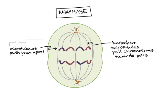
Anaphase. The sister chromatids separate from one another and are pulled towards opposite poles of the cell. The microtubules that are not attached to chromosomes push the two poles of the spindle apart, while the kinetochore microtubules pull the chromosomes towards the poles.
In anaphase, the sister chromatids separate from each other and are pulled towards opposite ends of the cell.
The protein “glue” that holds the sister chromatids together is broken down, allowing them to separate. Each is now its own chromosome. The chromosomes of each pair are pulled towards opposite ends of the cell.
Microtubules not attached to chromosomes elongate and push apart, separating the poles and making the cell longer.
All of these processes are driven by motor proteins, molecular machines that can “walk” along microtubule tracks and carry a cargo. In mitosis, motor proteins carry chromosomes or other microtubules as they walk.

Telophase. The spindle disappears, a nuclear membrane re-forms around each set of chromosomes, and a nucleolus reappears in each new nucleus. The chromosomes also start to decondense.
In telophase, the cell is nearly done dividing, and it starts to re-establish its normal structures as cytokinesis (division of the cell contents) takes place.
The mitotic spindle is broken down into its building blocks.
Two new nuclei form, one for each set of chromosomes. Nuclear membranes and nucleoli reappear.
The chromosomes begin to decondense and return to their “stringy” form.
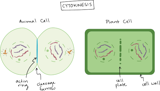
Cytokinesis in animal and plant cells.
Cytokinesis in an animal cell: an actin ring around the middle of the cell pinches inward, creating an indentation called the cleavage furrow.
Cytokinesis in a plant cell: the cell plate forms down the middle of the cell, creating a new wall that partitions it in two.
Cytokinesis, the division of the cytoplasm to form two new cells, overlaps with the final stages of mitosis. It may start in either anaphase or telophase, depending on the cell, and finishes shortly after telophase.
In animal cells, cytokinesis is contractile, pinching the cell in two like a coin purse with a drawstring. The “drawstring” is a band of filaments made of a protein called actin, and the pinch crease is known as the cleavage furrow. Plant cells can’t be divided like this because they have a cell wall and are too stiff. Instead, a structure called the cell plate forms down the middle of the cell, splitting it into two daughter cells separated by a new wall.

When division is complete, it produces two daughter cells. Each daughter cell has a complete set of chromosomes, identical to that of its sister (and that of the mother cell). The daughter cells enter the cell cycle in G1.
When cytokinesis finishes, we end up with two new cells, each with a complete set of chromosomes identical to those of the mother cell. The daughter cells can now begin their own cellular “lives,” and – depending on what they decide to be when they grow up – may undergo mitosis themselves, repeating the cycle.
5 notes
·
View notes
Note
challenge: how many cell organelles can you can you name in the pic you posted
golgi apparatus
endoplasmic reticulum (rough and smooth)
lyosomes
centrioles
mitochondria
cell membrane
nucleus
cytoplasm
😎
i did anatomy 😎
3 notes
·
View notes
Text
Centriole Building
The centriole – an organelle vital for the faithful separation into each daughter cell when a cell divides – is composed of an assembly of proteins. Using expansion microscopy, this study correlates the spatial location of 24 of those proteins with structural features of the human centriole, and then models the molecular architecture of the centriole as it assembles (shown here)
Read the published research article here
Video from work by Marine H. Laporte and colleagues
University of Geneva, Department of Molecular and Cellular Biology, Faculty of Sciences, Geneva, Switzerland
Video originally published with a Creative Commons Attribution 4.0 International (CC BY-NC 4.0)
Published in Cell, April 2024
You can also follow BPoD on Instagram, Twitter and Facebook
#science#biomedicine#biology#cell division#centrioles#centrosome#computer modeling#expansion microscopy
6 notes
·
View notes
Text
Scientists reconstruct assembly of the human centriole, image by image, for the first time [ Biology ]
Scientists reconstruct assembly of the human centriole, image by image, for
the first time [Highlights]
Cells contain various specialized structures—such as the nucleus,
mitochondria or peroxisomes—known as “organelles.
Centriole biogenesis, as in most organelle assemblies, involves the
sequential recruitment of sub-structural elements that will support its…
The largest repository of validated,…

View On WordPress
0 notes
Text
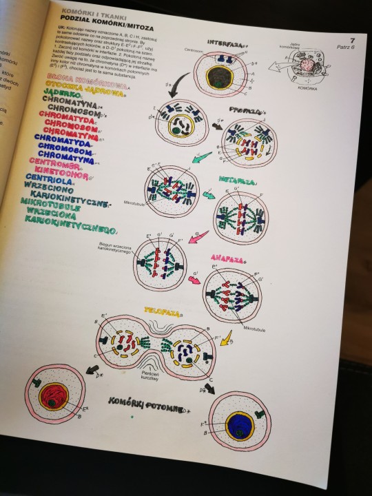
KOMÓRKI I TKANKI cz. 2/9
PODZIAŁ KOMÓRKI / MITOZA
Zdolność do reprodukcji jest cechą charakterystyczną wszystkich żywych organizmów. Komórki rozmnażają się w wyniku podziału pośredniego jądra komórkowego, czyli mitozy. W jądrze komórki chromatyna (substancja zbudowana z DNA i białek) ulega kondensacji i przekształca się w 46 chromosomów, które dzielą się na połowę. W wyniku tego procesu powstają 92 chromatydy, które wędrują do przeciwległych biegunów komórki, z których powstaje 46 chromosomów w każdej z dwóch nowych komórek potomnych. By zachować przejrzystość, pokazujemy tylko cztery pary chromatyd i chromosomów.
Interfaza - faza między podziałami komórki; najdłuższa część cyklu reprodukcyjnego. W tej fazie następuje replikacja (podwojenie) ilości DNA w chromatynie. Rozluźniona chromatyna (D*) jest siecią fibryli, niewidocznych gołym okiem, włókienkowatych struktur w nukleoplazmie. W tej fazie jądro komórkowe i jąderko pozostają nienaruszone, a w centrosomie dzielą się połączone w pary centriole.
Profaza - rozluźniona chromatyna (D*) zaczyna się kondensować, skracać i skręcać w chromosomy (D1*). Każdy chromosom jest zbudowany z dwóch chromatyd (E) i (F) połączonych centromerem (G). Chromatydy tego samego chromosomu mają identyczną sekwencję DNA. W tej fazie następuje rozpad i zanik błony jądrowej oraz jąderka. Centriole się rozdzielają i wędrują do przeciwnych biegunów komórki, gdzie z mikrotubul wrzeciona (włókien białka) formuje się wrzeciono kariokinetyczne (podziałowe). Na centromerze (przewężeniu chromosomu) formuje się kinetochor (G1).
Metafaza - włókna wrzeciona kariokinetycznego (mikrotubule) łączą się z centromerami chromosomów (na kinetochorze), umożliwiając "wędrówkę" chromatyd do przeciwległych biegunów komórki (46 chromatyd na każdym biegunie). Błona jądrowa ulega rozpadowi.
Anafaza - siostrzane centromery (G1; kinetochory) każdej z chromatyd przemieszczają się do biegunów komórki wzdłuż wrzeciona kariokinetycznego. Tam z chromatyd powstają chromosomy. Anafaza się kończy, gdy chromosomy potomne znajdują się na przeciwległych biegunach (46 chromosomów po każdej ze stron).
Telofaza - chromosomy na dwóch przeciwległych biegunach tworzą dwie komórki potomne; każda jest identyczna z komórką macierzystą (pod warunkiem że nie występują żadne mutacje). Cytoplazma i organelle komórkowe, wcześniej zduplikowane, dzielą się, tworząc dwie nowe komórki. Powstają dwa jądra komórkowe oraz otoczki jądrowe. W każdej nowej komórce powstaje jąderko, struktura chromosomów w chromatynie ulega rozluźnieniu, a centrometr zanika. Następuje rozdzielenie komórki macierzystej na komórki potomne, zawierające taki sam materiał, który kończy proces mitozy. Każda komórka potomna wchodzi w etap interfazy i cały proces zaczyna się od nowa.
0 notes
Text
🗒️💬
Les microtubules sont assemblés à partir de dimères d'α- et de β-tubuline. Ces sous-unités sont légèrement acides, avec un point isoélectrique compris entre 5,2 et 5,8[2]. Chacune a un poids moléculaire d'environ 50 kDa[3].
Pour former les microtubules, les dimères d'α- et de β-tubuline se lient au GTP et s'assemblent sur les extrémités (+) des microtubules lorsqu'ils sont à l'état lié au GTP[4]. La sous-unité de β-tubuline est exposée sur l'extrémité plus du microtubule, tandis que la sous-unité d'α-tubuline est exposée sur l'extrémité moins. Après l'incorporation du dimère dans le microtubule, la molécule de GTP liée à la sous-unité β-tubuline finit par s'hydrolyser en GDP par le biais de contacts interdimères le long du protofilament du microtubule[5]. La molécule de GTP liée à la sous-unité α-tubuline n'est pas hydrolysée pendant tout le processus. Le fait que le membre β-tubuline du dimère de tubuline soit lié au GTP ou au GDP influence la stabilité du dimère dans le microtubule. Les dimères liés au GTP ont tendance à s'assembler en microtubules, tandis que les dimères liés au GDP ont tendance à se désagréger ; ce cycle du GTP est donc essentiel pour l'instabilité dynamique du microtubule.
Microtubules bactériens
modifier
Des homologues de l'α- et de la β-tubuline ont été identifiés dans le genre de bactéries Prosthecobacter[6]. Ils sont désignés BtubA et BtubB pour les identifier comme des tubulines bactériennes. Toutes deux présentent une homologie avec les α- et β-tubulines[7]. Bien que leur structure soit très similaire à celle des tubulines eucaryotes, elles présentent plusieurs caractéristiques uniques, notamment un repliement sans chaperon et une faible dimérisation[8]. Des études in vitro montrent que les BtubA/B forment des « mini-microtubules » à quatre brins[9], contrairement aux microtubules eucaryotes, qui en contiennent généralement 13.
La tubuline possède 3 sites de liaison, qui sont les cibles de médicaments anticancéreux ; le site du Taxol, de la Vinblastine et de la colchicine. La colchicine et la Vinblastine se lient à la tubuline et inhibent sa polymérisation, c'est-à-dire la formation de microtubules, immobilisant les neutrophiles et abaissant l'inflammation. Le Taxol inhibe la dépolymérisation des microtubules.
🗒️💬
Centrosome
Le centre organisateur des microtubules, abrégé COMT ou MTOC, est une structure présente dans les cellules eucaryotes d'où émergent les microtubules. Dans les cellules animales, il est composé de matériel péricentriolaire et d'un centrosome, lui-même composé de deux centrioles.
Dans les cellules animales, le centre organisateur des microtubules est composé d'un centrosome, organite non membrané qui se compose d'une paire de centrioles, et d'un nuage de matériel amorphe appelé matériel péricentriolaire[1]. Un centriole est composé de neuf triplets de microtubules (avec treize protofilaments entre chaque microtubule), formant la paroi d'un cylindre. C'est à partir de cet ensemble que s'effectue la nucléation des microtubules grâce à la présence, à sa surface, d'anneaux de tubuline γ, homologue de la protéine ARP pour l'actine. Il n'existe pas de continuité entre les centrioles et les microtubules cytosoliques, qui polymérisent autour des anneaux de tubuline γ. Les microtubules polymérisent à partir de ce centre organisateur qui représente le point de ralliement des microtubules, lui donnant alors un rôle primordial dans le trafic intracellulaire. Le centre organisateur des microtubules a un rôle dans l'orientation des cellules et est à l'origine des cils et des flagelles.
Durant l'interphase, le centrosome est responsable de la nucléation microtubulaire. Le centrosome se duplique au cours de la phase de synthèse (pendant l'interphase) et, pendant la mitose, se sépare pour former les deux pôles du fuseau mitotique (appareil mitotique). Il y a donc deux paires de centrioles appelées chacune « diplosome », c'est de ces deux pôles que seront nucléés les microtubules du fuseau mitotique.
Dans les cellules végétales, il n'y a pas de centrosome, mais le gamma-tubulin ring complex y est conservé et permet la nucléation de nouveaux microtubules. Cette nucléation est microtubules dépendante, c'est-à-dire qu'elle a lieu le long de microtubules déjà existants. En absence de centrosome et de centriole, dans les cellules végétales on parle généralement d'un centre organisateur de microtubule diffus. L'absence de centrosome n’empêche pas la division cellulaire d'avoir lieu, les cellules végétales ne sont d'ailleurs pas les seules à ne pas avoir de centrosome, les ovocytes en sont également dépourvus, lors de la division les pôles du fuseau sont simplement moins focalisés[2].
Les levures ne possèdent pas de centrosome, mais ont un centre organisateur des microtubules, situé en périphérie du noyau, mais aussi le long de microtubules déjà existants, cette structure est à la base de la formation des microtubules[3].
Les neurones n'ont pas de centrosome.
Les cellules cancéreuses contiennent un ou plusieurs centrosomes supplémentaires, mais peuvent néanmoins se reproduire. Ceci est une caractéristique propre, connue depuis le début du xxe siècle, qui pourrait peut-être permettre de mieux cibler ces cellules par de nouveaux médicaments anti-cancéreux que l'on cherche à développer[4]. L'exposition de certaines cellules au bisphénol A pourrait perturber les centrosomes et peut-être expliquer un risque accru de cancer de la prostate chez les hommes exposés à cette molécule (qui est aussi un perturbateur endocrinien)[5].
Le centrosome est une entité très complexe dont le fonctionnement reste quelque peu mystérieux.
🗒️💬
Mitose
🗒️💬
Protofilament
🗒️💬
In Vivo
🗒️💬
In vitro
In vitro (en latin : « sous verre ») s'applique à toute activité expérimentale réalisée sur micro-organismes, organes ou cellules en dehors de leur contexte naturel (en dehors de l'environnement, de l'organisme vivant ou de la cellule) et en conditions définies et contrôlées. Un exemple est la fécondation in vitro (FIV)
Le terme « in vitro » provient du latin qui signifie « sous verre ». Il est à mettre en association avec les termes « in vivo » et « in silico ».
In vivo (en latin : « dans le vivant ») signifie une approche au sein d'un environnement complexe (plus proche des conditions naturelles). Par opposition, les investigations réalisées in vitro sont menées en dehors du milieu naturel, de l'organisme vivant ou de la cellule initiale.
In silico (qui est un néologisme d'inspiration latine), se traduit par des méthodes physiques et/ou mathématiques permettant des modélisations totalement soustraites des conditions naturelles.
In vitro (à la différence de in silico) ne veut pas forcément dire en dehors du vivant puisque des cultures de cellules vivantes peuvent se faire en dehors de leur environnement naturel.
Il existe donc une gradation entre ces trois termes, qui suggère un éloignement plus ou moins marqué des conditions naturelles.
De nombreuses disciplines utilisent des approches in vitro, telles que :
De nombreuses approches expérimentales dans le domaine de la recherche biologique et biotechnologique s’appuient sur des techniques in vitro comme la culture cellulaire[1] ou la culture de végétaux vasculaires[2].
Industrie outils de production médicale et cosmétique
modifier
La production de masse de certains composés et médicaments sont aujourd'hui réalisés grâce aux techniques de production in vitro comme la production d'insuline qui fut la première utilisation de bactéries et levures pour produire une protéine humaine d’intérêt[3],[4] ou de composés cosmétiques[5].
Industrie et outils de production agroalimentaire
modifier
L'utilisation de fermenteur va permettre la synthèse de composés et enzymes pour l’industrie agroalimentaire comme la synthèse de la lactase ou de ferments destinés à la transformation des produits laitiers, la brasserie, la viticulture. De plus, dans le domaine agricole, la multiplication de plantes de consommation et d'ornement est parfois réalisée par micropropagation (culture de végétaux vasculaire) afin de produire rapidement et à grande échelle certaines plantes d’intérêt[6].
🗒️💬
Polymère
🗒️💬
Polarisé
🗒️💬
Protéines :
Il y as des protéines associés aux microtubules (MAP) qui se lient aux microtubules et leur confèrent des fonctions.
Protéines de séquestration :
Dans les microtubules on associe aux monomères d'actine G liée au GTP (vide supra). On associe aux dimères de tubuline libres, des protéines de séquestration appelées stathmines.
Elles ont une double fonction :
principalement elles fixent les dimères de tubuline en forme G libre pour en empêcher la polymérisation ;
mais elles sont impliquées aussi dans la présentation optimale des dimères libres à l’extrémité + des microtubules (stimulation de la polymérisation).
Ces protéines maintiennent une concentration faible des formes G libres (équilibre).
Ces protéines favorisent la dépolymérisation.
🗒️💬
Protéines de fragmentation :
Katanine : notamment pendant le cycle de mitose au moment de la cytodiérèse, il rompt les microtubules en petites fragments qui se dépolymérisent en dimères. Ils peuvent de réassembler.
MCAK : Supprime les dimères des extrémités et entraîne un raccourci du microtubule.
MIDD1 : Elle s'accumule directement proge des microtubules dans les cellules, et in vitro, il s'y lie directement.
🗒️💬
Protéines stabilisatrices :
🗒️💬
Protéines motrice :
🗒️💬
Kinésine :
🗒️💬
GTP :
Le guanosine triphosphate (GTP) est une coenzyme de transfert de groupements phosphate.
Les propriétés du guanosine triphosphate et de ses dérivés, guanosine diphosphate et guanosine monophosphate, sont identiques à celles de l'adénosine triphosphate (et ses dérivés). C'est un donneur de phosphate. Il est hydrolysé par toute une série d'enzymes appelées les GTPases. Ces protéines alternent entre deux conformations : active liée au GTP et inactive liée au GDP. L'équilibre entre ces deux conformations est régulé par des facteurs d'échange (GEF) permettant l'échange du GDP par le GTP, des protéines GAP catalysant l'hydrolyse du GTP, et enfin des protéines GDI inhibant la dissociation du GDP.
🗒️💬
GDP :
La guanosine diphosphate, abrégée en GDP, est un nucléotide. C'est une coenzyme de transfert de groupements phosphate. Elle résulte de l'hydrolyse de la GTP. Le groupement phosphate libéré peut être transféré sur une protéine.
🗒️💬
GTPases :
Les GTPases sont une classe importante d'enzymes qui catalysent l'hydrolyse de la guanosine triphosphate (GTP) pour donner une guanosine diphosphate (GDP) et un ion phosphate. La fixation du GTP est effectuée par un domaine très conservé dans l'évolution, appelé domaine G, caractéristique de l'ensemble de cette superfamille. Cette hydrolyse est en général couplée à un autre processus biologique, comme la transduction du signal dans la cellule. Les GTPases appartiennent à la catégorie EC 3.6.5 de la nomenclature internationale des enzymes.
🗒️💬
Dynéines :
Molécule de kinésine progressant le long d'un microtubule : cette protéine fonctionne comme une nanomachine.
Les moteurs moléculaires sont des ATPases :
Les kinésines, moteurs moléculaires liés à des éléments figurés qui se déplacent vers l'extrémité positive (+) des microtubules; on parle de transport antérograde.
Les dynéines, moteurs moléculaires liés à des éléments figurés qui se déplacent vers l'extrémité négative (-) des microtubules; on parle de transport rétrograde.
🗒️💬
Protéines stabilisatrice :
🗒️💬
MAP2
🗒️💬
Tau
🗒️💬
Tau : elle assure le même rôle que MAP2 mais cette fois ci principalement dans les axones des cellules nerveuses. Lors du dysfonctionnement de cette protéine, on parle de tauopathie, étant l'une des causes (ou conséquences ?) des maladies neurodégénératives comme la maladie d'Alzheimer par exemple.
Dans la maladie d'Alzheimer, la protéine Tau est abondante et anormalement phosphorylée dans les neurones. Ce qui entraine une anomalie dans l'organisation des microtubules qui génère des amas neurofibrillaires avec filaments introduisant une dégénérescence neuronale.
🗒️💬
TUBB, TUBB1, TUBB2A, TUBB2B, TUBB2C, TUBB3, TUBB4, TUBB4Q, TUBB6 et TUBB8.
0 notes
Text
A Midcellmmer Night’s Dream
Our cell membranes press against each other, and I know I’m in love. Accurate to their name, G2trude is in the G2 phase of the cell cycle. Through their semipermeable cell membrane, I can see enzymes checking that their genetic information is all correct. If I had cheeks and blood to blush with right now, I would. Unlike G2trude, I am only in the G1 phase. I’m still growing, developing organelles, and other things that probably seem super juvenile to G2trude. Hey, someday they’ll be back to where I am though. Kinda. Soon enough, they’ll go into mitosis and after that they’ll be 2 daughter cells in G1. Will they both be G2trude then? Or just one? Or are they completely their own cells? Who really knows, philosopher cells don’t come around these parts. Or any parts, they don’t exist.
It would be easier if we were gametes. We wouldn’t go thru mitosis, and we could be together and be together forever. However, we are skin cells, stuck in the epidermis, stuck in the cell cycle.
We, us, G2trude and Trevor
stuck in cell cycle forever
G1, S, G2, mitosis
believing you love me is psychosis
Something happens! I watch centrioles head to opposite sides of the cell. Oh man, mitosis already? Maybe our phase gap isn’t that bad after all! I watch in silence, for I am a cell and have nothing to make noise with, as G2trude starts prophase. From the centrioles spring forty spindle fibers. The nuclear envelope breaks down, and the chromosomes line up in the middle. I watch in fear and fascination. I can’t believe I’m witnessing this coming of phase. The spindle fibers grab onto the centromeres and pull them apart. I CAN’T WATCH!!! But I do anyway. Oh golly gee, G2trude isn’t their usual shape anymore! They look like two suns, still attached at the side. If only they e could merge together like that! Only gametes merge though. As said before, we are skin cells stuck in the epidermis.
We, us, G2trude and Trevor
stuck in th’epiderm forever
If we were gametes we’d be one
Mr. and Mrs. Trevor, why, that’s fun!
The nuclear envelopes are forming again, around the chromatids. Then, I watch as G2trude is no more. If I had tears to cry with, I would cry. No. SOB. I now know that neither of them is really my G2trude. They’re their own cells. I’ll name them Epinand Dermis.
G2trude isn’t the only one who’s changed however. I’m now in S phase. Synthesizing phase. My DNA is getting replicated into perfect copies. Soon, I’ll be in G2 phase just like G2trude was. Then in M phase: Then G1 again. That’s cell cycle for you. Never ending, never escapable. Predetermined.
This is the story of G2trude and Trevor,
Now it’s time to say cellut forever
#cell#cell cycle#biology#crack fic#fanfiction#cell cycle fanfiction#yearning#this is a summative grade for my biology class#no i am not joking#writblr#writerblr#creative writing#writers of tumblr#writing community
1 note
·
View note
Last Seen Blogs
delusionalteenagewhispers
Randomness

kitsu-smiec
;3 But Ironically
smartys-diary
Ada
mightbeaweeaboo
Gung
l-a-l-o-u
l a l o u