#Atomic Force Microscopy
Text
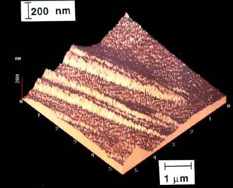
Steel, austenitised at 1200 centigrade for 120 s and then transformed isothermally at 350 centigrade for 2000 s before cooling to room temperature.
Composition
Fe, C 0.24, Si 2.18, Mn 2.32, Ni 1.05 (wt%)
[...]
Processing
The sample was metallographically polished flat prior to transformation. For details see E. Swallow & H. Bhadeshia, Materials Science and Technology 12 (1996) 121
[...]
Sample preparation
Polished flat prior to transformation. All visible relief is due to phase change - the sample is unetched.
Technique
Atomic force microscopy (AFM)
Length bar
1 μm
Further information
Steel, austenitised at 1200 centigrade for 120 s and then transformed isothermally at 350 centigrade for 2000 s before cooling to room temperature. The specimen was polished prior to transformation. This atomic force microscope image shows the displacements caused by the formation of bainite.
Contributor
Prof H K D H Bhadeshia
Organisation
Department of Materials Science and Metallurgy, University of Cambridge
Source.
#Materials Science#Science#Steel#Magnified view#Atomic force microscopy#University of Cambridge#DoITPoMS
6 notes
·
View notes
Text
A Journey into the World of Microscopy: From Humble Beginnings to High-Tech Magnification
The science of looking into the hidden invisible Microscopy has transformed our understanding of the world around us. It can explore the universe beyond the reach of our naked eyes, with complex cellular structures, red blood cells, viruses and other viruses and microorganisms taking on amazing perspectives
The history of the microscope is a fascinating story of human curiosity, scientific genius, and relentless exploration. From the humble beginnings of simple magnifying glasses to the sophistication of modern electronic microscopes, the invention of microscopes has shaped our understanding of the microscopic world
In the 1600s, Dutch opticians such as Hans and Zachary Janssen are credited with inventing the first microscope. Known for this hybrid microscope, many lenses were used to magnify objects up to 30 times.At the end of the 17th century, Antony van Leeuwenhoek, Dutch draper some changed our perception of thumbnails. Armed with a well-made single-lens microscope, and explored the hidden reaches of nature. In 1674, Leeuwenhoek discovered microorganisms in lake water, which he aptly named “animalcules”. His discovery laid the foundations of biology and inspired generations of scientists. This incredible feat allowed him to uncover a hidden universe – the first sightings of bacteria, red blood cells, and other microorganisms.
Formation of the scientific environment (17th-19th centuries): Leeuwenhoek’s discoveries boosted scientific research. Robert Hooke, an English scientist, established these developments. In 1665, his book "Micrographia" recorded his observations with a compound microscope. Notably, the term "cell" was coined by Hooke when he examined cork tissue, laying the foundation for cell biology.Microscope systems flourished throughout the 18th and 19th centuries Joseph Lister and other scientists addressed the limitations of the early lenses, introducing improvements that reduced image distortion.
Beyond the Limits of Light: The Beginning of the New Age (19th-20th century): As the 19th century progressed, the limitations of optical microscopy became apparent and scientists yearned for a tool which can go deeper into cells. This research culminated in the development of the electron microscope in the 1930s. The 20th century was revolutionary with the invention of the electron microscope. Unlike light microscopes, which use visible light, electron microscopes use electron beams to achieve much higher magnification.Formation of the scientific environment (17th-19th centuries): Leeuwenhoek’s discoveries boosted scientific research. Robert Hooke, an English scientist, established these developments. In 1665, his book "Micrographia" recorded his observations with a compound microscope. Notably, the term "cell" was coined by Hooke when he examined cork tissue, laying the foundation for cell biology.Microscope systems flourished throughout the 18th and 19th centuries Joseph Lister and other scientists addressed the limitations of the early lenses, introducing improvements that reduced image distortion.
Beyond the Limits of Light: The Beginning of the New Age (19th-20th century): As the 19th century progressed, the limitations of optical microscopy became apparent and scientists yearned for a tool which can go deeper into cells. This research culminated in the development of the electron microscope in the 1930s. The 20th century was revolutionary with the invention of the electron microscope. Unlike light microscopes, which use visible light, electron microscopes use electron beams to achieve much higher magnification.
In the 1930s, German experts Max Knoll and Ernst Ruska made the first electron microscope. This tool let us see tiny things like cells and even atoms by using electron beams, not light, getting images many times bigger. This cool invention showed us the tiny parts inside cells, viruses, and stuff too small to see before. The 1900s brought even more cool microscopes. New kinds like phase-contrast and confocal microscopy let scientists look at live cells without using stuff that could hurt them. Now, the world of looking at tiny things is getting even better. Today, we have high-tech microscopes that use computers and lasers. These let us see and even change tiny things in ways we never could before.
Modern Microscopy's Diverse Arsenal - Today, the field of microscopy boasts a diverse range of specialized instruments, each tailored to address specific scientific needs. Here's a glimpse into some remarkable examples:
Scanning Electron Microscope (SEM): Imagine a high-tech camera that captures images using a beam of electrons instead of light. That's the essence of a SEM. By scanning the surface of a sample with a focused electron beam, SEMs generate detailed information about its topography and composition. This makes them ideal for studying the intricate structures of materials like insect wings, microchips, and even pollen grains.
Transmission Electron Microscope (TEM): While SEMs provide exceptional surface detail, TEMs take us a step further. They function by transmitting a beam of electrons through a very thin sample, allowing us to observe its internal structure. TEMs are the go-to instruments for visualizing the intricate world of viruses, organelles within cells, and macromolecules like proteins.
Confocal Microscopy: Ever wished to focus on a specific layer within a thick biological sample and blur out the rest? Confocal microscopy makes this possible. It utilizes a laser beam to precisely illuminate a chosen plane within the sample, effectively eliminating information from out-of-focus regions. This allows researchers to create sharp, three-dimensional images of cells, tissues, and even small organisms.
Atomic Force Microscopy (AFM): This technique takes a completely different approach, venturing into the realm of physical interaction. AFM employs a tiny cantilever, akin to a microscopic feeler, to physically scan the surface of a sample. By measuring the minute forces between the cantilever and the sample's surface, AFM can map its topography at an atomic level. This provides invaluable insights into the properties of materials at an unimaginable scale, making it crucial for research in fields like nanotechnology and surface science.
Fluorescence Microscopy: Imagine illuminating a sample with specific wavelengths of light and observing it glowing in response. That's the essence of fluorescence microscopy. This technique utilizes fluorescent molecules or tags that bind to specific structures within a cell or tissue. When excited by light, these tags emit their own light, highlighting the target structures with remarkable clarity. This allows researchers to visualize specific proteins, DNA, or even pathogens within biological samples.
Super-resolution Microscopy (SRM): Overcoming the limitations imposed by the wavelength of light, SRM techniques like STED (Stimulated Emission Depletion) and PALM (Photoactivated Localization Microscopy) achieve resolutions surpassing the diffraction limit. This allows researchers to visualize structures as small as 20 nanometers, enabling the observation of intricate cellular machinery and the dynamics of individual molecules within living cells.
Cryo-Electron Microscopy (Cryo-EM): This powerful technique takes a snapshot of biological samples in their near-life state. Samples are rapidly frozen at ultra-low temperatures, preserving their native structure and minimizing damage caused by traditional fixation methods. Cryo-EM has been instrumental in determining the three-dimensional structures of complex molecules like proteins and viruses, providing crucial insights into their function and potential drug targets.
Correlative Microscopy: Combining the strengths of multiple microscopy techniques, correlative microscopy offers a comprehensive view of biological samples. For instance, researchers can utilize fluorescence microscopy to identify specific structures within a cell and then switch to electron microscopy to examine those structures in high detail. This integrated approach provides a deeper understanding of cellular processes and their underlying mechanisms.
Light Sheet Microscopy (LSM): Imagine illuminating a thin slice of a sample within a living organism. LSM achieves this feat by focusing a laser beam into a thin sheet of light, minimizing photobleaching and phototoxicity – damaging effects caused by prolonged exposure to light. This allows researchers to observe dynamic processes within living organisms over extended periods, providing valuable insights into cellular behavior and development.
Expansion Microscopy (ExM): This innovative technique physically expands biological samples by several folds while preserving their structural integrity. This expansion allows for better resolution and visualization of intricate cellular structures that would otherwise be difficult to distinguish using traditional microscopy methods. ExM holds immense potential for studying the organization and function of organelles within cells.
Scanning Near-Field Optical Microscopy (SNOM): This innovative technique pushes the boundaries of resolution by utilizing a tiny probe that interacts with the sample at an extremely close range. SNOM can not only image the surface features of a sample with exceptional detail but also probe its optical properties at the nanoscale. This opens doors for research in areas like material science and photonics, allowing scientists to study the behavior of light at the interface between materials.
X-ray Microscopy: Stepping outside the realm of light and electrons, X-ray microscopy offers unique capabilities. By utilizing high-energy X-rays, this technique can penetrate deep into samples, making it ideal for studying the internal structure of dense materials like bones and minerals. Additionally, it allows for the visualization of elements within a sample, providing valuable information about their distribution and composition.
From revealing the building blocks of life to aiding in the development of new medicines, the microscope has played an undeniable role in shaping our scientific understanding. As technology continues to evolve, one can only imagine the future breakthroughs this remarkable invention holds in unveiling the secrets of our universe, both seen and unseen. These advancements hold the potential to revolutionize our understanding of biological processes, develop new materials with extraordinary properties, and ultimately pave the way for breakthroughs in medicine, nanotechnology, and countless other fields. As we continue to refine and develop novel microscopy techniques and the future holds immense promise for further groundbreaking discoveries that will undoubtedly revolutionize our perception of the world around us.
#science sculpt#life science#science#molecular biology#biology#biotechnology#artists on tumblr#microscopy#microscope#Scanning Electron Microscope#Transmission Electron Microscope#Confocal Microscopy#Atomic Force Microscopy#Fluorescence Microscopy#Expansion Microscopy#X-ray Microscopy#Super-resolution Microscopy#Light Sheet Microscopy#illustration#illustrator#illustrative art#education#educate yourself#techniques in biotechnology#scientific research#the glass scientists#scientific illustration#scientific advancements
6 notes
·
View notes
Text
Seeing antibiotics in action
Unveiling the activity of some antibiotics in development using high-speed atomic force microscope. With this machine, I observe interactions between individual molecules in real time.
#biophysics#physics#biology#biochemistry#stem#antimicrobial#antibiotics#research#science#molecules#bacteria#atomic force microscopy#afm#high speed imaging#microscopy#women in science#postdoc#original content
53 notes
·
View notes
Text
This is an actual photograph of a molecule taken by IBM created by using a technique called Atomic Force Microscopy (AFM).
Photo: IBM
0 notes
Video
20230612_F0001: AFM probe holder closeup by Wei-Feng Xue
Via Flickr:
- Scientific equipment seen over #10YearsAgo. This is the closeup of a probe holder for the classic MultiMode SPM/AFM microscope with a probe inserted.
#atomic force microscopy#microscopy#AFM#probe#cantilever#metal#instrument#science#scientific#technology#laboratory#lab#equipment#macro#flickr
0 notes
Text
Best Park NX12 Atomic Force Microscope Supplier

The Park NX12 is an Atomic Force Microscopy platform specifically tailored to address the needs of analytical and electrochemistry researchers as well as those working in shared use facilities. It provides a versatile solution for SPM based characterization of chemical and electrochemical properties and surface characterization in both air and liquid media for a broad range of opaque and transparent materials.
To Learn About More Details On Park NX12 Atomic Force Microscope Please Come And Visit Our Website..!
#Park NX12 Atomic Force Microscope#Park Systems Atomic Force Microscopy#Park NX12 - Atomic Force Microscope#Park NX12 - Park Systems#Park NX12
0 notes
Text
Atomic force microscopy, or AFM, is a widely used technique that can quantitatively map material surfaces in three dimensions, but its accuracy is limited by the size of the microscope's probe. A new AI technique overcomes this limitation and allows microscopes to resolve material features smaller than the probe's tip.
The deep learning algorithm developed by researchers at the University of Illinois Urbana-Champaign is trained to remove the effects of the probe's width from AFM microscope images. As reported in the journal Nano Letters, the algorithm surpasses other methods in giving the first true three-dimensional surface profiles at resolutions below the width of the microscope probe tip.
Continue Reading.
47 notes
·
View notes
Text
How lovecraftian would we seem to machine life
Imagine that there exists a planet populated entirely by mechanical life, where "plants" are power stations and ore harvesters, where creatures have motors for muscles and iron rods for bones.
Imagine then an intelligent alien of this world coming to Earth. When analyzing our life, they think of discrete components, parts doing their jobs in obvious ways. And there are some, bones obviously provide support and joints act like hinges and...
What the f are all those bags of slime doing in the center of the body? Where is the motor, the wires, the sensors? WHY IS IT SO WET?
Our alien scientist might then pull out a microscope to examine a piece of tissue. First they see the larger structure, the blood vessels, the nerves, the blobs of fat. This doesn't explain anything. Then they may see cells, and this is where the horror starts. Cells are small, unbelivably small. The smallest component in an average electromotor is a wire which forms the armature. The average wire is 30 cells wide. What could a thing that small possibly do?
At the current zoom level the cell just has some stuff floating in it, moves a bit, and thats about it. And then, one of them starts twitching, moving about, and splits.
How? Why? What? After many hours of high power microscopy and chemical analysis, the alien figures out what the cells do and how: they are a bag of goop, with other smaller bags of goop in it. It lives, and grows, and does its job as long as the surrounding fluid has the right chemicals and gasses and temperature. And in it, lives The Dance.
The Dancers are myriad. Their shapes are more diverse than entire ecosystems back home, their form ever changing. There are small dancers, of mere dozen or so atoms. They run and spin and collide wildly, their moves ruled only by the mad forces of entropy and chance.
Then the larger dancers assemble. They form and break and form constantly, mostly in special places when many come to become one, where form and shape are copied and translated and changed. The dance of the larger dancers is still wild, the tumblings of a hot solution slamming them into each other many tines a second, and only sometimes do they actually merge, or change one another, or split apart like a dandelion being blown apart in the wind.
Tens of thousands of dancers dance, and they in the dance build and move and change, playing with chemistry itself as a hyperactive toddler plays with his toys. And all this madness, guides the wet, pulsating blobs of hot goo, and they form structures ever larger, until a bird takes flight, or a plant opens its flower.
The life of this world is a dance, one designed by madmen, one which can move and think and see and hear and live...
And the dancers dance into the dark, ever more chaotic, until all form and rules are forgotten and the music stops and the last dancer turns off the lights, to wait until they are eaten or drunk or breathed in, and the dance once more returns.
#biology#ramblings#lovecrafian#long reads#microbiology#exobiology#aliens#essay#chemistry#biochemistry#i will turn you into a biologist#not a reblog
179 notes
·
View notes
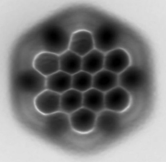
Photo

High-resolution images of terminals in water chains.
Shiotari, A., Sugimoto, Y. Ultrahigh-resolution imaging of water networks by atomic force microscopy. Nat Commun 8, 14313 (2017). https://doi.org/10.1038/ncomms14313
153 notes
·
View notes
Text
Types of Microscopes
1. Simple Microscope
2. Compound Microscope
3. Phase Contrast Microscope
4. Fluorescence Microscope
5. Electron Microscope
6. Scanning Electron Microscope (SEM)
7. Transmission Electron Microscope (TEM)
8. Dark Field Microscope
9. Dissecting Microscope (Stereo Microscope)
10. Digital Microscope
11. Scanning Probe Microscope (SPM)
12. Atomic Force Microscope (ATM)
13. Inverted Microscope
14. Acoustic Microscope
15. X-Ray Microscope
16. Polarizing Microscope
17. Metallurgical Microscope
18. Pocket Microscope
19. USB Microscope
20. Confocal Microscope
21. Laser Scanning Microscope
22. Differential Interference Contrast Microscope (DIC)
23. Near-field Scanning Optical Microscope (NSOM)
24. Raman Microscope
25. Super-resolution Microscope
26. Cryo-electron Microscope
27. Time-lapse Microscope
There is a wide range of microscopy techniques and instruments used in various fields of science and research.
#forensic#forensics#criminology#forensic science#evidence#criminalistic#forensic field#crime#forensic science notes#crime scene investigation#electron microscope#microscope
6 notes
·
View notes
Text
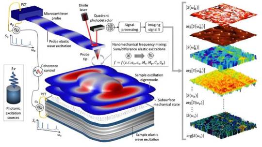
Subsurface nanometrology: Probing hidden materials via atomic force microscopy
A new nanoscience study led by a researcher at the Department of Energy's Oak Ridge National Laboratory takes a big-picture look at how scientists study materials at the smallest scales.
The paper, published in Science Advances, reviews leading work in subsurface nanometrology, the science of internal measurement at the nanoscale level, and suggests quantum sensing could become the foundation for the field's next era of discoveries. Potential applications could range from mapping intracellular structures for targeted drug delivery to characterizing quantum materials and nanostructures for the advancement of quantum computing.
"Our goal was to define the state of the art and to consider what's been done and where we need to go," said Ali Passian, an ORNL senior research scientist and senior author of the study.
Read more.
#Materials Science#Science#Nanotechnology#Atomic force microscopy#Materials Characterization#Surfaces
12 notes
·
View notes
Photo
Described by the researchers as an “experimental tour de force,” the study used a newly optimized form of liquid-phase transmission electron microscopy (TEM) to gain unprecedented insights into the self-assembly process. Before this work, researchers have used microscopy to watch micron-sized colloids — which are 10 to 100 times larger than nanoparticles — self-assemble into crystals. They also have used X-ray crystallography or electron microscopy to visualize single layers of atoms in a crystalline lattice. But they were unable to watch atoms individually move into place.
https://www.nanotechnologyworld.org/post/watch-nanoparticles-grow-into-crystals
#nanotechnology #materialsscience #engineering #chemistry #innovation #electronics #energy
24 notes
·
View notes
Photo

So there’s this bug in the software of the atomic force microscope (AFM) that I use.
You image your sample - cellular membrane here - and than you can indent it with a tiny tip to measure its stiffness and elasticity. Sometimes, the image dissapears the moment you switch to the indentations so you are blind on where to push and indent. This is the best solution I thought of today to mark where the membrane was. 😆
#physics#experiment#postdoc#academia#biophysics#biology#stem#cell membrane#women in science#fast solutions#handy#software bug#Atomic Force Microscopy#afm#original content
2 notes
·
View notes
Note
🌵
cactus ⇢ something you’re currently learning (about)?
Fun answer: woodworking! I'm working on building myself a bench so I can start building myself some simple furniture and boxes. So far it mostly seems to boil down to "sharpen your chisel", but its a lot of fun!
Boring answer: mortgages. Might be buying a house soon.
Extremely specific technical answer: photo-induced atomic force microscopy; I'm potentially starting on a project for a conference next year, so I need to figure out the key differences between PIAFM and scanning probe microscopy.
Thanks for the ask!
2 notes
·
View notes
Video
20230611_F0001: AFM probe holder by Wei-Feng Xue
Via Flickr:
- Scientific equipment seen over #10YearsAgo. This is a probe holder the classic MultiMode SPM/AFM microscope.
#atomic force microscopy#microscopy#AFM#probe#cantilever#metal#instrument#science#scientific#technology#laboratory#lab#equipment#macro#flickr
0 notes
Text
@ people asking questions on the group chat at 9pm causing me to jump off the bed to check the answer because I get anxious can you please stop. It's not the time to check atomic force microscopy techniques to study biopolimer's bending stiffness
#because I'm not so sure you could use it for that i mean with afm you will find Young's modulus and the shear one#which guess what. are different.#so you can't use kf=YΨ because you can't hypothesize isotropy!#and now I won't sleep
1 note
·
View note
Last Seen Blogs

sultedsults-blog
sultedsult

ittasteslikecoconut
All of the below

crguang
are there still beautiful things?

petriquors
with everything i am