#Expansion Microscopy
Text

Blown-up in Blood
An atlas of development of the malaria parasite as it proliferates in its blood cell stage generated using expansion microscopy – a means of physically inflating tissue to enhance the microscopic view
Read the published research article here
Image from work by Benjamin Liffner and Ana Karla Cepeda Diaz, and colleagues
Department of Pharmacology and Toxicology, Indiana University School of Medicine, Indianapolis, IN, USA
Image originally published with a Creative Commons Attribution 4.0 International (CC BY 4.0)
Published in eLife, December 2023
You can also follow BPoD on Instagram, Twitter and Facebook
#science#biomedicine#immunofluorescence#biology#malaria#expansion microscopy#blood cells#erythrocytes#life cycle#parasites
13 notes
·
View notes
Text

A Journey into the World of Microscopy: From Humble Beginnings to High-Tech Magnification
The science of looking into the hidden invisible Microscopy has transformed our understanding of the world around us. It can explore the universe beyond the reach of our naked eyes, with complex cellular structures, red blood cells, viruses and other viruses and microorganisms taking on amazing perspectives
The history of the microscope is a fascinating story of human curiosity, scientific genius, and relentless exploration. From the humble beginnings of simple magnifying glasses to the sophistication of modern electronic microscopes, the invention of microscopes has shaped our understanding of the microscopic world
In the 1600s, Dutch opticians such as Hans and Zachary Janssen are credited with inventing the first microscope. Known for this hybrid microscope, many lenses were used to magnify objects up to 30 times.At the end of the 17th century, Antony van Leeuwenhoek, Dutch draper some changed our perception of thumbnails. Armed with a well-made single-lens microscope, and explored the hidden reaches of nature. In 1674, Leeuwenhoek discovered microorganisms in lake water, which he aptly named “animalcules”. His discovery laid the foundations of biology and inspired generations of scientists. This incredible feat allowed him to uncover a hidden universe – the first sightings of bacteria, red blood cells, and other microorganisms.
Formation of the scientific environment (17th-19th centuries): Leeuwenhoek’s discoveries boosted scientific research. Robert Hooke, an English scientist, established these developments. In 1665, his book "Micrographia" recorded his observations with a compound microscope. Notably, the term "cell" was coined by Hooke when he examined cork tissue, laying the foundation for cell biology.Microscope systems flourished throughout the 18th and 19th centuries Joseph Lister and other scientists addressed the limitations of the early lenses, introducing improvements that reduced image distortion.
Beyond the Limits of Light: The Beginning of the New Age (19th-20th century): As the 19th century progressed, the limitations of optical microscopy became apparent and scientists yearned for a tool which can go deeper into cells. This research culminated in the development of the electron microscope in the 1930s. The 20th century was revolutionary with the invention of the electron microscope. Unlike light microscopes, which use visible light, electron microscopes use electron beams to achieve much higher magnification.Formation of the scientific environment (17th-19th centuries): Leeuwenhoek’s discoveries boosted scientific research. Robert Hooke, an English scientist, established these developments. In 1665, his book "Micrographia" recorded his observations with a compound microscope. Notably, the term "cell" was coined by Hooke when he examined cork tissue, laying the foundation for cell biology.Microscope systems flourished throughout the 18th and 19th centuries Joseph Lister and other scientists addressed the limitations of the early lenses, introducing improvements that reduced image distortion.
Beyond the Limits of Light: The Beginning of the New Age (19th-20th century): As the 19th century progressed, the limitations of optical microscopy became apparent and scientists yearned for a tool which can go deeper into cells. This research culminated in the development of the electron microscope in the 1930s. The 20th century was revolutionary with the invention of the electron microscope. Unlike light microscopes, which use visible light, electron microscopes use electron beams to achieve much higher magnification.
In the 1930s, German experts Max Knoll and Ernst Ruska made the first electron microscope. This tool let us see tiny things like cells and even atoms by using electron beams, not light, getting images many times bigger. This cool invention showed us the tiny parts inside cells, viruses, and stuff too small to see before. The 1900s brought even more cool microscopes. New kinds like phase-contrast and confocal microscopy let scientists look at live cells without using stuff that could hurt them. Now, the world of looking at tiny things is getting even better. Today, we have high-tech microscopes that use computers and lasers. These let us see and even change tiny things in ways we never could before.
Modern Microscopy's Diverse Arsenal - Today, the field of microscopy boasts a diverse range of specialized instruments, each tailored to address specific scientific needs. Here's a glimpse into some remarkable examples:
Scanning Electron Microscope (SEM): Imagine a high-tech camera that captures images using a beam of electrons instead of light. That's the essence of a SEM. By scanning the surface of a sample with a focused electron beam, SEMs generate detailed information about its topography and composition. This makes them ideal for studying the intricate structures of materials like insect wings, microchips, and even pollen grains.
Transmission Electron Microscope (TEM): While SEMs provide exceptional surface detail, TEMs take us a step further. They function by transmitting a beam of electrons through a very thin sample, allowing us to observe its internal structure. TEMs are the go-to instruments for visualizing the intricate world of viruses, organelles within cells, and macromolecules like proteins.
Confocal Microscopy: Ever wished to focus on a specific layer within a thick biological sample and blur out the rest? Confocal microscopy makes this possible. It utilizes a laser beam to precisely illuminate a chosen plane within the sample, effectively eliminating information from out-of-focus regions. This allows researchers to create sharp, three-dimensional images of cells, tissues, and even small organisms.
Atomic Force Microscopy (AFM): This technique takes a completely different approach, venturing into the realm of physical interaction. AFM employs a tiny cantilever, akin to a microscopic feeler, to physically scan the surface of a sample. By measuring the minute forces between the cantilever and the sample's surface, AFM can map its topography at an atomic level. This provides invaluable insights into the properties of materials at an unimaginable scale, making it crucial for research in fields like nanotechnology and surface science.
Fluorescence Microscopy: Imagine illuminating a sample with specific wavelengths of light and observing it glowing in response. That's the essence of fluorescence microscopy. This technique utilizes fluorescent molecules or tags that bind to specific structures within a cell or tissue. When excited by light, these tags emit their own light, highlighting the target structures with remarkable clarity. This allows researchers to visualize specific proteins, DNA, or even pathogens within biological samples.
Super-resolution Microscopy (SRM): Overcoming the limitations imposed by the wavelength of light, SRM techniques like STED (Stimulated Emission Depletion) and PALM (Photoactivated Localization Microscopy) achieve resolutions surpassing the diffraction limit. This allows researchers to visualize structures as small as 20 nanometers, enabling the observation of intricate cellular machinery and the dynamics of individual molecules within living cells.
Cryo-Electron Microscopy (Cryo-EM): This powerful technique takes a snapshot of biological samples in their near-life state. Samples are rapidly frozen at ultra-low temperatures, preserving their native structure and minimizing damage caused by traditional fixation methods. Cryo-EM has been instrumental in determining the three-dimensional structures of complex molecules like proteins and viruses, providing crucial insights into their function and potential drug targets.
Correlative Microscopy: Combining the strengths of multiple microscopy techniques, correlative microscopy offers a comprehensive view of biological samples. For instance, researchers can utilize fluorescence microscopy to identify specific structures within a cell and then switch to electron microscopy to examine those structures in high detail. This integrated approach provides a deeper understanding of cellular processes and their underlying mechanisms.
Light Sheet Microscopy (LSM): Imagine illuminating a thin slice of a sample within a living organism. LSM achieves this feat by focusing a laser beam into a thin sheet of light, minimizing photobleaching and phototoxicity – damaging effects caused by prolonged exposure to light. This allows researchers to observe dynamic processes within living organisms over extended periods, providing valuable insights into cellular behavior and development.
Expansion Microscopy (ExM): This innovative technique physically expands biological samples by several folds while preserving their structural integrity. This expansion allows for better resolution and visualization of intricate cellular structures that would otherwise be difficult to distinguish using traditional microscopy methods. ExM holds immense potential for studying the organization and function of organelles within cells.
Scanning Near-Field Optical Microscopy (SNOM): This innovative technique pushes the boundaries of resolution by utilizing a tiny probe that interacts with the sample at an extremely close range. SNOM can not only image the surface features of a sample with exceptional detail but also probe its optical properties at the nanoscale. This opens doors for research in areas like material science and photonics, allowing scientists to study the behavior of light at the interface between materials.
X-ray Microscopy: Stepping outside the realm of light and electrons, X-ray microscopy offers unique capabilities. By utilizing high-energy X-rays, this technique can penetrate deep into samples, making it ideal for studying the internal structure of dense materials like bones and minerals. Additionally, it allows for the visualization of elements within a sample, providing valuable information about their distribution and composition.
From revealing the building blocks of life to aiding in the development of new medicines, the microscope has played an undeniable role in shaping our scientific understanding. As technology continues to evolve, one can only imagine the future breakthroughs this remarkable invention holds in unveiling the secrets of our universe, both seen and unseen. These advancements hold the potential to revolutionize our understanding of biological processes, develop new materials with extraordinary properties, and ultimately pave the way for breakthroughs in medicine, nanotechnology, and countless other fields. As we continue to refine and develop novel microscopy techniques and the future holds immense promise for further groundbreaking discoveries that will undoubtedly revolutionize our perception of the world around us.
#science sculpt#life science#science#molecular biology#biology#biotechnology#artists on tumblr#microscopy#microscope#Scanning Electron Microscope#Transmission Electron Microscope#Confocal Microscopy#Atomic Force Microscopy#Fluorescence Microscopy#Expansion Microscopy#X-ray Microscopy#Super-resolution Microscopy#Light Sheet Microscopy#illustration#illustrator#illustrative art#education#educate yourself#techniques in biotechnology#scientific research#the glass scientists#scientific illustration#scientific advancements
6 notes
·
View notes
Photo
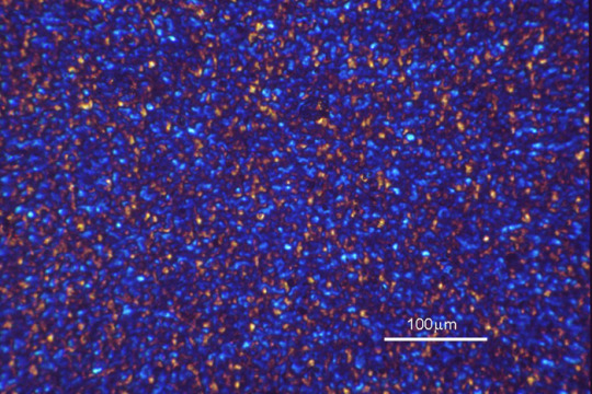
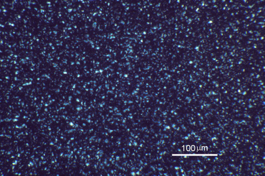

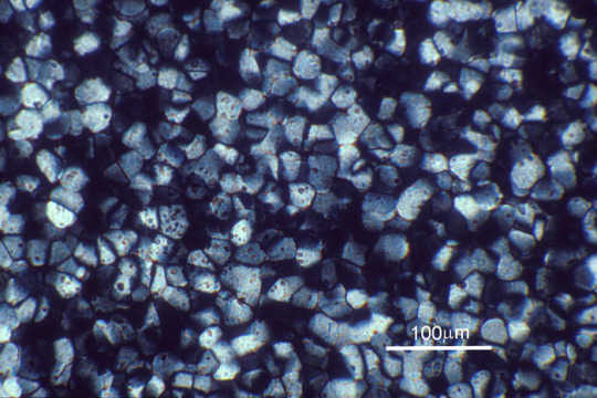

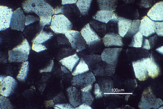
Glass ceramic
System
LAS: Li2O-Al2O3-SiO2 Composition
[...]
Processing
Heat treated at 750°C [top two] / 840°C [middle two] / 900°C [bottom two] for 3 hours
Applications
LAS glass ceramics are principally used for domestic cooker hobs and cookware. LAS and other glass ceramics can also be used for microwave radomes, vacuum and laser envelopes, telescope mirrors, and in bioceramic applications.
Sample preparation
Thin section
Technique
Transmitted polarised light microscopy, [with sensitive tint plate - left images]
Length bar
100 μm
Further information
Glass ceramics are materials that are cooled from the melt in the form of a glass, and then heat treated to induce controlled crystallisation of the glass. Heterogeneous nucleation is carried out at a temperature to maximise the nucleation rate (common nucleating agents include TiO2 and ZrO2), and the temperature is then raised sufficiently to cause the nuclei formed to grow rapidly.
Glass ceramics are strong, reasonably tough, transparent to IR radiation, have a low coefficient of thermal expansion, a high resistance to thermal shock and a low thermal conductivity, which makes them very useful in domestic applications such as cookware and cooker hobs.
The most common glass ceramic system is LAS (Li2O-Al2O3-SiO2), but others include MgO-Al2O3-SiO2, Na2O-BaO-Al2O3-SiO2, and Li2O-MgO-Al2O3-SiO2.
Contributor
Dr K M Knowles
Organisation
Department of Materials Science and Metallurgy, University of Cambridge
Sources: ( 1 ) ( 2 ) ( 3 ) ( 4 ) ( 5 ) ( 6 )
#Materials Science#Science#Glass ceramics#Glass#Ceramics#Heat treatment#Crystals#Oxides#Lithium#Aluminum#Silicon#Alumina#Silica#University of Cambridge#Optical microscopy#Polarization#DoITPoMS#Magnified view
47 notes
·
View notes
Text
The Significance of Piezo Stack Actuators
In the world of precision engineering, where accuracy and control reign supreme, the advent of piezo stack actuators has heralded a new era. These remarkable devices, born from the marvel of piezoelectricity, have swiftly become indispensable tools across industries that demand unparalleled precision and responsiveness.

In this article, we delve into the multifaceted importance of piezo stack actuators, shedding light on their mechanics, applications, and transformative impact.
For More Information Please visit, piezo stacks
The Marvel of Piezo Stack Actuators
At the core of piezo stack actuators lies a stack of meticulously crafted piezoelectric ceramic layers. This assembly is a testament to scientific ingenuity, as each layer is polarized with alternating positive and negative charges. The resulting electromechanical magic occurs when an electric voltage is applied. The layers expand or contract in perfect harmony, delivering linear motion with a level of precision that has redefined the boundaries of possibility.
Precision Redefined: Control at the Sub-Micrometer Level
The defining attribute of piezo stack actuators is their unparalleled precision. This precision is not merely a catchphrase; it's a testament to the inherent characteristics of piezoelectric materials. When voltage courses through the stack, the response is immediate and exact, generating forces that translate into controlled linear motion. This level of fine-tuned movement, often reaching sub-micrometer scales, is nothing short of awe-inspiring and is particularly vital in applications where infinitesimal adjustments can make all the difference.
A World of Applications Unveiled
Piezo stack actuators have woven themselves seamlessly into an array of industries, leaving a trail of transformative impacts in their wake. In the realm of optics, these actuators play a pivotal role in laser systems, facilitating the precise focusing of laser beams. This precision translates into enhanced efficiency in material processing, improved medical procedures, and more efficient communication technologies.
Microscopy, an arena where detail is king, has been revolutionized by piezo stack actuators. These devices enable the delicate movement of microscope lenses, affording researchers the ability to capture intricate images of minuscule structures with unparalleled clarity.
Beyond Earth: Piezo Stacks in Aerospace and Beyond
It's not just on Earth where piezo stack actuators have left their mark. In aerospace, these devices are instrumental in stabilizing flight control surfaces, making minute adjustments that contribute to the safety and precision of airborne vehicles. Even beyond our planet, in the vast expanse of space, piezo stack actuators play a role in satellite stabilization and the deployment of mechanisms in space probes, ensuring accuracy and functionality in the most challenging environments.
Navigating Challenges and Harnessing Potential
While the potential of piezo stack actuators is immense, certain considerations accompany their integration. Thermal effects can influence their performance, demanding careful temperature management. Moreover, the need for a driving voltage to maintain position may be a factor in power-sensitive applications. However, these challenges pale in comparison to the benefits these devices bring to industries that value and rely on precision.

Conclusion: Precision Pinnacle Achieved
In the grand tapestry of precision engineering, piezo stack actuators stand as a pinnacle of achievement. Their ability to translate electrical energy into controlled, precise motion has opened doors to advancements that were once mere dreams. From medical imaging to aerospace navigation, their impact reverberates across disciplines, promising a future where precision is not just a possibility but a reality. As we continue to explore and harness the significance of piezo stack actuators, we propel ourselves toward a world where precision is the norm, and the boundaries of possibility continue to expand.
2 notes
·
View notes
Text
Covering strategy as well as gift wrapping elevation work together to modify actual physical exposures through manual pallet having to wrap.
Complete thyroidectomy established the actual papillary carcinoma thyroid gland. Post-operatively, she was given radioactive iodine (I-131) ablation therapy for 7 a few years ended up being asymptomatic during this period; nonetheless, during the last 12 months, she's already been whining involving puffiness from the shoulder, which usually failed to answer palliative radiotherapy and rapidly increased in proportions. Disarticulation of the shoulder complex has been done, that confirmed anaplastic carcinoma about histopathological examination. Anaplastic transformation regarding papillary carcinoma in the metastatic websites will be well documented from the novels and is also uncommon. Nevertheless, the identical has not been noted in the make and also through Of india just before. Though soft tissues sarcomas are usually most popular on this site, nonetheless, the possibility of anaplastic transformation should be saved in the particular differential diagnosis of quickly enlarging unpleasant muscle size inside a identified case of metastatic thyroid gland carcinoma to stop incorrect diagnosis.TaCN levels have been placed making use of metal-organic chemical-vapor buildup pertaining to software because metal entrance electrodes within p-type metal-oxide-semiconductor (pMOS) products. The films ended up created by simply winter breaking down of tertiary-amylimido-tris(dimethylamido)tantalum (TAIMATA (Ur)) between 500 LEE011 along with 600 levels D. The structure was influenced by the growth temperature along with increasing H along with reducing In written content in higher heat. Motion pictures produced below 400 levels D have been nearly amorphous and have become weakly polycrystalline with a cubic composition at higher expansion heat. The covering thickness looked like 7.One g/cm(Three), about 50 % of with the TaCN bulk thickness. Grazing-incidence x-ray diffraction as well as tranny electron microscopy established that the films contain little polycrystalline whole grains in a amorphous matrix. The particular resistivity is discovered to lower together with raising development temp. Least expensive resistivity values ended up around One meters Omega cm pertaining to movies developed with Six-hundred certifications C. The films produced the just like 4 nm thicker protecting surface area oxide, which ends up in the width dependence from the motion picture resistivity. Air have also been located to be able to dissipate slowly and gradually in the volume metal, which leads to a resistivity getting older influence. The particular effective work objective of the TaCN motion pictures is discovered to get Four.Eight eV upon HfSiO(4) as well as HfSiON and change weakly by simply substantial cold weather price range annealing toward the particular Supposrr que valence music group, reaching Four.Being unfaithful eV on HfSiO(Some).Since platelet service has an important position throughout bodily hemostasis as well as pathological thrombosis, it is crucial inside the total hemocompatibility evaluation of new health-related products along with biomaterials to evaluate their own results upon platelet operate. Nonetheless, there are zero extensively recognized in vitro examination techniques to execute this specific review. In an effort to produce efficient platelet tests regarding prospective utilization in health care device evaluation, this research in contrast your sensitivity involving platelet reactions to be able to shear anxiety arousal of individual and also bovine body utilizing numerous platelet service guns.
4 notes
·
View notes
Text
Oxidative Stress Assay Market In-depth Insights, Revenue Details, Regional Analysis to 2024-2033
The Oxidative Stress Assay Market size was USD 1.3 Billion in 2022 and is anticipated to reach USD 2.8 Billion in 2032, growing at a rate of 8.0% from 2023 to 2032.
Oxidative stress is a condition in which the body’s cells are damaged by free radicals. Free radicals are unstable molecules that can damage cells, causing them to age prematurely. The body produces free radicals as a byproduct of normal metabolism. However, exposure to environmental toxins, such as cigarette smoke and pollution, can also increase the levels of free radicals in the body.
To Know More@ https://www.globalinsightservices.com/reports/oxidative-stress-assay-market/?utm_id=Pranalip
Key Trends
There are several key trends in oxidative stress assay technology.
One is the development of more sensitive and specific assays. This is important because it allows researchers to more accurately measure levels of oxidative stress in cells and tissues.
Another key trend is the development of new methods for measuring oxidative stress. This is important because it allows researchers to more accurately measure levels of oxidative stress in cells and tissues.
Finally, there is an increasing interest in using oxidative stress assays to screen for new drugs and therapies. This is important because it allows researchers to identify potential new treatments for diseases associated with oxidative stress.
Key Drivers
There are several key drivers of the oxidative stress assay market.
First, oxidative stress is a major factor in many chronic diseases, including cardiovascular disease, cancer, and diabetes.
Second, oxidative stress levels can be measured relatively easily and cheaply, making it an attractive target for research and clinical applications.
Third, there is a growing body of evidence linking oxidative stress to a variety of other health conditions, such as Alzheimer’s disease, Parkinson’s disease, and age-related macular degeneration.
Request Sample@ https://www.globalinsightservices.com/request-sample/GIS21084/?utm_id=Pranalip
Research Objectives
Estimates and forecast the overall market size for the total market, across product, service type, type, end-user, and region
Detailed information and key takeaways on qualitative and quantitative trends, dynamics, business framework, competitive landscape, and company profiling
Identify factors influencing market growth and challenges, opportunities, drivers and restraints
Identify factors that could limit company participation in identified international markets to help properly calibrate market share expectations and growth rates
Trace and evaluate key development strategies like acquisitions, product launches, mergers, collaborations, business expansions, agreements, partnerships, and R&D activities
Thoroughly analyze smaller market segments strategically, focusing on their potential, individual patterns of growth, and impact on the overall market
To thoroughly outline the competitive landscape within the market, including an assessment of business and corporate strategies, aimed at monitoring and dissecting competitive advancements.
Identify the primary market participants, based on their business objectives, regional footprint, product offerings, and strategic initiatives
Request Customization@ https://www.globalinsightservices.com/request-customization/GIS21084/?utm_id=Pranalip
Market Segments
The oxidative stress array market is segmented by product, technology, end-user, and region. By product, the market is classified into consumables, kits, and others. On the basis of technology, it is bifurcated into flow cytometry, microscopy, and others. Based on end-user, it is divided into clinical laboratories, CROs, cosmetic companies, and others. Region-wise, the market is segmented into North America, Europe, Asia Pacific, and Rest of the World.
Key Players
The global oxidative stress array market includes players such as Abcam PLC, AMS Biotechnology, BioVision Inc, Merck and Co Inc, Cell Biolabs Inc, Promega Corporation, Enzo Biochem, Oxford Biomedical Research, Qiagen N.V., Sigma-Aldrich Corporation, and others.
Buy your copy here@ https://www.globalinsightservices.com/checkout/single_user/GIS21084/?utm_id=Pranalip
Research Scope
Scope – Highlights, Trends, Insights. Attractiveness, Forecast
Market Sizing – Product Type, End User, Offering Type, Technology, Region, Country, Others
Market Dynamics – Market Segmentation, Demand and Supply, Bargaining Power of Buyers and Sellers, Drivers, Restraints, Opportunities, Threat Analysis, Impact Analysis, Porters 5 Forces, Ansoff Analysis, Supply Chain
Business Framework – Case Studies, Regulatory Landscape, Pricing, Policies and Regulations, New Product Launches. M&As, Recent Developments
Competitive Landscape – Market Share Analysis, Market Leaders, Emerging Players, Vendor Benchmarking, Developmental Strategy Benchmarking, PESTLE Analysis, Value Chain Analysis
Company Profiles – Overview, Business Segments, Business Performance, Product Offering, Key Developmental Strategies, SWOT Analysis.
With Global Insight Services, you receive:
10-year forecast to help you make strategic decisions
In-depth segmentation which can be customized as per your requirements
Free consultation with lead analyst of the report
Infographic excel data pack, easy to analyze big data
Robust and transparent research methodology
Unmatched data quality and after sales service
Contact Us:
Global Insight Services LLC
16192, Coastal Highway, Lewes DE 19958
E-mail: [email protected]
Phone: +1-833-761-1700
Website: https://www.globalinsightservices.com/
About Global Insight Services:
Global Insight Services (GIS) is a leading multi-industry market research firm headquartered in Delaware, US. We are committed to providing our clients with highest quality data, analysis, and tools to meet all their market research needs. With GIS, you can be assured of the quality of the deliverables, robust & transparent research methodology, and superior service.
0 notes
Text
Rising Demand For Advanced Ophthalmic Imaging Systems To Fuel Growth Of The Global Optical Instrument And Lens Market

The global optical instrument and lens market involves a wide range of precision instruments and specialty lenses that are used in applications ranging from microscopy to ophthalmology. Optical instruments help in examining, measuring and inspecting minute details and structures that cannot be perceived through naked eyes. Meanwhile, optical lenses have become integral components across various consumer electronic devices as well as industrial and medical equipment. Technological advancements have enabled the development of novel optical instruments and high-precision lenses that offer improved resolution, accuracy and functionality. The growing need for early disease diagnosis and treatment is propelling demand for advanced ophthalmic imaging systems among eye care professionals globally.
The Global Optical Instrument And Lens Market Size Is Estimated To Be Valued At US$ 29.8 Bn In 2024 And Is Expected To Exhibit A CAGR Of 4.7% Over The Forecast Period 2024-2031.
Key Takeaways
Key players operating in the global optical instrument and lens market are Carl Zeiss Ag, Hoya Corporation, Canon Inc., Nidek Co., Ltd., Topcon Corporation, Intelligent Retinal Imaging Systems, Inc., Kowa Company Ltd., Optomed, Vision Equipment Inc., Clarity Medical Systems, Inc., Medimaging Integrated Solution Inc., S4OPTIK LLC., Shenzhen Thondar Technology Co., Ltd., CenterVue SpA, Alton Vision LLC, LENSTECH OPTICALS, Alcon Inc. These players are focusing on developing advanced optical instruments and lenses through investments in R&D.
The global market is driven by the rising geriatric population and increasing prevalence of ocular diseases. According to WHO, at least 2.2 billion people have vision impairment or blindness globally. This is spurring demand for ophthalmic diagnostic equipment and lenses.
Technological advancements are allowing manufacturers to develop more compact, high-resolution and precision optical instruments integrated with advanced imaging and analytical capabilities. This is supporting applications in diverse fields including life sciences, industrial inspection and healthcare.
Market Trends
Some of the key trends observed in the global optical instrument and lens market include rising adoption of hybrid imaging systems and multifunctional optical instruments. Manufacturers are integrating capabilities of different imaging modalities into single platforms to improve workflow and diagnostic capabilities. Another trend gaining traction is the emergence of 3D printed ophthalmic lenses with customized prescriptions and designs. 3D printing technology enables mass-personalization of lenses as per the specific vision requirements of individuals.
Market Opportunities
The market players have opportunity to tap into the growing demand for tele-ophthalmology and remote patient monitoring solutions. There is a need for portable diagnostic devices integrated with telecommunication capabilities. Another opportunity lies in developing nations in Asia Pacific and Latin America driven by improving healthcare infrastructure and increasing healthcare spending. Expansion of product offerings catering to veterinary and industrial applications also presents lucrative growth prospects.
Get more insights on this topic: https://www.pressreleasebulletin.com/global-optical-instrument-and-lens-market-growth-size-and-demand/
#Global Optical Instrument and Lens Market Trend#Global Optical Instrument and Lens Market Growth#Global Optical Instrument and Lens Market Size#Global Optical Instrument and Lens Market Analysis
0 notes
Text
Optical Lens Market Demand Overview, Growth Innovation, Latest Trends till 2030

****Everything You Need to Know About Optical Lens everything is Here....!
The Comprehensive study on Optical Lens Market includes historical data as well as share, size, and projection information for the major players, geographies, applications, and product categories for the years 2024 to 2030. The Market study includes comprehensive insights on the competitive environment, description, broad product portfolio of key players, SWOT analysis, and significant business strategy implemented by rivals, revenue, Porters Five Forces Analysis, and sales projections. The report also features an impact analysis of the market dynamics, highlighting the factors currently driving and limiting market growth, and the impact they could have on the short, medium, and long-term outlook. The main goal of the paper is to further illustrate how the latest scenario, the economic slowdown, and war events affect the market for Optical Lens.
Optical Lens Market is growing at a +7.4% CAGR during the forecast period 2024-2030. The increasing interest of the individuals in this industry is that the major reason for the expansion of this market.
The Top Key Players profiled in the report:
Carl Zeiss AG, Olympus Corporation, Nikon, Bausch & Lomb Incorporated, Cosina Co., Ltd., Meade Instruments Corp., Thorlabs Inc., MENICON CO., LTD.
Click the link to get a free sample copy of the report :
(*If you have any special requirements, please let us know and we will provide you with the report as you wish.)
Optical Lens Market Segmentation:
Optical Lens Market by Type, 2020-2029, (USD Million) (Thousand Units)
Converging Lenses
Diverging Lenses
Optical Lens Market By Application, 2020-2029, (USD Million) (Thousand Units)
Ophthalmic
Microscopy
Laser Processing
Imaging
Optical Lens Market by End User, 2020-2029, (USD Million) (Thousand Units)
Consumer Electronics
Defense
Healthcare
Life Sciences
Other
Based on geography, the global market for Optical Lens and Disruptions has been segmented as follows:
North America (United States, Canada, Mexico)
South America (Brazil, Argentina, Ecuador, Chile)
Asia Pacific (China, Japan, India, Korea)
Europe (Germany, UK, France, Italy)
Middle East Africa (Egypt, Turkey, Saudi Arabia, Iran) and more.
Strategic Points Covered in Optical Lens Market Directory:
To study and analyze the global market size (value & volume) by company, key regions/countries, products and application, history data, and forecast to 2030.
To understand the structure of market by identifying its various sub segments.
To share detailed information about the key factors influencing the growth of the market (growth potential, opportunities, drivers, industry-specific challenges and risks).
Focuses on the key global manufacturers, to define, describe and analyze the sales volume, value, market share, market competition landscape, SWOT analysis and development plans in next few years.
To analyze the growth trends, future prospects, and their contribution to the total market.
To project the value and volume of submarkets, with respect to key regions (along with their respective key countries).
To analyze competitive developments such as expansions, agreements, new product launches, and acquisitions in the market.
To strategically profile the key players and comprehensively analyze their growth strategies.
The report provides insights on the following pointers:
Market Penetration: Comprehensive information on the product portfolios of the top players in the Optical Lens
Product Development/Innovation: Detailed insights on the upcoming technologies, R&D activities, and product launches in the market.
Competitive Assessment: In-depth assessment of the Optical Lens market strategies, geographic and business segments of the leading players in the market.
Market Development: Comprehensive information about emerging markets. This report analyzes the market for various segments across geographies.
Market Diversification: Exhaustive information about new products, untapped geographies, recent developments, and investments in the Optical Lens
Take a look at the full report with detailed TOC here:
Some of the key questions scrutinized in the study are:
Which companies are expanding litanies of products with the aim to diversify product portfolio?
Which companies have drifted away from their core competencies and how have those impacted the strategic landscape of the Optical Lens market?
Which companies have expanded their horizons by engaging in long-term societal considerations?
Which firms have bucked the pandemic trend and what frameworks they adopted to stay resilient?
What are the marketing programs for some of the recent product launches?
We offer customization on the Optical Lens market report based on specific client requirements:
20% free customization.
Five Countries can be added as per your choice.
Five Companies can add as per your choice.
Free customization for up to 40 hours.
After-sales support for 1 year from the date of delivery.
Get More: https://exactitudeconsultancy.com/primary-research/
Thank you for your interest in the Optical Lens Market research publications; you can also get individual chapters or regional/country report versions such as Germany, France, China, Latin America, GCC, North America, Europe or Asia……
Other Reports:
3D Print Photopolymer Parts market
Molecular Weight Marker market
Radiotherapy market
Alexandrite Lasers market
Chromatography Instruments market
About Us:
Exactitude Consultancy is a Market research & consulting services firm which helps its client to address their most pressing strategic and business challenges. Our professional team works hard to fetch the most authentic research reports backed with impeccable data figures which guarantee outstanding results every time for you. So, whether it is the latest report from the researchers or a custom requirement, our team is here to help you in the best possible way.
Contact: Exactitude Consultancy
PHONE NUMBER +1(704) 266-3234
EMAIL ADDRESS: [email protected]
#Optical Lens Market#Optical Lens Market growth#Optical Lens Market size#Optical Lens Market analysis 2024#Optical Lens Market CAGR#Optical Lens Growth#Optical Lens Market 2024#Optical Lens Market analysis by types#Optical Lens Market data#Optical Lens Market Outlook#Optical Lens Market Size and Share#Optical Lens Strategic Industry Latest News#Top Companies Analysis By Optical Lens Market#Optical Lens Market Trends#Optical Lens Market Forecast#Optical Lens Industry
0 notes
Text
Exploring Vacuum Technology: A Comprehensive Overview of Vacuum Gate Valves and High Vacuum Gate Valves
In the expansive world of vacuum technology, where precision and reliability intersect to create environments free from air and other gases, two pivotal components play critical roles: Vacuum Gate Valves and High Vacuum Gate Valves. These devices are not just mere accessories but are fundamental in controlling the flow of gases, ensuring that vacuum systems operate efficiently and effectively. This article delves into the essence of these valves, their unique features, and their indispensable role in various industrial applications.
The Heart of Vacuum Systems:
At its core, a Vacuum Gate Valve functions as a gatekeeper, regulating the passage of gases in vacuum systems. It operates by moving a flat, circular gate perpendicular to the path of the gas flow. This movement either opens the valve, allowing gas to pass through or closes it, creating a hermetic seal that prevents any gas movement. The design of these valves is ingeniously simple yet highly effective, making them a popular choice for systems requiring minimal leakage and maximum flow control.
The application of vacuum gate valves spans numerous sectors, including semiconductor manufacturing, where creating a controlled environment is crucial, and the aerospace industry, where precision and reliability are paramount. Their versatility and efficiency make them indispensable tools in the pursuit of technological advancement and industrial productivity.
Elevating Performance: The Role of High Vacuum Gate Valves
As we delve deeper into the vacuum technology realm, the High Vacuum Gate Valves emerge as the elite performers, explicitly designed for systems that operate under shallow pressures, often referred to as high vacuum environments. These valves are engineered with precision to ensure they offer superior sealing capabilities and minimal outgassing, thus maintaining the integrity of the high vacuum.
The construction of high vacuum gate valves often involves materials that are less prone to outgassing and can withstand the rigours of high vacuum conditions. Stainless steel, with its high resistance to corrosion and minimal outgassing properties, is a commonly used material. The meticulous design and material selection make these valves the cornerstone of high vacuum applications, such as in particle accelerators and electron microscopy, where any contamination or leakage can compromise the entire system's performance.
Conclusion:
The journey through the world of vacuum technology, from the versatile vacuum gate valves to the sophisticated high vacuum gate valves, highlights the importance of these components in creating and maintaining controlled environments. As technology advances and the demand for precision and reliability grows, the role of these valves becomes ever more critical.
For those looking to explore further or to procure high-quality vacuum gate valves, vacuumchamber.com stands as a beacon, offering a comprehensive range of products tailored to meet the diverse needs of the vacuum technology community.
0 notes
Text

Mapping the Changes
Expansion microscopy 'blows up' tissue samples in 3D enabling easy visualisation of small structures. Here a new tool called GelMap – an embedded fluorescent reference grid – calibrates the field of view and corrects for any resulting deformations
Read the published research paper here
Image from work by Hugo G. J. Damstra and Josiah B. Passmore, and colleagues
Cell Biology, Neurobiology and Biophysics, Department of Biology, Faculty of Science, Utrecht University, Utrecht, The Netherlands
Image originally published with a Creative Commons Attribution 4.0 International (CC BY 4.0)
Published in Nature Methods, September 2023
You can also follow BPoD on Instagram, Twitter and Facebook
#science#biomedicine#immunofluorescence#expansion microscopy#super resolution microscopy#STED#biology
13 notes
·
View notes
Text
Diagnostic Testing of STDs Market is Estimated to Witness High Growth Owing to Rising Prevalence of STDs

Diagnostic testing of STDs involves laboratory testing of samples such as blood, urine, or other body fluids to detect the presence of pathogens that cause sexually transmitted diseases (STDs). It helps identify the disease-causing microorganisms and provide effective treatment options.
Market Dynamics:
The growth of the diagnostic testing of STDs market is driven by the rising prevalence of sexually transmitted diseases and increasing awareness about their diagnosis. It is estimated that over 1 million cases of chlamydia, gonorrhea and syphilis are reported in the United States annually. Rapid diagnosis helps in timely treatment and control of the infection from spreading further. Furthermore, advancements in diagnostic technologies such as polymerase chain reaction (PCR) assays have made STD diagnosis easy, quick and affordable. This is positively impacting the market growth.
Increasing prevalence of STDs globally
The global prevalence of sexually transmitted diseases (STDs) like chlamydia, gonorrhea, syphilis and HIV has been on the rise over the past few decades. According to the World Health Organization (WHO), worldwide there are over 1 million new cases of STDs daily. The rising incidences can be attributed to factors like risky sexual behavior among youth, lack of sexual health education and screening programs, and increasing resistance to antibiotics used for treating certain STDs. The growing STD patient pool is driving higher demand for diagnostic testing services to confirm infections and initiate appropriate treatment. Sexually active individuals are increasingly opting for regular testing to stay informed about their health status.
Access to more innovative and advanced diagnostics
Vendors in the diagnostic testing for STDs market have been investing heavily in research and development of novel diagnostic technologies. Conventional testing methods based on microscopy analysis and culturing are being replaced by newer techniques like polymerase chain reaction (PCR), flow cytometry, rapid point-of-care tests, and biochips among others. The advanced diagnostics allow for sensitive and accurate detection of infections within a short turnaround time. Features like portability and ease of use are propelling the adoption of point-of-care STD testing. Companies are also developing multiplex panels that can simultaneously screen for multiple pathogens, providing comprehensive testing results with just one sample. Increased accessibility to high-performance diagnostics is positively impacting market growth.
Rising healthcare costs constraining market expansion
While the growing disease incidence and technological advancement in diagnostics are fueling opportunities, the rising costs of STD healthcare are acting as a market restraint. The prices of diagnostic tests and treatment therapies for STDs have witnessed a continuous upward trend over the past decade. Managing infectious diseases like HIV/AIDS requires long-term treatment stretching over months or years and follow-up testing, leading to high cumulative costs. Additionally, advanced diagnostics while being effective are also highly priced. The costs are further weighing on patients in developing countries lacking comprehensive medical insurance and adequate public health programs. The financial burden on consumers as well as national healthcare budgets is limiting wider access and uptake of diagnostic services.
Opportunity for home-based self-testing kits
With the rise in Internet penetration and online purchasing, the STD diagnostics market is witnessing a growing demand for home-based self-testing options. People are increasingly looking for private, discreet ways of testing themselves without visiting clinics or laboratories. Vendors see a huge opportunity in developing easy-to-use, mail-order self-testing kits that can be administered in the comfort of one's home. The at-home samples can then be mailed to authorized labs for examination. Companies are investing in research to design simplified tests suited for home settings without compromising on diagnostic sensitivity and accuracy. The self-sampling kits provide greater testing coverage by encouraging populations that are otherwise hesitant to get clinically tested.
Growing focus on HPV screening and vaccination programs
Human papillomavirus (HPV) has emerged as a major public health concern credited to be the primary cause of cervical cancer. It is estimated that HPV affects over 80% of the sexually active population. Diagnostic testing and prevention of HPV is among the fastest growing segments within the overall STD testing market. Governments and health agencies are launching extensive national HPV immunization programs targeting adolescents for vaccinations before onset of sexual activity. Regular HPV screening through tests like Pap smear is being promoted as part of routine health check-ups. Advanced HPV testing based on molecular diagnostic methods is gaining widespread adoption. The intensive focus on combating HPV through comprehensive screening, vaccination and public health initiatives will propel the market growth in the coming years.
0 notes
Photo
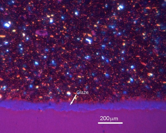


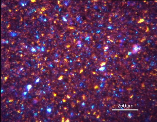

Oven-to-tableware [with surface glaze, image 1]
Sample preparation
Thin section
Technique
Transmitted polarised light microscopy, [with sensitive tint plate on images 1, 2, and 4]
Length bar
200 μm / 100 µm / 250 µm
Further information
Oven-to-tableware ceramics have a low coefficient of thermal expansion, and therefore a high resistance to thermal shock, which makes them suitable for use at a wide range of temperatures.
Contributor
Dr K M Knowles
Organisation
Department of Materials Science and Metallurgy, University of Cambridge
Sources: ( 1 ) ( 2 ) ( 3 ) ( 4 ) ( 5 )
#Materials Science#Science#Ceramics#Optical microscopy#Polarization#Magnified view#University of Cambridge#DoITPoMS
15 notes
·
View notes
Text
Market Outlook for High-Resolution 3D X-Ray Microscopy Market Services
Market Overview –
The High-Resolution 3D X-Ray Microscopy Market is experiencing substantial growth due to the increasing demand for advanced imaging techniques in various scientific and industrial applications. High-resolution 3D X-ray microscopy, also known as X-ray tomography or computed tomography (CT), enables detailed three-dimensional imaging of internal structures with superior resolution and clarity.
The 3D X-ray Microscopy market is thriving as industries and research sectors demand high-resolution imaging solutions. These advanced systems offer detailed insights into the internal structures of various materials, aiding in research, quality control, and failure analysis. With continuous technological advancements, the market for 3D X-ray microscopy is poised for further growth.
Key drivers of market growth include advancements in imaging technology, such as higher spatial resolution and faster image acquisition, which allow for precise visualization of microstructures in diverse samples. Additionally, the expanding applications of high-resolution 3D X-ray microscopy across multiple industries, including materials science, life sciences, electronics, and geology, fuel market expansion.
The market offers a wide range of high-resolution 3D X-ray microscopy systems, including laboratory-based and synchrotron-based systems, tailored to specific research and industrial requirements. These systems enable researchers and scientists to analyze complex samples, such as biological tissues, composite materials, and geological specimens, with unparalleled detail and accuracy.
Furthermore, collaborations between research institutions, academia, and industry players drive innovation and technological advancements in high-resolution 3D X-ray microscopy, further stimulating market growth.
Moreover, the market benefits from increasing investment in research and development activities, supportive government initiatives, and growing awareness about the advantages of high-resolution 3D X-ray microscopy in scientific research, quality control, and product development.
Despite the market's positive outlook, challenges such as high initial costs, limited accessibility to advanced imaging facilities, and data analysis complexities may hinder market growth. Nonetheless, ongoing efforts to enhance system capabilities, improve user-friendliness, and expand application areas are expected to drive continued adoption of high-resolution 3D X-ray microscopy in the coming years.
Over the projection period of 2022-2030, the high-resolution 3D x-ray microscopy market is expected to grow at an 8.8% annual rate to reach USD 2487.83 million by 2030.
Segmentation –
The global high resolution 3D X-ray microscopy market is segmented into type, applications, end user, and region. The type is segmented into Sub-micron XRM, Nanoscale XRM and others. The applications are segmented into advanced package development, Mineralogy Discrimination, Failure analysis, Surface measurements and others. The end users are segmented into Oil & Gas, Material Science, Semiconductors, Metrology, Life Science, Healthcare and others. The market is spanned across regions including North America, Europe, Asia Pacific, and rest of the world.
Regional Analysis –
The High-Resolution 3D X-Ray Microscopy Market demonstrates distinct regional dynamics shaped by factors such as technological advancements, research and development capabilities, and industrial applications. North America leads the market, driven by a strong presence of key market players, advanced R&D infrastructure, and widespread adoption of cutting-edge imaging technologies across various industries.
The region's robust healthcare and industrial sectors contribute significantly to the demand for high-resolution 3D X-ray microscopy systems, thus holding a substantial market share. Similarly, Europe portrays a lucrative market landscape, characterized by a strong focus on technological innovation, stringent quality standards, and a well-established industrial base. Adoption of high-resolution 3D X-ray microscopy in automotive, aerospace, and materials science sectors further fuels market growth in the region. In Asia Pacific, the market is witnessing rapid expansion due to increasing investments in research and development, rising industrialization, and growing applications in fields like electronics and semiconductors.
Countries like China, Japan, and South Korea are driving market growth with their expanding manufacturing sectors and rising demand for advanced imaging solutions. Latin America and the Middle East & Africa regions present opportunities for market penetration, driven by growing industrialization and investments in scientific research. However, challenges such as limited awareness and infrastructure may impact market growth in these regions. Overall, the High-Resolution 3D X-Ray Microscopy Market displays promising growth prospects across diverse regions, driven by the increasing demand for precise imaging solutions in various industries.
Key Players –
High-resolution 3D X-ray microscopy key players include Zeiss, Rigaku Corporation, Bruker Corporation, Thermo Fisher Scientific Inc., GE Measurement & Control Solutions, National Resource for Automated Molecular Microscopy, Phenom-World BV, TESCAN, Matsusada Precision Inc., and Octopus Imaging Software.
Related Reports –
Meningitis Diagnosis and Treatment
Endodontic Devices
Fertility Drug and Surgery
Opioids
For more information visit at MarketResearchFuture
#High-Resolution 3D X-Ray Microscopy Market#High-Resolution 3D X-Ray Microscopy Market Size#High-Resolution 3D X-Ray Microscopy Market Share#High-Resolution 3D X-Ray Microscopy Market Trends
0 notes
Text
Expansion microscopy reveals unique ultrastructural features of pathogenic budding yeast species
BioRxiv: http://dlvr.it/T34WZR
0 notes
Text
Bio-orthogonal Glycan Imaging of Culture Cells and Whole Animal C. elegans with Expansion Microscopy
http://dlvr.it/T2Dtyy
0 notes
Text
Imaging method reveals new cells and structures in human brain tissue
New Post has been published on https://thedigitalinsider.com/imaging-method-reveals-new-cells-and-structures-in-human-brain-tissue/
Imaging method reveals new cells and structures in human brain tissue

Using a novel microscopy technique, MIT and Brigham and Women’s Hospital/Harvard Medical School researchers have imaged human brain tissue in greater detail than ever before, revealing cells and structures that were not previously visible.
Among their findings, the researchers discovered that some “low-grade” brain tumors contain more putative aggressive tumor cells than expected, suggesting that some of these tumors may be more aggressive than previously thought.
The researchers hope that this technique could eventually be deployed to diagnose tumors, generate more accurate prognoses, and help doctors choose treatments.
“We’re starting to see how important the interactions of neurons and synapses with the surrounding brain are to the growth and progression of tumors. A lot of those things we really couldn’t see with conventional tools, but now we have a tool to look at those tissues at the nanoscale and try to understand these interactions,” says Pablo Valdes, a former MIT postdoc who is now an assistant professor of neuroscience at the University of Texas Medical Branch and the lead author of the study.
Edward Boyden, the Y. Eva Tan Professor in Neurotechnology at MIT; a professor of biological engineering, media arts and sciences, and brain and cognitive sciences; a Howard Hughes Medical Institute investigator; and a member of MIT’s McGovern Institute for Brain Research and Koch Institute for Integrative Cancer Research; and E. Antonio Chiocca, a professor of neurosurgery at Harvard Medical School and chair of neurosurgery at Brigham and Women’s Hospital, are the senior authors of the study, which appears today in Science Translational Medicine.
Making molecules visible
The new imaging method is based on expansion microscopy, a technique developed in Boyden’s lab in 2015 based on a simple premise: Instead of using powerful, expensive microscopes to obtain high-resolution images, the researchers devised a way to expand the tissue itself, allowing it to be imaged at very high resolution with a regular light microscope.
The technique works by embedding the tissue into a polymer that swells when water is added, and then softening up and breaking apart the proteins that normally hold tissue together. Then, adding water swells the polymer, pulling all the proteins apart from each other. This tissue enlargement allows researchers to obtain images with a resolution of around 70 nanometers, which was previously possible only with very specialized and expensive microscopes such as scanning electron microscopes.
In 2017, the Boyden lab developed a way to expand preserved human tissue specimens, but the chemical reagents that they used also destroyed the proteins that the researchers were interested in labeling. By labeling the proteins with fluorescent antibodies before expansion, the proteins’ location and identity could be visualized after the expansion process was complete. However, the antibodies typically used for this kind of labeling can’t easily squeeze through densely packed tissue before it’s expanded.
So, for this study, the authors devised a different tissue-softening protocol that breaks up the tissue but preserves proteins in the sample. After the tissue is expanded, proteins can be labelled with commercially available fluorescent antibodies. The researchers then can perform several rounds of imaging, with three or four different proteins labeled in each round. This labelling of proteins enables many more structures to be imaged, because once the tissue is expanded, antibodies can squeeze through and label proteins they couldn’t previously reach.
“We open up the space between the proteins so that we can get antibodies into crowded spaces that we couldn’t otherwise,” Valdes says. “We saw that we could expand the tissue, we could decrowd the proteins, and we could image many, many proteins in the same tissue by doing multiple rounds of staining.”
Working with MIT Assistant Professor Deblina Sarkar, the researchers demonstrated a form of this “decrowding” in 2022 using mouse tissue.
The new study resulted in a decrowding technique for use with human brain tissue samples that are used in clinical settings for pathological diagnosis and to guide treatment decisions. These samples can be more difficult to work with because they are usually embedded in paraffin and treated with other chemicals that need to be broken down before the tissue can be expanded.
In this study, the researchers labeled up to 16 different molecules per tissue sample. The molecules they targeted include markers for a variety of structures, including axons and synapses, as well as markers that identify cell types such as astrocytes and cells that form blood vessels. They also labeled molecules linked to tumor aggressiveness and neurodegeneration.
Using this approach, the researchers analyzed healthy brain tissue, along with samples from patients with two types of glioma — high-grade glioblastoma, which is the most aggressive primary brain tumor, with a poor prognosis, and low-grade gliomas, which are considered less aggressive.
“We wanted to look at brain tumors so that we can understand them better at the nanoscale level, and by doing that, to be able to develop better treatments and diagnoses in the future. At this point, it was more developing a tool to be able to understand them better, because currently in neuro-oncology, people haven’t done much in terms of super-resolution imaging,” Valdes says.
A diagnostic tool
To identify aggressive tumor cells in gliomas they studied, the researchers labeled vimentin, a protein that is found in highly aggressive glioblastomas. To their surprise, they found many more vimentin-expressing tumor cells in low-grade gliomas than had been seen using any other method.
“This tells us something about the biology of these tumors, specifically, how some of them probably have a more aggressive nature than you would suspect by doing standard staining techniques,” Valdes says.
When glioma patients undergo surgery, tumor samples are preserved and analyzed using immunohistochemistry staining, which can reveal certain markers of aggressiveness, including some of the markers analyzed in this study.
“These are incurable brain cancers, and this type of discovery will allow us to figure out which cancer molecules to target so we can design better treatments. It also proves the profound impact of having clinicians like us at the Brigham and Women’s interacting with basic scientists such as Ed Boyden at MIT to discover new technologies that can improve patient lives,” Chiocca says.
The researchers hope their expansion microscopy technique could allow doctors to learn much more about patients’ tumors, helping them to determine how aggressive the tumor is and guiding treatment choices. Valdes now plans to do a larger study of tumor types to try to establish diagnostic guidelines based on the tumor traits that can be revealed using this technique.
“Our hope is that this is going to be a diagnostic tool to pick up marker cells, interactions, and so on, that we couldn’t before,” he says. “It’s a practical tool that will help the clinical world of neuro-oncology and neuropathology look at neurological diseases at the nanoscale like never before, because fundamentally it’s a very simple tool to use.”
Boyden’s lab also plans to use this technique to study other aspects of brain function, in healthy and diseased tissue.
“Being able to do nanoimaging is important because biology is about nanoscale things — genes, gene products, biomolecules — and they interact over nanoscale distances,” Boyden says. “We can study all sorts of nanoscale interactions, including synaptic changes, immune interactions, and changes that occur during cancer and aging.”
The research was funded by K. Lisa Yang, the Howard Hughes Medical Institute, John Doerr, Open Philanthropy, the Bill and Melinda Gates Foundation, the Koch Institute Frontier Research Program, the National Institutes of Health, and the Neurosurgery Research and Education Foundation.
#2022#aging#antibodies#approach#Arts#Biological engineering#Biology#Biomolecules#blood#blood vessels#Brain#Brain and cognitive sciences#brain research#brain tumors#Cancer#cell#cell types#Cells#chemical#chemicals#Design#diagnostics#Diseases#education#electron#engineering#form#Foundation#Future#genes
0 notes
Last Seen Blogs

j0hn4
Untitled

jojikann
jojika_n

clickboxagency
ClickBox Agency

christianserrano1404
“All About Me”

cloverstellar
nobody talk to me about dc i'll go feral