#electron microscope
Photo
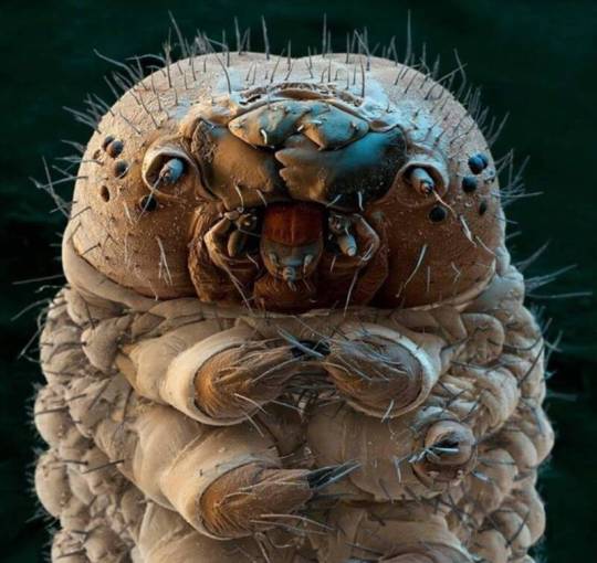
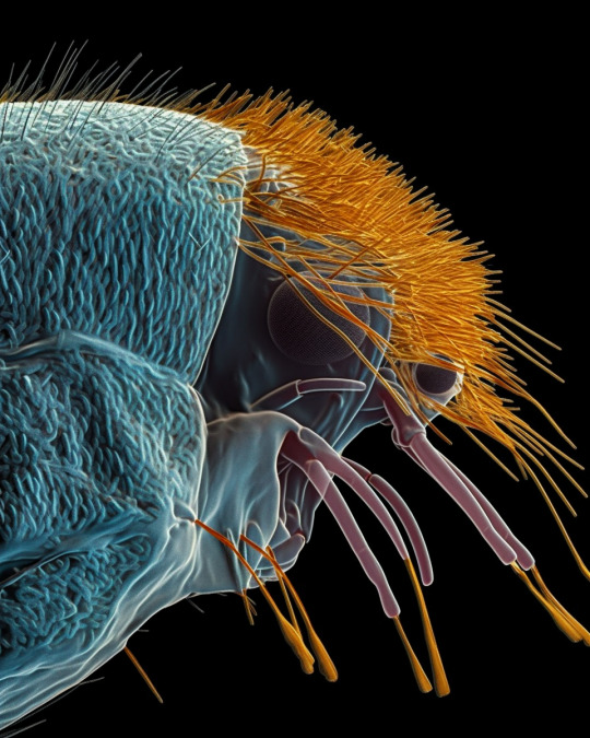
Silk moth caterpillar at 31:1 magnification.
Science Source
#science source#photographer#silk moth caterpillar#micro photography#insect#nature#electron microscope
75 notes
·
View notes
Photo
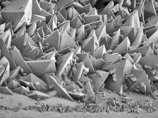
(via Kidney stone surface as seen in an electron microscope : oddlyterrifying)
ow
62 notes
·
View notes
Text
Colorizing Electron Microscope Photos Deceptively/Incorrectly
Some 'science' photo libraries are…questionable.

This photo claims to be "Ant carrying aphid egg, SEM" colorized to highlight the "egg" but look closely!
It's NOT an egg. That's just part of the ant's mouth! To understand what's wrong, take a look at the second set of photos by Hugo Darras. You can see the "egg" is part of her maxillae, the part she uses to collect liquid.
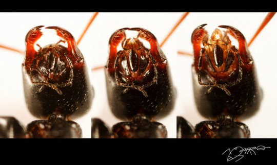
How did they decide it was an aphid egg?
Most aphids don't even lay eggs...they give live birth!
#ants#antposting#lies#electron microscope#ant photos#ant mouth#mouth parts#aphids#aphid eggs#maxillae#ant body#anatomy#ant anatomy#antblr#bug#bugblr#insects#invertebrates#ant#myrmecology#antkeeping#bugs#bug photo#insect photography#macro photography
10 notes
·
View notes
Photo

Environmental scanning electron microscope image of partially melted ice crystal (Wikimedia Commons)
81 notes
·
View notes
Text

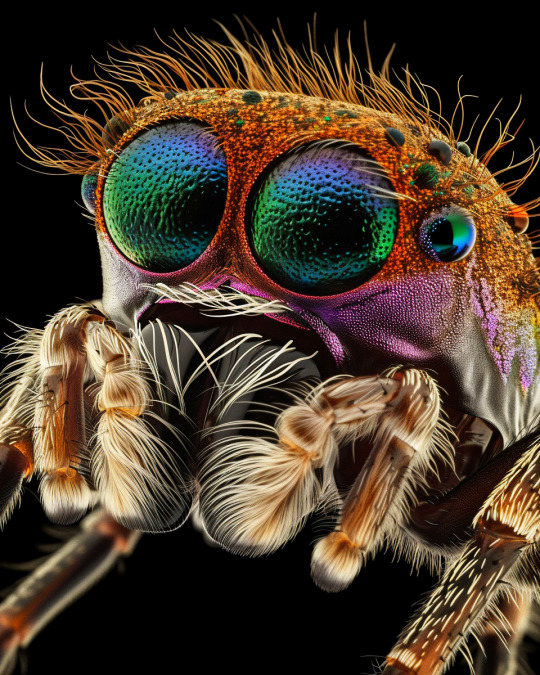


/𝗶𝗺𝗮𝗴𝗶𝗻𝗲 𝗽𝗿𝗼𝗺𝗽𝘁:
A coloured scanning electron micrograph + head of a Jumping Spider (family Salticidae) + common housefly (Musca domestica) + cat flea (Ctenocephalides felis + head of a maggot or the larva of a bluebottle fly (Protophormia sp.) with tiny teeth-like fangs extending from its mouth + head of a soldier turtle ant (Cephalotes sp.) from the Amazonian rainforest + head of a honey bee
#nature#midjourney#midjourney art#midjourney ai#artificial intelligence#electron microscope#insects#electron microscopy
33 notes
·
View notes
Text
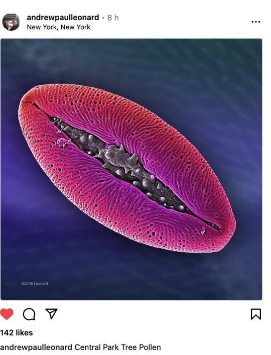
Kiss Kiss! This is what some pollen grains look like under the scanning electron microscope. The exact species wasn't listed in the original post but many plants, ranging from lilies to redbuds, that have pollen grains that look similar to this.
From Wikipedia:
Pollen is a powdery substance produced by flowers of seed plants. It consists of pollen grains (highly reduced microgametophytes), which produce male gametes (sperm cells). Pollen grains have a hard coat made of sporopollenin that protects the gametophytes during the process of their movement from the stamens to the pistil of flowering plants, or from the male cone to the female cone of gymnosperms. If pollen lands on a compatible pistil or female cone, it germinates, producing a pollen tube that transfers the sperm to the ovule containing the female gametophyte. Individual pollen grains are small enough to require magnification to see detail. The study of pollen is called palynology and is highly useful in paleoecology, paleontology, archaeology, and forensics. Pollen in plants is used for transferring haploid male genetic material from the anther of a single flower to the stigma of another in cross-pollination. In a case of self-pollination, this process takes place from the anther of a flower to the stigma of the same flower.
Source: Andrew Paul Lenonard's Instagram page
#katia plant scientist#botany#plant biology#plant science#plants#naturecore#nature#nature photography#microscope#electron microscope#tiny things#pollen#reproduction#mad science#science#biology#flowers#pink lips#juicy lips#luscious lips#beautiful lips#lipstick#kiss kiss
12 notes
·
View notes
Text
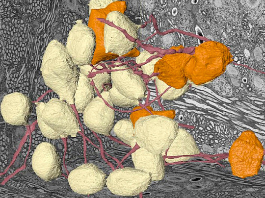
Bush Telegraphy
Analysing and modelling cells of the brain's cochlear nuclei called globular bushy cells using volume electron microscopy to better understand how sound is perceived
Read the published research paper here
Image from work by George A Spirou and Paul B Manis, and colleagues
Department of Medical Engineering, University of South Florida, Tampa, FL and Department of Otolaryngology/Head and Neck Surgery, University of North Carolina at Chapel Hill, Chapel Hill, NC, USA
Image originally published with a Creative Commons Attribution 4.0 International (CC BY 4.0)
Published in eLife, June 2023
You can also follow BPoD on Instagram, Twitter and Facebook
9 notes
·
View notes
Text
Types of Microscopes
1. Simple Microscope
2. Compound Microscope
3. Phase Contrast Microscope
4. Fluorescence Microscope
5. Electron Microscope
6. Scanning Electron Microscope (SEM)
7. Transmission Electron Microscope (TEM)
8. Dark Field Microscope
9. Dissecting Microscope (Stereo Microscope)
10. Digital Microscope
11. Scanning Probe Microscope (SPM)
12. Atomic Force Microscope (ATM)
13. Inverted Microscope
14. Acoustic Microscope
15. X-Ray Microscope
16. Polarizing Microscope
17. Metallurgical Microscope
18. Pocket Microscope
19. USB Microscope
20. Confocal Microscope
21. Laser Scanning Microscope
22. Differential Interference Contrast Microscope (DIC)
23. Near-field Scanning Optical Microscope (NSOM)
24. Raman Microscope
25. Super-resolution Microscope
26. Cryo-electron Microscope
27. Time-lapse Microscope
There is a wide range of microscopy techniques and instruments used in various fields of science and research.
#forensic#forensics#criminology#forensic science#evidence#criminalistic#forensic field#crime#forensic science notes#crime scene investigation#electron microscope#microscope
5 notes
·
View notes
Text

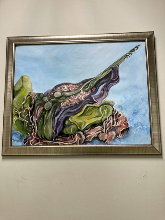
I donated this painting to my highschool. The reference was an electron microscope image of a detached bee stinger :D.
If the frame seems weirdly sized the reason is that we had to pick a new frame last minute because the one I had picked out got stolen 💀.
#bees#traditional art#painting#my art#biology#electron microscope#science#art#fine arts#my painting#art donation#high school
5 notes
·
View notes
Photo

Dogpile
#abstractart#deep dream#deep dream generator#dream#abstract#macro#macrophotography#microscopic#texture#microphotography#electron microscope#abstract art#lock screen#wallpaper#background#inspiration#weird art#cool lockscreen#trippy art
6 notes
·
View notes
Text
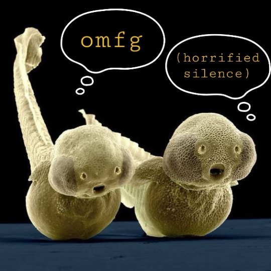
#meme#zebrafish#electron microscope#horrified silence#omfg#speechless#horror#the horrors persist#every time i get online
1 note
·
View note
Photo
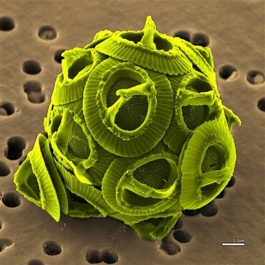
The algae Gephyrocapsa oceanica.
Wikimedia Commons
#wikimedia commons#photographer#algae#gephyrocapsa oceanic#micro photography#electron microscope#nature
50 notes
·
View notes
Text
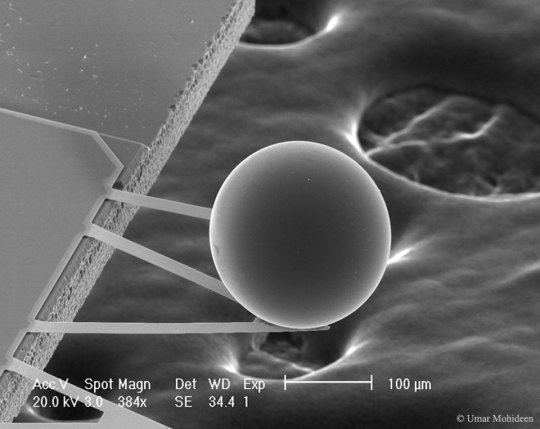
A Force from Empty Space: The Casimir Effect
Image Credit & Copyright: Umar Mohideen (U. California at Riverside)
Explanation: This tiny ball provides evidence that the universe will expand forever. Measuring slightly over one tenth of a millimeter, the ball moves toward a smooth plate in response to energy fluctuations in the vacuum of empty space. The attraction is known as the Casimir Effect[...]
#casimir effect#apod#umar mohideen#electron microscope#2015 December 6#apod 2015 December 6#nasa#photography#sphere#science#physics#quantum mechanics
1 note
·
View note
Text
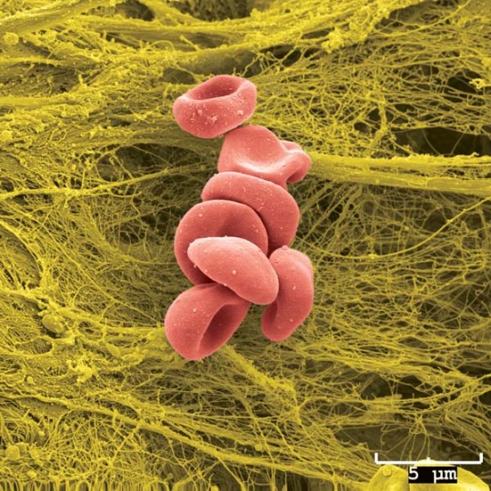
Electron microscope image of red blood cells.
source: https://cmrf.research.uiowa.edu/scanning-electron-microscopy-images
1 note
·
View note
Text

A Scanning Electron Microscope (SEM) image of Pollen from a variety of common plants:
Sunflower (Helianthus annuus, small spiky sphericals, colorized pink), morning glory (Ipomoea purpurea, big sphericals with hexagonal cavities, colorized mint green), hollyhock (Sildalcea malviflora, big spiky sphericals, colorized yellow), lily (Lilium auratum, bean shaped, colorized dark green), primrose (Oenothera fruticosa, tripod shaped, colorized red) and castor bean (Ricinus communis, small smooth sphericals, colorized light green). The image is magnified some x500, so the bean shaped grain in the bottom left corner is about 50 μm long.
0 notes
Text

Cross-sectional transmission electron microscopy (TEM) of a 3-nm UTB MOSFET.
0 notes
Last Seen Blogs

siochaile
I talk to myself in the Tags

angel-blitz
🩷🍭🤡🍭🩷

irrefutablyhysterical
i won't let you let me be

lihat
Untitled
angel-blitz
🩷🍭🤡🍭🩷