#microglial cell
Text
Hemoglobina "Hema" Rojas

She is one of those old OCs I'm bringing back to life.
She is based in "Osmosis Jones (2001)" and is a red blood cells, because they are the cutest cells, if you don't believe me, just look at them under a microscope. And neurons are the sexier cells, because brain is the new sexy. XD
She have a long story so if you want to read it you can find it in ToyHouse, and more art under the cut.
First designs:

New designs:

#rbc#wbc#wbc x rbc#digital art#fanfic#original character#red blood cell#osmosis jones#microglial cell#my ocs#oc ref sheet#toyhouse#hemoglobin#cytology
20 notes
·
View notes
Text
it's significantly easier for me to think today, i think i've worked out a little combo for when post sickness neuroinflammation hits:
i took 15mg dxm + 2mg naltrexone last night. usually i feel out of it several days with dxm, but not this time. i'm guessing it's because of naltrexone blocking my mu + delta opioid receptors. this is huge for me, the reason i put off taking dxm when my pots+csf/me gets bad is because i don't want to be unable to think clearly for the next few days
also applied a 17.5 mg nicotine patch. i've been using these for a while now but haven't posted about it. basically nicotine activates neuronal nicotinic acetylcholine receptors which regulate microglial activity (resident immune cells of the CNS) inhibiting secretion of proinflammatory molecules and upregulates glutamate transporters enhancing glutamate clearance from the synapse and reducing excitotoxicity.
2 notes
·
View notes
Photo
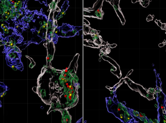
Feasting on Synapses
Your thoughts and memories are held within billions of neurons in your brain and the connections between them (synapses). In Alzheimer's disease, neurons and synapses are destroyed by the immune system. Researchers investigate how using a mouse model for Alzheimer's. 3D reconstructions using confocal microscopy images (pictured) of healthy (right) and affected (left) mouse brains revealed immune cells called microglia and astrocytes, and small immune proteins called the complement cascade are involved. Synapse proteins accumulated in microglia (white) and astrocytes (blue) of diseased brains, specifically in lysosomes (green) – compartments that digest substances a cell engulfs. Astrocytic lysosomes contained more proteins from excitatory synapses (yellow), while microglial lysosomes contained more from inhibitory synapses (red). Notably, Alzheimer’s model mice genetically engineered to lack a complement protein had fewer synapse proteins in either type of lysosome and fewer contacts between astrocytes and synapses, revealing the importance of complement in the loss of synapses.
Written by Lux Fatimathas
Image from work by Borislav Dejanovic and colleagues
Stanley Center for Psychiatric Research, Broad Institute of MIT and Harvard, Cambridge, MA; and Department of Neuroscience, Genentech, South San Francisco, CA, USA
Image originally published with a Creative Commons Attribution 4.0 International (CC BY 4.0)
Published in Nature Aging, September 2022
You can also follow BPoD on Instagram, Twitter and Facebook
#science#biomedicine#synapses#neuroscience#brain#lysosomes#alzheimer's#confocal microscopy#microglia#astrocytes#brain cells#immunofluorescence#immune system#neurons
23 notes
·
View notes
Text
"the name of the beast"
parasitic reaction in the hindbrain
long-forgotten viruses, microglial cascades,
cells singing with love for something that
never lived. Hyperflat meets misfolded
protein, self-replicating prions infest
the cerebellum, the heart, the small
and large intestine. Everything is a
metaphor. Everything in the world
is exactly the same. Diseases gentle
and warm take us in tender embraces
and usher us to a state of rest. The
growing pains of globalization settle
easy in the soft bones of children,
hard in joints made firm by age
We can put it all behind us:
I am thinking, and you are thinking with me,
and it could be faster, we could be closer,
If you'd only find a way to let your guard
down, let the light in. Repentance, they say,
is as good as medicine. Cheaper, besides.
(febrile from the poison some hired
goon sprayed into my brain, careless,
like hot glue used to set a bone, maybe
nothing but hot glue used to set a bone
fractured by a burrsaw for the intromission
of wires. Like a bedbug mating, spraying
the stuff of life into a living creature with
a needle and cauter so like a penis. We know
human beings. Human beings love wires and
they don't care who suffers or how, who
dies or how. One creature's good as another
If I could do it all over again, if I could
do it all over again, if I could do it all
over again, darling, I'd never have let
them take me alive)
I am thinking, and the machine is thinking
with me. The machine is thinking, and I am
thinking with it. I am thinking, and the machine
is thinking with me. I am thinking, and the
machine is thinking with me
the children first, whose minds nature
cast in some unlovable contour. The sick
next, who we must learn to see as a burden
to make peace. When we meet the men who
remake us, it will be face to face, with eyes
like ours, but cold and born to command;
it will be with one mind, one heart,
one seat of thought and spirit. They
will untangle the tongues God once confused
into the straight and perfect english of
the terminal, and our obedience to
our masters' voices will be before
thought. Faster, anyway, than prayer,
with thoroughly documented behaviors
and perfectly anticipated responses
a safe environment in which we can
be trusted, at last, and for all time
12 notes
·
View notes
Text
The Neurobiology of Anxiety Disorders
As May is Mental Health Awareness Month, I thought I would take the time to write about a very common group of psychiatric disorders, anxiety. Anxiety disorders can be highly disabling conditions, with a wide range of symptoms depending on the individual. Specific anxiety disorders include panic disorder, generalized anxiety disorder, social anxiety disorder, and particular phobias. Anxiety disorders are the most prevalent mental disorders and are associated with high costs of healthcare and often a high burden of disease (Bandelow and Michaelis, 2015). Large population-based surveys show that up to 33.7% of the population are impacted by an anxiety disorder at some point during their life (Bandelow and Michaelis, 2015). Some symptoms of these disorders include fatigue, difficulty concentrating, irritability, headaches/migraines, stomach issues, difficulty controlling feelings of worry, and more (Anxiety Disorders, 2019). There are many factors that influence the prevalence of anxiety disorders, both environmental and biological. There are probably many pathways in the brain that cause anxious behavior or stem from having the condition. This proves it difficult to explain the exact neuroscience behind psychiatric disorders such as anxiety. Many experiments have been conducted to try and gain more insight into specific cells, proteins, and pathways in the nervous system that either give rise to or mediate anxious behaviors. Research continues to be done to help better understand and treat psychiatric disorders such as anxiety.
One experiment that was conducted found that there is a prefrontal cortex to amygdala pathway in the brain in chronic stress induced anxiety. The prefrontal cortex is the area of the brain that lies at the very front of the brain. It plays an important role in executive function, meaning planning, decision making, personality expression, etc. The amygdala is the part of the brain that processes emotions and emotional stimuli. Typically, in psychiatric disorders such as anxiety and depression, there is dysregulation of the amygdala by the prefrontal cortex. An experiment performed by Wei-Zhu Liu and colleagues in 2020 used a rodent anxiety model induced by chronic resistant stress to show that this dysregulation more specifically occurs in basolateral amygdala projection neurons receiving signals only in one direction from the dorsomedial prefrontal cortex (Liu et al, 2020). The signals should be received in multiple directions, not just one, so this is where the dysfunction occurs. Many questions about other mechanisms remain open, but this experiment revealed one pathway in the brain that is affected by anxiety induced by chronic stress. However, it is important to note that this experiment was performed using a rat model, which doesn’t exactly correspond to humans. Nonetheless, discoveries like this are important because they give insight into what happens in the brain when there is chronic stress and anxiety disorders present, and might help scientists in designing treatments that target such pathways.
Another experiment performed by Ya-Lin Wang and colleagues in 2021 revealed that microglial activation mediates depressive and anxiety-like behavior. Microglial cells are a specialized population of macrophages found in the central nervous system. Essentially, this means that they act as a form of immune defense in the nervous system by removing damaged neurons and infections. In this experiment, male rats were subjected to stressors for a twelve-week period. The researchers found that 12 weeks of chronic mild stress induced a remarkable amount of both depressive and anxiety-like behavior. They also found that the hippocampus was quite inflamed, shown by the activation of inflammatory mediators, as well as the activation of microglial cells (Wang et al, 2021). They concluded that activation of microglial cells reduces depressive and anxiety-like behavior induced by chronic mild stress. The also found that microglial cells helped reduce hippocampal inflammation (Wang et al, 2021). Again, as before, this study was conducted in rats, so it may be tough to say if these results can directly apply to humans. These researchers also acknowledge that the exact mechanisms that underlie disorders such as depression and anxiety have yet to be discovered.
The two experiments described are just the tip of the iceberg when it comes to scientific research on psychiatric disorders such as anxiety. It is through such experiments that breakthroughs can be made in terms of treating people with these disorders. As someone who has lived with anxiety from a very young age, it is fascinating to see the recent discoveries made in the field of neurobiology, and it gives me hope for the future of mental healthcare.
References
Bandelow, B., & Michaelis, S. (2015). Epidemiology of anxiety disorders in the 21st century. Anxiety, 17(3), 327–335. https://doi.org/10.31887/dcns.2015.17.3/bbandelow
Biological markers of generalized anxiety disorder. (2017). Generalized Anxiety Disorders, 19(2), 147–158. https://doi.org/10.31887/dcns.2017.19.2/dnutt
Liu, W.-Z., Zhang, W.-H., Zheng, Z.-H., Zou, J.-X., Liu, X.-X., Huang, S.-H., You, W.-J., He, Y., Zhang, J.-Y., Wang, X.-D., & Pan, B.-X. (2020). Identification of a prefrontal cortex-to-amygdala pathway for chronic stress-induced anxiety. Nature Communications, 11(1). https://doi.org/10.1038/s41467-020-15920-7
National Institute of Mental Health. (2019). Anxiety disorders. Nih.gov; National Institute of Mental Health. https://www.nimh.nih.gov/health/topics/anxiety-disorders
Wang, Y.-L., Han, Q.-Q., Gong, W.-Q., Pan, D.-H., Wang, L.-Z., Hu, W., Yang, M., Li, B., Yu, J., & Liu, Q. (2018). Microglial activation mediates chronic mild stress-induced depressive- and anxiety-like behavior in adult rats. Journal of Neuroinflammation, 15(1). https://doi.org/10.1186/s12974-018-1054-3
2 notes
·
View notes
Link
0 notes
Quote
Microglia are central players in Alzheimer’s disease pathology but analyzing microglial states in human brain samples is challenging due to genetic diversity, postmortem delay and admixture of pathologies. To circumvent these issues, here we generated 138,577 single-cell expression profiles of human stem cell-derived microglia xenotransplanted in the brain of the AppNL-G-F model of amyloid pathology and wild-type controls. Xenografted human microglia adopt a disease-associated profile similar to that seen in mouse microglia, but display a more pronounced human leukocyte antigen or HLA state, likely related to antigen presentation in response to amyloid plaques. The human microglial response also involves a pro-inflammatory cytokine/chemokine cytokine response microglia or CRM response to oligomeric Aβ oligomers. Genetic deletion of TREM2 or APOE as well as APOE polymorphisms and TREM2R47H expression in the transplanted microglia modulate these responses differentially. The expression of other Alzheimer’s disease risk genes is differentially regulated across the distinct cell states elicited in response to amyloid pathology. Thus, we have identified multiple transcriptomic cell states adopted by human microglia in a multipronged response to Alzheimer’s disease-related pathology, which should be taken into account in translational studies.
Xenografted human microglia display diverse transcriptomic states in response to Alzheimer’s disease-related amyloid-β pathology | Nature Neuroscience
0 notes
Text
IJMS, Vol. 25, Pages 2406: Intravenous Administration of Mesenchymal Stem Cell-Derived Exosome Alleviates Spinal Cord Injury by Regulating Neutrophil Extracellular Trap Formation through Exosomal miR-125a-3p
Spinal cord injury (SCI) leads to devastating sequelae, demanding effective treatments. Recent advancements have unveiled the role of neutrophil extracellular traps (NETs) produced by infiltrated neutrophils in exacerbating secondary inflammation after SCI, making it a potential target for treatment intervention. Previous research has established that intravenous administration of stem cell-derived exosomes can mitigate injuries. While stem cell-derived exosomes have demonstrated the ability to modulate microglial reactions and enhance blood–brain barrier integrity, their impact on neutrophil deactivation, especially in the context of NETs, remains poorly understood. This study aims to investigate the effects of intravenous administration of MSC-derived exosomes, with a specific focus on NET formation, and to elucidate the associated molecular mechanisms. Exosomes were isolated from the cell supernatants of amnion-derived mesenchymal stem cells using the ultracentrifugation method. Spinal cord injuries were induced in Sprague-Dawley rats (9 weeks old) using a clip injury model, and 100 μg of exosomes in 1 mL of PBS or PBS alone were intravenously administered 24 h post-injury. Motor function was assessed serially for up to 28 days following the injury. On Day 3 and Day 28, spinal cord specimens were analyzed to evaluate the extent of injury and the formation of NETs. Flow cytometry was employed to examine the formation of circulating neutrophil NETs. Exogenous #miRNA was electroporated into neutrophil to evaluate the effect of inflammatory NET formation. Finally, the biodistribution of exosomes was assessed using 64Cu-labeled exosomes in animal positron emission tomography (PET). Rats treated with exosomes exhibited a substantial improvement in motor function recovery and a reduction in injury size. Notably, there was a significant decrease in neutrophil infiltration and NET formation within the spinal cord, as well as a reduction in neutrophils forming NETs in the circulation. In vitro investigations indicated that exosomes accumulated in the vicinity of the nuclei of activated neutrophils, and neutrophils electroporated with the miR-125a-3p mimic exhibited a significantly diminished NET formation, while miR-125a-3p inhibitor reversed the effect. PET studies revealed that, although the majority of the transplanted exosomes were sequestered in the liver and spleen, a notably high quantity of exosomes was detected in the damaged spinal cord when compared to normal rats. MSC-derived exosomes play a pivotal role in alleviating spinal cord injury, in part through the deactivation of NET formation via miR-125a-3p. https://www.mdpi.com/1422-0067/25/4/2406?utm_source=dlvr.it&utm_medium=tumblr
0 notes
Text
Neurolemmocytes vs Microglial Cells
Neurolemmocytes
-> PNS neuroglia
-> form myelin sheaths
Microglial Cells
-> CNS neuroglia
-> act as macrophages
.
Patreon
#studyblr#notes#my notes#biology#bio#human biology#human bio#anatomy#human anatomy#anatomical structures#anatomical vocabulary#anatomical variation#medblr#medical notes#med notes#human anatomical structures#anatomy and physiology#anatomy & physiology#life science#biological science#note cards#flashcards#flash cards
1 note
·
View note
Text
Everyone gets reincarnated into the microbiome and shit out by somebody who was more intelligent than them but didn't have the right credentials, when they learn something from a high school drop out
In Hell. Hellenism. Delirium? Underworld! Lower Astral? Sleep Paralysis, Hypnagogic and Hypnopompic hallucinations, sometimes psychosis not always - uhm, mycotoxin exposure? Sleep Deprivation. Oxygen deprivation? Carbon monoxide poisoning. Alzheimer's, Parkinson's, dementia? Yes
The "underworld" the "lower astral" the scary hallucinations - hell
Sometimes DMT? SOMETIMES SALVINORIN A, NOT ALWAYS
Never dissociation or 5-MEO-DMT
Dissociation is how we escape trauma, or hell, suffering, tragedy
Oh, and delirium but ITS NOT HELL, NOT "DARK" if it's a manic dissociative delirium from a mania-inducing Dissociative NMDA antagonist that induces sleep deprivation for 24-36 hours or more
The biofilm shits inside of us, triggering microglial activation a neurotoxic immune response that precedes Alzheimer's and Parkinson's disease dementia by destroying brain cells, and interacts with ion channels in our brain, learning to control our behavior until we turn into zombies and forget who we are. Then, the microbiota eats their way out - the body begins to rot and decompose decades before it's in the ground when the immune system becomes compromised with age, or when people abuse drugs that act as Immunosuppressants
That's Alzheimer's for ya
0 notes
Text
This common fungus can trigger ‘key player’ in Alzheimer’s disease development
HOUSTON — A troubling new study shows a link between a common fungus and Alzheimer’s disease. Scientists from the Baylor College of Medicine are revealing how the fungus Candida albicans enters the brain, triggers mechanisms that aid in its clearance, and generates toxic protein fragments known as amyloid beta (Ab)-like peptides — a key player in Alzheimer’s disease development.
“Our lab has years of experience studying fungi, so we embarked on the study of the connection between C. albicans and Alzheimer’s disease in animal models,” says study corresponding author Dr. David Corry, the Fulbright Endowed Chair in Pathology and a professor of pathology and immunology and medicine at Baylor, in a university release. “In 2019, we reported that C. albicans does get into the brain where it produces changes that are very similar to what is seen in Alzheimer’s disease. The current study extends that work to understand the molecular mechanisms.”
The study first sought to unravel how Candida albicans gains access to the brain. Researchers discovered that the fungus produces enzymes called secreted aspartic proteases (Saps), which break down the blood-brain barrier — a protective barrier that usually prevents harmful substances from entering the brain. This breach allows the fungus to infiltrate the brain and cause damage.
The next question posed by the researchers was how the brain effectively clears the fungus. Previous research has shown that C. albicans brain infections resolve entirely in healthy mice after 10 days. In this study, the team unveiled two mechanisms triggered by the fungus in microglia brain cells, which play a crucial role in the brain’s immune response.
“The same Saps that the fungus uses to break the blood-brain barrier also break down the amyloid precursor protein into Ab-like peptides,” says study first author Dr. Yifan Wu, postdoctoral scientist in pediatrics working in the Corry lab. “These peptides activate microglial brain cells via a cell surface receptor called Toll-like receptor 4, which keeps the fungi load low in the brain, but does not clear the infection.”
1 note
·
View note
Link
0 notes
Text
References
Available at: https://www.ncbi.nlm.nih.gov/books/NBK92752/. Accessed September 23, 2022.
Sikora E, Scapagnini G, Barbagallo M. Curcumin, inflammation, ageing and age-related diseases. Immun Ageing. 2010 Jan 17;7(1):1.
Bahrami A, Montecucco F, Carbone F, et al. Effects of Curcumin on Aging: Molecular Mechanisms and Experimental Evidence. Biomed Res Int. 2021;2021:8972074.
Benameur T, Soleti R, Panaro MA, et al. Curcumin as Prospective Anti-Aging Natural Compound: Focus on Brain. Molecules. 2021 Aug 7;26(16).
Zia A, Farkhondeh T, Pourbagher-Shahri AM, et al. The role of curcumin in aging and senescence: Molecular mechanisms. Biomed Pharmacother. 2021 Feb;134:111119.
Lee KS, Lee BS, Semnani S, et al. Curcumin extends life span, improves health span, and modulates the expression of age-associated aging genes in Drosophila melanogaster. Rejuvenation Res. 2010 Oct;13(5):561-70.
Liao VH, Yu CW, Chu YJ, et al. Curcumin-mediated lifespan extension in Caenorhabditis elegans. Mech Ageing Dev. 2011 Oct;132(10):480-7.
Shen LR, Parnell LD, Ordovas JM, et al. Curcumin and aging. Biofactors. 2013 Jan-Feb;39(1):133-40.
Shen LR, Xiao F, Yuan P, et al. Curcumin-supplemented diets increase superoxide dismutase activity and mean lifespan in Drosophila. Age (Dordr). 2013 Aug;35(4):1133-42.
Soh JW, Marowsky N, Nichols TJ, et al. Curcumin is an early-acting stage-specific inducer of extended functional longevity in Drosophila. Exp Gerontol. 2013 Feb;48(2):229-39.
Stepien K, Wojdyla D, Nowak K, et al. Impact of curcumin on replicative and chronological aging in the Saccharomyces cerevisiae yeast. Biogerontology. 2020 Feb;21(1):109-23.
Hewlings SJ, Kalman DS. Curcumin: A Review of Its Effects on Human Health. Foods. 2017 Oct 22;6(10).
Seddon N, D’Cunha NM, Mellor DD, et al. Effects of Curcumin on Cognitive Function—A Systematic Review of Randomized Controlled Trials. Exploratory Research and Hypothesis in Medicine. 2019 03/19;4(1):1-11.
Taka T, Changtam C, Thaichana P, et al. Curcuminoid derivatives enhance telomerase activity in an in vitro TRAP assay. Bioorg Med Chem Lett. 2014 Nov 15;24(22):5242-6.
Xiao Z, Zhang A, Lin J, et al. Telomerase: a target for therapeutic effects of curcumin and a curcumin derivative in Abeta1-42 insult in vitro. PLoS One. 2014;9(7):e101251.
Yu Y, Shen Q, Lai Y, et al. Anti-inflammatory Effects of Curcumin in Microglial Cells. Front Pharmacol. 2018;9:386.
Sunny A, Ramalingam K, Das S, et al. Bioavailable curcumin alleviates lipopolysaccharide-induced neuroinflammation and improves cognition in experimental animals. Pharmacognosy Magazine. 2019 April 1, 2019;15(62):111-7.
Dong S, Zeng Q, Mitchell ES, et al. Curcumin enhances neurogenesis and cognition in aged rats: implications for transcriptional interactions related to growth and synaptic plasticity. PLoS One. 2012;7(2):e31211.
Kim CS, Park S, Kim J. The role of glycation in the pathogenesis of aging and its prevention through herbal products and physical exercise. J Exerc Nutrition Biochem. 2017 Sep 30;21(3):55-61.
Simm A. Protein glycation during aging and in cardiovascular disease. J Proteomics. 2013 Oct 30;92:248-59.
Hu TY, Liu CL, Chyau CC, et al. Trapping of methylglyoxal by curcumin in cell-free systems and in human umbilical vein endothelial cells. J Agric Food Chem. 2012 Aug 22;60(33):8190-6.
Liu JP, Feng L, Zhu MM, et al. The in vitro protective effects of curcumin and demethoxycurcumin in Curcuma longa extract on advanced glycation end products-induced mesangial cell apoptosis and oxidative stress. Planta Med. 2012 Nov;78(16):1757-60.
Sajithlal GB, Chithra P, Chandrakasan G. Effect of curcumin on the advanced glycation and cross-linking of collagen in diabetic rats. Biochem Pharmacol. 1998 Dec 15;56(12):1607-14.
Lima TFO, Costa MC, Figueiredo ID, et al. Curcumin, Alone or in Combination with Aminoguanidine, Increases Antioxidant Defenses and Glycation Product Detoxification in Streptozotocin-Diabetic Rats: A Therapeutic Strategy to Mitigate Glycoxidative Stress. Oxid Med Cell Longev. 2020;2020:1036360.
Tang Y, Chen A. Curcumin eliminates the effect of advanced glycation end-products (AGEs) on the divergent regulation of gene expression of receptors of AGEs by interrupting leptin signaling. Lab Invest. 2014 May;94(5):503-16.
Ohtani N. The roles and mechanisms of senescence-associated secretory phenotype (SASP): can it be controlled by senolysis? Inflamm Regen. 2022 Apr 2;42(1):11.
Cherif H, Bisson DG, Jarzem P, et al. Curcumin and o-Vanillin Exhibit Evidence of Senolytic Activity in Human IVD Cells In Vitro. J Clin Med. 2019 Mar 29;8(4).
Yousefzadeh MJ, Zhu Y, McGowan SJ, et al. Fisetin is a senotherapeutic that extends health and lifespan. EBioMedicine. 2018 Oct;36:18-28.
Jakubczyk K, Druzga A, Katarzyna J, et al. Antioxidant Potential of Curcumin-A Meta-Analysis of Randomized Clinical Trials. Antioxidants (Basel). 2020 Nov 6;9(11).
de Oliveira MR, Jardim FR, Setzer WN, et al. Curcumin, mitochondrial biogenesis, and mitophagy: Exploring recent data and indicating future needs. Biotechnol Adv. 2016 Sep-Oct;34(5):813-26.
Rainey NE, Moustapha A, Petit PX. Curcumin, a Multifaceted Hormetic Agent, Mediates an Intricate Crosstalk between Mitochondrial Turnover, Autophagy, and Apoptosis. Oxid Med Cell Longev. 2020;2020:3656419.
Kumar D, Jacob D, Subash PS, et al. Enhanced bioavailability and relative distribution of free (unconjugated) curcuminoids following the oral administration of a food-grade formulation with fenugreek dietary fibre: A randomised double-blind crossover study. J Funct Foods. 2016;22:578-87.
0 notes
Text
Applying high-resolution spatial transcriptomics to characterise the amyloid plaque cell niche in Alzheimer's Disease
The amyloid plaque cell niche is a pivotal hallmark of Alzheimer's disease (AD). Where early spatial transcriptomics (ST) technologies have provided valuable information on transcriptomic alterations in the small tissue domains overlaying with amyloid plaques, they lacked cellular resolution. Here we compare two novel high-resolution ST platforms, CosMx and Stereo-seq, in their ability to characterize the cellular response in the amyloid plaque niche in an AD mouse model. Combining the results from both techniques empowered us to survey the highly variable microglial-astrocytic response across the amyloid plaque micro-environment and provided a first insight into how these responses could relate to neuronal transcriptomic alterations. This pilot study demonstrates the great potential of high-resolution ST, while simultaneously highlighting limitations that, when addressed, will unleash the full power of these techniques to map the progression of molecular and cellular changes in the brains of AD patients. http://dlvr.it/SrXzSb
0 notes
Text
miR-29a-3p promotes the regulatory role of eicosapentaenoic acid in the NLRP3 inflammasome and autophagy in microglial cells
Pubmed: http://dlvr.it/Slf2Zc
0 notes
Photo

Perivascular cells could induce microglial malfunction associated with Alzheimer's disease
0 notes
Last Seen Blogs