#Volcano heat signature
Text
NASA Earth Observatory: Another Eruption in Iceland
FromEarthOrbit, #PlanetEarth, #OurHomeInSpace, #TheOverviewEffect
Hot stuff! 🌋
Satellites measured the infrared heat signature of three recent volcanic eruptions in southwestern Iceland. The first two lava flows appear black, while last week’s eruption radiates heat, which appears red/orange in this #Landsat image - #NASAEarthObservatory
#Iceland Volcano#NASA Earth Observatory#SW Iceland#Volcano heat signature#Landsat Image#Reykjanes Peninsula#EarthObservatory.NASA.gov
0 notes
Text

Started putting booksby's signature on the monstiary pages because it seemed fun.
"Geologica capetra (Noggin) average around 4’5”-1.3m tall. Noggin are bipedal and mostly made of stone with fleshy appendages. They have strong jaws made for cracking rocks. Like most monsters who are primarily made of rock they have a kiln for a digestive tract.(Kilns in monster anatomy are a modified version of the "convert everything to calories" organs other monsters have, they work via magically producing heat that also absorbs nutrients from things, but mainly serves to melt rock)
Their bodies themselves are made of three main parts, the main cylindrical rocky body, the aforementioned fleshy limbs, and their jaw. The limbs are directly attached to the body, but are counted as a different part for their organic makeup. The jaw is locked in place by two protrusions on the inside of it that push into dents in the body allowing it to swivel open and closed.
The makeup of their bodies depends on what rocks they eat, a noggin who eats mafic rocks will be darker in color, and vice versa. Their skin tone also depends on outside factors and can change, in areas of high sunlight they will get darker as they develop more melanin, and paler as they are exposed to less. With their jaws being detached from their bodies, it's assumed they grow magically, each different rock type has its own signature of the earth element allowing the jaw to be made of the same rock as the body despite it not being formed from minerals the noggin consumes. Most commonly their stone is a medium gray, and their skin is a tan tone.
With their stone plate growth they have to grate down their forms regularly. They take dust baths and sit in streams to erode their rock. Or they carve themselves, some who do that decide to get more decorative with their visage and carve themselves into unique shapes. Some may even implant other stones and metals into their bodies for a little extra flair, often in most noggin-centric groups this is seen as posh and eccentric behavior."
Variants

"G. capetra therma (Primal noggin, magma noggin, or thermal noggin): These noggins are native to earth island and near solely live there. They are mafic and always dark in color. Their skin is covered in rocky scales. Their kilns burn hotter than average, causing them to be warm to the touch and partially glow at the mouth. They all live in a large social group in the bowels of the Second Volcano and have a unique culture. They worship the earth colossal and Igeo (torrt, if you remember the old name I changed it after coming to the conclusion that earth monsters don't get to be named after gemstones), being one of the only groups of monsters who widely think the celestials are real. They believe they are directly descended from the first noggins to exist with very little trace of other elements in their non-existent blood. No one has any way of proving this, but at least currently they appear to be mostly earth, which is what will happen if you live in an elemental sink.
Their mouths are made of stony digits instead of the usual solid chunk in the common variation. Their claws are for digging in the much harder solid stone that makes up their home. In their old age they may grow horns from their backs and sides below their hands.
As for more about the social group, called the magma refuge(thus the name magma noggins), came into existence when the cataclysm occurred. A group of noggins hid in the deep caves of the second volcano and lived there for the years leading up to the present. During that time they evolved into their unique forms and developed the skills necessary to survive in that harsh environment. They also became mostly carnivorous in their organic diet, with the only organic food in the cave being the weird cave dwelling lizards and bats that could handle the heat down there.
When they rediscovered that the surface was safe, they found out the large herbivores of the earth lands, now earth island, still existed, and wanted to eat them. They learned to make weapons to hunt the critters, and hunting became a part of their culture. To be considered an adult there you have to take down a rock monitor.
When the first monsters began awakening from their statues the magma refuge helped them rebuild monster society and get used to having to all live directly off the land again, which I am so glad I didn’t have to do. I woke up to a nice functional civilization where everyone already knew what they were doing and had all sorts of systems up and running. And it also wasn’t freaking earth island, no giant lizards trying to kill you.
They also HATE hyehehe, they compete for the same (organic) food, and their general nature annoys them, so it’s bound to be at least a rivalry. They even have the hide of one actually! They used to use it for ritualistic purposes and intimidation originally, but after it was found out that hyehehe were sapient they felt bad and retired the use of it. Kept it for sentimental reasons though."
Ecology and behavior
"Noggins live in areas with rocks, which are pretty much everywhere. They are primarily geovoreous with a small amount of organic food in their diets to support the organic parts of their body. They get rock to eat by digging them up with their feet, by biting them off of rocky surfaces, or just eating them off the ground. You can tell when noggins live nearby when there’s a noticeable lack of pebbles or stones on the ground. Their foraging behavior and removal of large stones in the earth makes them very good for plant growth. For organic things they are opportunistic and eat anything they can find.
Sometimes they use their rocky appearance as camouflage, as they are usually made of the most common rock in their area so they blend in perfectly, they are small and easily overpowered by critters similar in size to them, and their limbs are made of meat and edible. Their high mineral content probably makes them not the most appetizing thing in the world, but some critters can be desperate enough to go for them. They also use this for hunting occasionally, most often for small critters that they wait to get close enough, and then quickly snapping them up in their jaws."

(the image is of a fairly old young noggin, like maybe a 9 year old in human years, they are usually smaller)
"Noggins, like most natural single elementals will hatch anywhere. Young noggins don’t hatch from their eggs and instead form from them. They are very wobbly and clumsy so they often sit rather than standing. Adult noggins will carry young ones on their heads to move them around. They do start walking quite late in development which is often annoying because they get super heavy very quickly so it’s difficult to carry them for that entire time, but it must be done as they will quickly devour all the rocks in an area due to their need to grow."
Just a fun note to finish this off, a group of noggins is called a quarry.
87 notes
·
View notes
Text

NASA’s Juno to Get Close Look at Jupiter’s Volcanic Moon Io on Dec. 30.
The orbiter has performed 56 flybys of Jupiter and documented close encounters with three of the gas giant’s four largest moons.
NASA’s Juno spacecraft will on Saturday, Dec. 30, make the closest flyby of Jupiter’s moon Io that any spacecraft has made in over 20 years. Coming within roughly 930 miles (1,500 kilometers) from the surface of the most volcanic world in our solar system, the pass is expected to allow Juno instruments to generate a firehose of data.
“By combining data from this flyby with our previous observations, the Juno science team is studying how Io’s volcanoes vary,” said Juno’s principal investigator, Scott Bolton of the Southwest Research Institute in San Antonio, Texas. “We are looking for how often they erupt, how bright and hot they are, how the shape of the lava flow changes, and how Io’s activity is connected to the flow of charged particles in Jupiter’s magnetosphere.”
A second ultra-close flyby of Io is scheduled for Feb. 3, 2024, in which Juno will again come within about 930 miles (1,500 kilometers) of the surface.
The spacecraft has been monitoring Io’s volcanic activity from distances ranging from about 6,830 miles (11,000 kilometers) to over 62,100 miles (100,000 kilometers), and has provided the first views of the moon’s north and south poles. The spacecraft has also performed close flybys of Jupiter’s icy moons Ganymede and Europa.
“With our pair of close flybys in December and February, Juno will investigate the source of Io’s massive volcanic activity, whether a magma ocean exists underneath its crust, and the importance of tidal forces from Jupiter, which are relentlessly squeezing this tortured moon,” said Bolton.
Now in the third year of its extended mission to investigate the origin of Jupiter, the solar-powered spacecraft will also explore the ring system where some of the gas giant’s inner moons reside.
Picture This
All three cameras aboard Juno will be active during the Io flyby. The Jovian Infrared Auroral Mapper (JIRAM), which takes images in infrared, will be collecting the heat signatures emitted by volcanoes and calderas covering the moon’s surface. The mission’s Stellar Reference Unit (a navigational star camera that has also provided valuable science) will obtain the highest-resolution image of the surface to date. And the JunoCam imager will take visible-light color images.
JunoCam was included on the spacecraft for the public’s engagement and was designed to operate for up to eight flybys of Jupiter. The upcoming flyby of Io will be Juno’s 57th orbit around Jupiter, where the spacecraft and cameras have endured one of the solar system’s most punishing radiation environments.
“The cumulative effects of all that radiation has begun to show on JunoCam over the last few orbits,” said Ed Hirst, project manager of Juno at NASA’s Jet Propulsion Laboratory in Southern California. “Pictures from the last flyby show a reduction in the imager’s dynamic range and the appearance of ‘striping’ noise. Our engineering team has been working on solutions to alleviate the radiation damage and to keep the imager going.”
More Io, Please
After several months of study and assessment, the Juno team adjusted the spacecraft’s planned future trajectory to add seven new distant Io flybys (for a total of 18) to the extended mission plan. After the close Io pass on Feb. 3, the spacecraft will fly by Io every other orbit, with each orbit growing progressively more distant: The first will be at an altitude of about 10,250 miles (16,500 kilometers) above Io, and the last will be at about 71,450 miles (115,000 kilometers).
The gravitational pull of Io on Juno during the Dec. 30 flyby will reduce the spacecraft’s orbit around Jupiter from 38 days to 35 days. Juno’s orbit will drop to 33 days after the Feb. 3 flyby.
After that, Juno’s new trajectory will result in Jupiter blocking the Sun from the spacecraft for about five minutes at the time when the orbiter is at its closest to the planet, a period called perijove. Although this will be the first time the solar-powered spacecraft has encountered darkness since its flyby of Earth in October 2013, the duration will be too short to affect its overall operation. With the exception of the Feb. 3 perijove, the spacecraft will encounter solar eclipses like this during every close flyby of Jupiter from now on through the remainder of its extended mission, which ends in late 2025.
Starting in April 2024, the spacecraft will carry out a series of occultation experiments that use Juno’s Gravity Science experiment to probe Jupiter’s upper atmospheric makeup, which provides key information on the planet’s shape and interior structure.
More About the Mission
JPL, a division of Caltech in Pasadena, California, manages the Juno mission for the principal investigator, Scott J. Bolton, of the Southwest Research Institute in San Antonio. Juno is part of NASA’s New Frontiers Program, which is managed at NASA’s Marshall Space Flight Center in Huntsville, Alabama, for the agency’s Science Mission Directorate in Washington. Lockheed Martin Space in Denver built and operates the spacecraft.
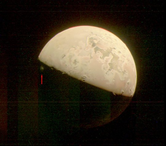
TOP IMAGE....This image revealing the north polar region of the Jovian moon Io was taken on October 15 by NASA’s Juno. Three of the mountain peaks visible in the upper part of image, near the day-night dividing line, were observed here for the first time by the spacecraft’s JunoCam. Credit: Image data: NASA/JPL-Caltech/SwRI/MSSS, Image processing by Ted Stryk
LOWER IMAGE....This JunoCam image of Jupiter’s moon Io captures a plume of material ejected from the (unseen) volcano Prometheus. Indicated by the red arrow, the plume is just visible in the darkness below the terminator (the line dividing day and night). The image was taken by NASA’s Juno spacecraft on October 15. Credit: NASA/JPL-Caltech/SwRI/MSSS
26 notes
·
View notes
Note
Top 5 planets!!!
😍 Thanks for the fun ask, aquaticpal! I'm going to modify this slightly because I already put Earth as my favorite celestial body of all in my previous post about that. I'll do my top 5 planets excluding Earth so I don't repeat myself.
Counting down!
5. Pluto
If you've been paying attention to some astronomy in the last decade or two, you probably know Pluto isn't being called a "Planet" officially anymore.
...I don't care.

The image above shows Pluto (lower right) and its large moon Charon (upper left) as imaged by the New Horizons probe. The frozen heart-shaped feature on Pluto's icy surface melted my own heart (and I wasn't alone).
The whole hullabaloo about Pluto not being a planet is just semantics. It's a celestial body with enough gravity to be round, and it orbits the Sun. I'll count it.
So, why does it make it to my favorites list?
Pluto is geologically active (like Earth - it has its own geology happening on its surface, like volcanoes, tectonics, and erosion), a reality many did not expect until the New Horizons spacecraft started returning close-up pictures of this distant world. The smoothness of Pluto's signature heart-shaped feature (particularly the left side) is a dead giveaway that geological activity is happening on its surface. Worlds where there's no active geology are covered in craters everywhere, not just in some places, and the more recently it's been active the fewer craters there are per area. So.... Pluto is a 'living world' with enough energy inside it to make its insides move.
Cool.
It might be the close orbit of its moon Charon causing it - the shifting gravity stirring up Pluto's innards (and Charon's!), causing friction, and thus heat.
Either way, there's enough geological activity on Pluto to create these gorgeous mountains:

It has seasons during which the frozen nitrogen at its surface becomes warm enough to create mists - nitrogen fog. Imagine if Earth were cold enough that our very air would turn into blocks of ice that create white vapor - kind of like dry ice does.
Pluto is fascinating: a world that defied our expectations. I love it.
4. Trappist-1f
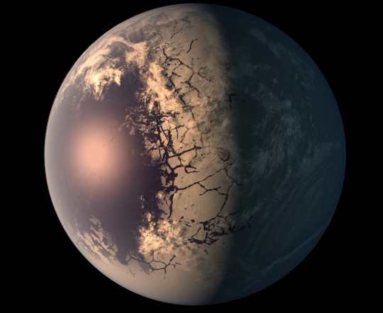
This artist's rendition shows what exoplanet Trappist-1f might look like if it has liquid surface water. It's too small and too far away to get an actual picture, but the evidence shows this planet exists in the habitable zone of its star.
The Trappist-1 star system lies about 40 light-years from Earth (very nearby in the grand scheme of things). Within it, seven planets orbit a small, cool red dwarf star (that's Trappist-1). The planets don't have 'given' names like Pluto or Earth. They're assigned letters b-h, with b the closest and h the furthest from the star. Planets e, f, and g are all in the star's habitable zone, which means they could have liquid water on their surfaces.
So, why did I specifically pick Trappist-1f? It's the planet most perfectly centered in the habitable zone, and its properties are all quite similar to Earth's: it's radius, mass, density, and surface gravity. In order to be hospitable to life as we know it, it's important for a planet to be able to keep itself warm (not just from the light from its Sun - it needs its own, internal heat source, too, to stay geologically active and produce a protective magnetic field - this keeps the atmosphere stable rather than letting its sun's radiative wind blow lots of it away, and also protects life on its surface from that same radiation that would split our complex organic components apart).
Trappist-1f is extremely close to its star, but the star is very cool, so it's unlikely to be very hot. It only takes the planet 9.2 days to orbit the star once. (Yes, you read that right - Trappist-1f's year is only 9.2 Earth days long! If you lived there, I bet you'd find some other way to celebrate birthdays. You'd be having a party just about every week).
I'm all for finding planets that might have water, and therefore might have life... or might be good for us to live on, some day.
The other cool thing about its place is ALL the planets are quite close to the star, so they'd appear huge in the sky regardless of which of the other planets you were standing on. This is a cool, artistic interpretation of that:

3. Proxima Centauri b
Speaking of places where we might be able to live...
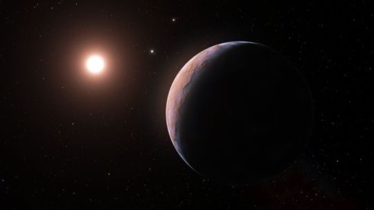
...welcome to Proxima Centauri b - the nearest exoplanet to Earth... and also in its star's habitable zone.
Proxima Centauri b is a small, cool, red dwarf star part of the Alpha Centauri system, a triple-star system consisting of two brighter stars (Alpha Centauri A and Alpha Centauri B, both similar to the Sun) which are close to each other, and the cool red dwarf Proxima Centauri b, still orbiting the others but much farther away.
And around that small cool dwarf star is a planet not too different from Earth.
I can't stress enough how incredible that is.
This is the very nearest star system to our own, only four light-years from here.
And in it, there is already a possible stepping stone for us to move outward into the Cosmos. And not just a stepping stone, but possibly somewhere habitable.
Wow.
We need to learn more about this planet. It may or may not be hospitable. It may or may not have an atmosphere. We just don't know yet. But it's hope. It's one of many signs that the galaxy is absolutely littered with planets, and there may be many worlds like ours.
2. Jupiter
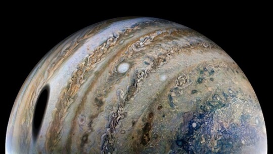
This image taken by the Juno Probe shows a lot of what makes Jupiter awe-inspiring: roiling masses of hydrogen and helium gas with traces of methane, ammonia, silica, and sulfur creating myriad colors in its clouds, storms which could swallow several Earths whole, and of course its huge mass which make it a gravitational well, holding on to a minimum of 80 moons. The huge shadow of its moon Ganymede is visible on the left-hand side in the picture.
First of all, Jupiter is, quite simply, beautiful. Just look at it.
All those colors, the swirls, its absurdly fast rotation (a 10-hour period!) pulling its clouds out into these thin bands. It's so striking. Add to that the shadows of its many moons crossing its surface, and you have a recipe for endless visual fascination.
But that alone isn't enough to make it one of my favorites.
I love Jupiter because it protects us.
Jupiter's gravity is so strong it tends to divert and even capture many objects which hurtle inward toward the Earth and the other inner planets, meaning fewer things actually hit the Earth than they otherwise would. Jupiter is the batter at plate, and we're the catcher. We really don't want any of those strikes thrown to cross the plate.
Thank-you, Jupiter.
I also love Jupiter because it provides a home for other worlds - the moons Europa and Ganymede in particular - which might be places to find life. Europa's the far more likely of the two, but I won't belabor that point. Suffice to say, Jupiter's gravity provides the energy that keeps Europa active, with an underground ocean, potentially a safe haven for organic life. I hope to live long enough to find out.
Mars
My pattern by this point is probably clear. I'm rooting for life and for places we could move to one day. The absolute tippy-top of that list is Mars.

This image shows many features of Mars which show past geological activity and the outline of what once was the shore of a truly vast ocean.
The more missions we send to Mars, the more likely we realize it is that Mars could once have had life on its surface, and could even harbor it right now below ground.
You can see the outlines of several ancient volcanoes in the left-hand side of this picture, and a truly massive TEAR in the planet's surface front-and-center. That thing is as long as the entire continent of North America. There are plenty of craters, which means it hasn't been geologically active ( at least not much) in a long time, but the signs of past activity on its surface are clear. It has dry riverbeds. Dry ocean beds. Dry lake beds. Dry glacial footprints. Ancient volcanoes. The robots we've landed to explore the surface have found clear, smoking-gun type evidence of past running water in these places (clay is a big one, and there's hematite, too), and the basic components for life are there. The one thing we haven't found yet is life itself.
We now understand Mars lost its once-watery exterior thanks to it being too small. Its interior cooled off, slowing and nearly stopping geological activity entirely, and stopping Mars' rotation from creating a magnetic dynamo like Earth has - so Mars lost its protective magnetic bubble stopping the Sun's radiation from striking its atmosphere and surface. The Sun basically blew Mars' air away into space, and irradiated its top soil. So.. the top two inches of the soil are entirely barren.
But RIGHT below that.... we have found water ice. And below that... there are underground rivers. Sinkholes and orbital measurements of density have shown that.
We might be able to send people there and have them survive underground. We'd need some way to deal with Mars' lost protective bubble at some point if we wanted to make the surface habitable. Dome cities might be okay, though those always creep me out. I just picture something puncturing it and causing problems. But.... it's so close. And it might have life, or once had it.
I have so much to say about all these places, but if I keep going this will just become interminable. So... I'll stop. Here's my list. I hope you enjoy!
#answering asks#top 5 planets#mars#jupiter#pluto#trappist-1f#proxima centauri b#astronomy#a bit of planetary geology#planets#what makes planets spiffy#the search for life#and livable places#thanks for the ask#long post
32 notes
·
View notes
Text

𝐒𝐔𝐏𝐄𝐑𝐏𝐎𝐖𝐄𝐑 𝐌𝐀𝐒𝐓𝐄𝐑𝐋𝐈𝐒𝐓
important notes: killer frost's powers are similar to how the lanterns construct their powers by sheer concentration and imagination, except her ice constructions don't break if she loses concentration. thanks to the metahuman training when exploited by T.A.B, louise had managed to master her powers throughout the years. it is said that she's a better power user than a martial artist. it has also been recorded she's unable to produce ice breath considering the fact she is weak against the cold. frost can only control her power via hands only. depending on her environment—her powers will become increasingly powerful or dampen, which is why the humid climate serves to strengthen her survival.
𝐏𝐎𝐖𝐄𝐑𝐒
heat absorption: is able to drain the sun's energy, survive being in a volcano, suck the moisture from the air, steal another person's body heat, absorb fire/lightning, destroy machinery and decrease explosions. it's been proven her heat is limitless
heightened thermosensation: is able to pick up other people's heat signatures within 3 metres and track their positions. can also sense if someone has been in a room if their heat signature is strong enough (ex: touching a couch and figuring out three people sat on it). this power is similar to how thermoception works except she like a living geiger counter instead
heat regeneration: she's able to slowly restore her injuries by using heat. without it stabling her condition she won't heal properly. it is a slow process give the fact she still needs medical attention if her injuries are too severe to heal. any attempt to heal it rapidly and she ends up being harmed. it is important to note that if louise remains unconscious and hurt it can threaten her metahuman condition to be in survival mode, consuming the environment until it's winter wonderland
body temperature manipulation: she's able to lower her own body temperature to the point her heart stops and avoid being detected from heat signature scans. can also share her body heat with anything or anyone at the cost of feeling dizzy and weak which she rarely ever does since it's too harmful on her own body
cryokinetic armor: is able to protect herself from hits that could knock her out or potentially kill her. can also freeze objects/bullets before it hits her and harm someone if they try to land a fatal blow
ice constructs: is able to form ice daggers, swords, platforms, bridges, spikes, shields, domes ect
ice storm creation: is able to create a ice storm with the snap of her fingers and fling anything or anyone at superspeed
cryokinetic surfing/flight: is able to create ice slides in midair and ice rocket boost herself up to impossible to reach places
molecular moisture inversion: is able to freeze someone's molecules from the inside out, but it would require the other person to be absolutely still
cryopreservation: is able to preserve people, animals or objects for long periods of time
3 notes
·
View notes
Text
More Underground Fakemon because I've been going insane.
Cucu
Normal/Flying
This tiny little bird is completely helpless. When their eggs are laid, their parents undergo long treks to ensure the eggs hatch in areas that are less dangerous than the Jungle Caverns where they were laid.
Urvaptor
Normal/Flying
It is highly intelligent compared to Cucu. Urvaptor run at high enough speeds that they begin to go airborn. Their fluffy plumage has fallen out, becoming replaced with proper feathers.
Herrerhacid
Fighting/Flying
These highly violent predators stalk the Jungle Caverns, hunting the prey that live there. Their sleek feathers have developed stripes to ensure its ambushes are even more successful. They are surprisingly doting parents, however, ensuring the survival of their offspring.
Sharcanic
Normal
These surprisingly placid Pokemon swim through the lava of the Volcano Caverns. As it swims in magma, it passively absorbs heat. This aspect makes it popular to use as a makeshift ice pack, absorbing the excess heat from fevers.
Bashark
Normal
While native to the Volcano Caverns, a Sharcanic that has come to care for a Remoraid from the Ocean Caverns will evolve into Bashark to nurture it. The heat energy it absorbs can be converted into healing energy, making it popular with nurses.
Ninjask (Cavern Form)
Bug/Ground
Nincada that evolve while still underground never properly shed their shells. Ninjask who evolve this way never learn to fly, but do wear their old shells as decoys.
Cumuloform
Normal
The sense of purpose and high amount of trust Castform received in the Underground allowed its power to reach new heights. Cumuloform changes the weather at will, without need of moves. They are essential for agriculture in the Underground.
Owlmaton
Steel/Psychic
A prototype of a guardian of the Lost City. The final design patrols the air surrounding the City, its routes having long since become obsolete. It is an automaton that can target based on heat signatures.
Corundrym
Ghost/Bug
An aspect of one of the Lost City's guardians. The Pokemon Corundrym is derived from moves endlessly through the bedrock of the Earth without stopping. It derives power from ancient veins of energy.
Lophimic
Dark/Rock
An offspring of the Pokemon that guards the Lost City. The parent is large enough to take up an entire cavern, its teeth acting as stalactites and stalagmites. Those who enter it searching for treasure are surely lost.
#Pokemon#Fakemon#Fan Region#The Underground#Castform#Ninjask#Yeah Herrerhacid is the Route 1 bird. What are you gonna do about it.
5 notes
·
View notes
Photo

Asteroid impacts might have created some of Mars’ sand | Science News
Sand on Earth is continuously being created by the slow erosion of rocks. But on Mars, violent asteroid impacts may play an important role in making new sand.
As much as a quarter of Martian sand is composed of spherical bits of glass forged in the intense heat of impacts, a new study shows. Since windblown sand sculpts the Martian landscape, this discovery reveals how asteroid impacts contribute to shaping Mars, even long after the collisions occur, Purdue University planetary scientist Briony Horgan and colleagues suggest. The team will present their results August 18 at the 85th Annual Meeting of the Meteoritical Society in Glasgow, Scotland.
Using data collected by spacecraft orbiting Mars, Horgan and collaborators looked at different wavelengths of visible and infrared light reflected from the planet’s surface to determine the minerals present in Martian sand. The team found signatures of glass all over the planet, particularly at higher latitudes.
One explanation for all that glass is volcanic eruptions, which are known to produce glass when magma mixes with water. But the most glass-rich swath of Mars — the planet’s northern plains — is conspicuously bereft of volcanoes, the researchers note. That rules out volcanic eruptions as the culprit in that location and instead suggests that far more cataclysmic events — asteroid impacts — might be involved. ...
10 notes
·
View notes
Text
Pat's Febu-Whump Day 13: Forced to Hurt A Loved One
“Masters, I still don’t believe this is a good idea.” Sinvulkt stood nervously before the Jedi Council.
“In great need of Jedi, we are. A great waste, your refusal to form your own Squadron, it is,” Yoda wheedled. Sinvulkt shifted on her feet, disturbed by Yoda as usual. She would never leave Aheka; that much was certain.
“Your potential as a Jedi Shadow is very valuable,” Mace Windu insisted. “I believe you are uniquely positioned to be our best infiltrator on this mission. As it stands, a direct assault will be far too costly with the target under heavy guard.”
Clearly uncomfortable at the “choice” laid out for her, she shakily responded, “Then I will take the mission.”
Windu gave a slight smile.
And I awoke with a jolt.
At least this time, my dream seemed helpful…
I awoke to a Temple awash in anxiety. It was all over the holonet—A Jedi Knight among us had turned to the Dark Side. Sinvulkt. Master, why would you do this?
Allegedly, she had been last sighted traveling to Mustafar in the extremely early hours of the morning.
Aheka and Rema both stared at me wordlessly over breakfast, not sure of what to say, or how I would act. Aheka finally spoke up. “Pat, I cannot express how sorry I am.”
I hid my face in my paws, torn between crying and yelling, and not fond of the idea of doing either.
Rema added, “You know we’ll do whatever it takes to keep you here, right? We’re not going to throw you out. No matter what.”
I nodded from beneath my paws. On the inside, I was screaming “Why isn’t anyone doing anything to save her? Or at the very least, save whoever’s in her path?”
Thanks to the alerts shared among Jedi for their own safety, I knew where she’d be, even without her Force signature being recognizable. After that, it was merely a matter of taking my new ship, the Beholden, straight over there, with enough Clones to— I paused. Of course I wasn’t sure it was the best idea. I opted to take a fighter instead. I may have been a new pilot, but with an R2 Unit, I was sure I could make it. The autopilot and droid assistant would help me stay more in tune with the Force as we traveled, anyhow.
As I left Hyperspace above Mustafar, I noticed two other flashes. I panicked at first. I was so careful with getting away stealthily! Who could these pilots be?
“Didn’t think you’d be on your own in this, did you?” Rema chided over the radio.
“Pat, officially, our orders were to stay away from here,” Aheka lectured tiredly. “But we’re not letting you do this alone. I’ll lead us down,” she finished. I fell into formation behind Aheka’s fighter.
I smiled for the first time since I’d gotten the horrible news. I should have believed I would never be alone.
We stealthily landed in the midst of a small grove of weather-beaten irontrees on the side of a dormant volcano. Sulfur stained the air as we disembarked our ships. Aheka led us up a rocky trail to the volcano’s mouth, hot wind buffeting our faces.
The old lava tube led us deep into the earth. The heat was oppressive, especially since I was covered in thick fur. The hissing and rumbling of gasses and underground streams made for a terrifying soundscape. The cavern began to widen.
Before anyone else noticed, I drew my lightsaber and slashed through a battle droid standing guard behind the rocks at the cavern’s mouth. My senses were heightened, and my Force sensitivity was even touchier than usual. A new, yet familiar presence filled my consciousness.
Stay away.
Master? I asked the new Force presence.
It didn’t reply, but I had to find where it was coming from. I had to know if it was true.
Aheka and Rema, startled at my sudden burst of speed, struggled to keep up with me as I entered a four-legged lope across the cavern floor. My fur grew streaked with sweat, and my lungs burned with exertion and soot. The cavern began to slope downwards, into a kind of basin shape with a massive purple-edged black crystal in the middle. Two people stood in its shadow. One was a Theelin, and the other one looked terribly familiar.
Who once was my Master looked nearly unrecognizable. She was hunched over in tiredness, her wings and tail almost seeming to trail her as she walked. Her downcast eyes glinted a horrible, sharp yellow. And her Force presence… I could only describe it as if it was crying, horrible retching sobs mixed with terrifying laughter. In this state, she hadn’t noticed me at all. A great repulsive force, similar to the spoken message earlier, told every fiber of my being to turn back. I refused.
“Master!” I yelled, trailing off in a wretched bout of coughing. “This isn’t you!”
Sinvulkt looked up at me with absolute terror. “You must leave!” She yelled, suddenly frantic.
“No—aaugh,” I spat, the volcanic ash working into my lungs with every second.
“He will do, my Apprentice,” the Theelin ordered. “Kill him, and I will be sure of your allegiance.”
“Go!” Sinvulkt yelled again.
“Doubting me, are you?” The Theelin taunted. “After all the convincing it took to make you fall… understandable, I suppose. But the time for action is now. Save yourself from those betrayers, and join me!”
I stepped forward, slowly and determinedly. She wouldn’t kill me, I just knew it. She couldn’t.
“S-stay away,” she warned again, her face becoming even more panicked. She drew her purple lightsaber, which burned frighteningly close to the color of the crystal.
“I’m willing to take the chance.” I simply stated, closing the gap between us.
Nothing could have prepared me for what happened next. It was the only thing worse than a lightsaber wound I could think of.
She struck me across the face with the lightsaber’s hilt.
Despite her seeming weakness, this was a hard hit. A desperate hit. My face spun sharply to the side and I caught sight of Aheka and Rema finally appearing over the basin’s edge.
“Leave!” she screamed, nearly begging.
Visions of an all-too-similar exchange between my father and I the day I was kicked out of my home flooded back to me, and I did my best to keep the panicked tears and hyperventilation at bay.
“Scélérat!” Aheka yelled. “This is over.”
“Oh! Are you finally back?” he spat. “It’s been over for a long time. But my replacement seems to be no better than I was! She fell far quicker than I did!”
Aheka herself looked wracked with guilt. “I thought you were dead,” she yelled, unable to hide the waver in her voice. “I’m sorry.”
The walls of the cavern began to rumble.
“I’m… not, not anymore,” he retorted venomously.
A squad of Droidekas rolled out from other caves and surrounded the mouth of the basin, their spinning action kicking up rocks that thudded painfully into our skin. Aheka and I drew our sabers and Rema readied her blaster.
“Rema, Pat, tactical retreat!” Aheka ordered.
I couldn’t believe what I had just heard.
I swung my saber and danced as the lethal droids swiped at me, one after the other. The odds were immense, but there was no way I was retreating now. Not after coming so close to rescuing Sinvulkt…
“Leave him,” Scélérat ordered the droids. “His kill still belongs to my new Apprentice.”
Rema and Aheka retreated, covering their backs with blaster shots and lightsaber whirls respectively. The Droidekas laid down heavy fire in their direction, before converting to rolling form to presumably chase them down and bowl them over.
In the brief moment I turned to look at my Master, I saw her expression change to one of resolution and calm. She looked more like herself, though her eyes were still glassy and yellow. I tried to send a questioning Force suggestion, but she wordlessly told me there was no time to explain.
“Let’s fly,” she replied instead, much more of her old Force presence back in my mind.
She quickly grabbed me by the arms and legs in our flying formation. “If I take off vertically, facing away from the others, Scélérat will hopefully believe I’m going to drop you for a few seconds,” she messaged. “Then we can get out of his range with a fast glide.”
With several wearied wingbeats, we lifted off, but it quickly became clear that vertical flight was out of the question. She was exhausted, and the glimpses I caught of her wings showed that they were caked with volcanic ash and bleeding in places from being scuffed on sharp rocks. At about 30 feet up, she shuddered, her left wing hardly opening at all, and we entered a spiral. But I could still land on my feet.
We landed on the volcanic gravel, the sharp and chalky stones plus Sinvulkt’s weight on my back slicing my paw pads to ribbons. I couldn’t hold back a scream of pain.
“I’m…” Sinvulkt wavered. “So sorry…” tears cut tracks through the soot covering her face.
“I’m not leaving you here,” I yelled. “Open your wings one more time.” I had caught sight of Aheka and Rema reaching the edge of the basin. After drawing all the Droidekas into a chase, Rema had dropped a perfectly-timed grenade, which fried all of them as they trod over it. More were coming, though; their whirring and the ear-splitting screech of metal on pumice already split the air.
“What?” Sinvulkt asked, bewildered.
“Just do it!”
I roughly opened her left wing into its soaring position for her and she bit back a screech of pain.
Next, I hoisted her by the stomach and threw her as hard as I could like a glider, adding every bit of Force push I could muster. She sailed in the direction of Aheka and Rema.
And lastly, I fell to the ground and blacked out.
A/N: Happy Birthday, @sinvulkt!! I'm eternally grateful for all you've done as both beta reader and friend! Awhile ago I realized I had the perfect opportunity to whump Sin's character today~ :D
@febuwhump
@formeralleycat
#taaoej#the amazing adventures of excentrics jedi#febuwhump2023#febuwhumpday13#febuwhump#whump writing#star wars fanfiction#star wars oc
3 notes
·
View notes
Text
The First Installation
When humans first set eyes on the colonies that emerged from the methane-filled volcanoes of the deep dark sea, their first thought wasn’t to name them, but to run. The names would come later. When they remembered certain familiar things about the number of legs and the humming in their heads and the hunger. Legions upon legions of hungry things with many legs and a constant buzzing. So when a name was needed the humans named them “Locust.” Vicious and biblical, they swarmed from the salty waters toward the land.
Before the legions emerged, the human mind would have struggled mightily to comprehend, much less name, the symbiotic beings-in-one-being that had been flung into the oceans so many years ago. Tucked away in places too hostile for something made of skin and crushable bone, the Locusts - more accurately called The Archetype - suffered and struggled and changed and adapted to their new home in the depths of the earth’s oceans. They reached out for food and chained themselves together like their cousins the siphonophores and threaded their thin filaments beyond the mud towers of hot methane.
They changed. And changed. And changed. And changed again. Until they could reach across every expanse of the great salt sea.
There, The Archetype learned to speak. Short electrical bursts at first. From the eels and the rays and the knifefish. They discovered the meaning of secrets from the eavesdropping sharptooth catfish and how to name things from the dolphins clicks. By the time they learned how the whales sing, they’d stretched their bioluminescent colonies across the ocean and turned their filaments toward the shore.
The earth’s air was as unkind as the crashing waves that broke their fine frames. But The Archetype persisted and created and changed and created again until it could reach its long tendrils beyond the sea and into the jungles and the marshes and the brackish bays.
With every reach, they touched, with every touch, they learned, but nothing changed until they touched the fine soft skin of a small flower clinging to a tree.
Then, everything changed.
It was a simple flower in appearance. Elegant in appearance though beauty had little use to The Archetype. They were far more interested in the elegance its evolution. It was a careful predator. One of opportunity, longevity and adaptability. It smiled as it ate. In all the time The Archetype had carefully - slowly, so slowly and patiently - explored the depths of the oceans and the edges of the land, only one other organism had spoken to them like the orchid.
The fungi had seemed the most promising as they reached their delicate tendrils from the oceans across the dirt. They were adaptable and long-living. Some were small. Some formed enormous networks and produced their own electrical currents and fields, spreading quick spikes and impulses across their webbed hyphae. The Archetype wanted to talk. Desperate to talk. But the fungi were volatile and hungry, producing acids that chewed through the slender reaching filaments the tapped gently at the door asking to be let in. Wounded, The Archetype pulled back and replenished, wondering if the whales had taught them the wrong songs.
Until they tried again and one slim little slither brushed the soft petal of a young orchid, and the entire colony paused.
WAIT.
The flower was not remarkable in appearance or external traits. It did not have an interesting heat signature and it did not sing. But when one of their thin watery tendril’s tapped the flower’s tiny leaves, it found a hungry heart within. Nearby fungi nurtured it. Served it. The Archetype paused and watched. The fungi fed the orchid and sometimes the flower reached down to the dark, dark, dark forest floor and fed its beleaguered companion a reward for its trouble. Several turns around the sun, and each relationship between flower and fungi differed. Now-healed tendrils reached out again, bolder now. Changed and adapted.
It looked for the orchids. Some had light and didn’t need fungi. The Archetype did not bother itself with these. Instead, it found and learned from the ones who needed light and did not have it and instead, gently installed themselves on other sources of energy. The ones that clung to trees and perched on the massive network in the soil below. Drawing everything it needed.
The Archetype realized how hungry it was.
Back in the salty sea, an idea tapped across long chambers and glowing chains of creatures young and old as they pushed themselves through the too-warm water. It started with the siphonophores and then moved quickly on. To the jellyfish and the lobsters and the fascinating, echoing octopus who showed them how to talk with its patterned skin. To the shapeless things in the dark with sharp teeth and glowing mouths.
The Archetype began to install itself. Like the orchid, it clung to what it needed. Then it changed. And changed again. And again. Until it’s host began to change too. Only a little at first. But The Archetype knew what it took to make colonies big and small. Knew to feed its oarsman and clean its sails and preen its feathers. It was not singular after all. Each iteration built from many, long before they were poured into the confusing liquid of the earth.
It adapted. It touched the orchids and the lampreys and the remoras and the cordyceps fungus. Learning and changing and it made new iterations of itself. New chains of organisms. Large and small. Simple and complex.
And then it was time. As the oceans grew too warm and the whalesongs more infrequent while dead coral leached into the water, The Archetype looked at the humans, ready to drag its many bodies from the ocean to fix the problem.
#The Butcher Bird#speculative fiction#evolution#fiction#snippet#alien invasion#surviving earth#dying earth#the beginning of the collapse#writeblr#writing#fiction writing#rough draft#worldbuilding#science fiction
3 notes
·
View notes
Text
Sintâ by Chef Ariel Manuel is Our New Favorite Fine Dining Restaurant in Tagaytay
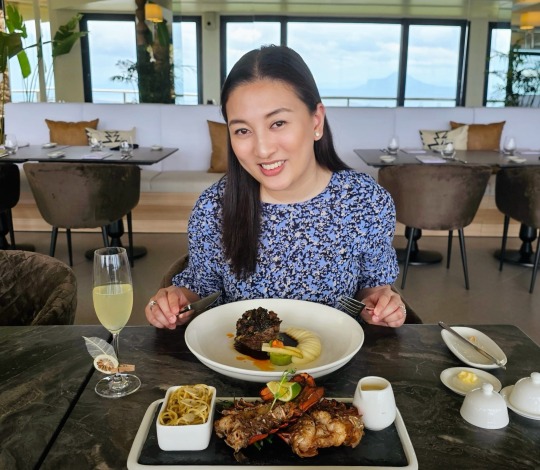
There's a new fine dining restaurant in Tagaytay that really impressed us and made our weekend trip all worth it. Sintâ is a charming and beautiful property where Culinary Artistry Meets Inspirational Views, especially with its fantastic landscape overlooking the majestic Taal Volcano and Taal Lake.
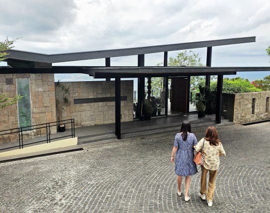
The Hungry Kat was invited to visit the new Sintâ restaurant a few weeks ago and it was a great way to escape the scorching summer heat in Manila. We had a leisurely Saturday drive all the way up to Sintâ which is located in a slightly different corner of Tagaytay. You can find it at 5385 Mystic Hills, Sitio Catmon, Ligaya Drive, which is closer to the Tagaytay Highlands and Picnic Grove side of the ridge. Just look for Sintâ Restaurant on Waze and you can't miss it. Traffic is actually much better here as most of the restaurants and tourists can be found on the other side near the Rotunda and Sky Ranch.

We were welcomed at the lobby reception by their very friendly and knowledgeable restaurant manager. Sintâ is an enchanting culinary haven helmed by the legendary Chef Ariel Manuel who is well known for his Lola Dad's and Bistro Manuel restaurants. Sinta emerges as a beacon of transformational dining, seamlessly blending sumptuous cuisine, captivating art, and awe-inspiring vistas to craft an unforgettable journey for the senses. Welcome drinks were served and we got to relax here a little bit after our long drive. Parking will not be a problem since they have a huge parking area right in front.

The place actually looks more like a hotel or resort lobby rather than just a restaurant. Formerly known as The Observatory Tagaytay, Sintâ brings exquisite art and food together to create a very lovely dining experience. It is open from Tuesday to Sundays from 10:00am to 3:00pm and from 4:00pm to 10:00pm.

What makes Sintâ different from the few other fine dining restaurants in Tagaytay is this majestic view. There are many casual dining restaurants in Tagaytay where you can just go and chill any time of the day. However, for those special occasions and important dates, the popular fine dining restaurants we know are mostly located outside the ridge and does not come with a backdrop like this.


One level down from the reception floor is Santâ's cocktail lounge. This al fresco dining area is a nice place to enjoy some drinks while waiting for your friends to arrive.

Sintâ's commitment to excellence extends beyond their culinary offerings to their meticulously curated beverage menu. They have a selection of handcrafted cocktails, premium wines, and artisanal spirits, thoughtfully chosen to complement their culinary creations and enhance your journey through flavors.

We ordered a few of their specialty beverages while enjoying the cool Tagaytay breeze. The Mt. Taal Sour (P320) is a signature cocktail that comes with a combination of whiskey, lemon super juice, tamarind, aquafaba, and aromatic bitters. It's a fruitier and more Asian version of the popular whiskey sour.


Another specialty beverage is the Nest and Mirror (P290). This one has rum-infused cinnamon white cacao, dry vermouth, sweet vermouth, orgeat, and cacao bitter. They also offer classic cocktails like the Aperol Spritz, Tequila Sunset, and Champagne cocktails. You can also have signature mocktails like the Pineapple Fresca, Cucumber Spritz, and more.
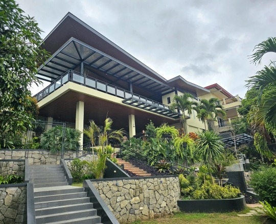
This structure is actually just the lobby and cocktail lounge. The main dining room is a bit further down the property. The garden can also be the venue for outdoor barbecues or other sunset events.
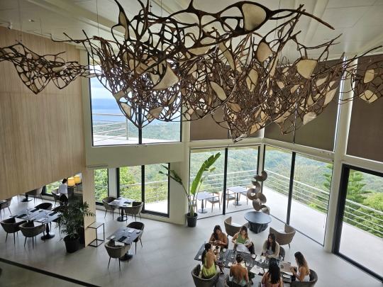
The main dining area is as gorgeous as you can get! But before we go down to the dining hall, let's explore some of the other areas around the restaurant.

What sets Sintâ apart is its commitment to curating a holistic dining experience, where every element - from the meticulously crafted dishes to the carefully selected art pieces adorning the walls - serves to stimulate the senses and nourish the soul. You can spot interesting art pieces all around the property and the restaurant manager will be more than happy to share the history and stories surrounding these.

Would you believe they even have hotel rooms right inside the property? For big events like weddings, Sintâ offers two big suites for the bride and groom plus their families. These come with panoramic views of Taal Volcano as well.
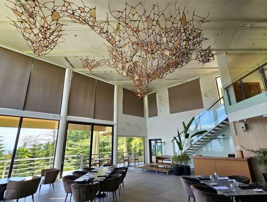
Now let's go down to the dining hall. You will immediately notice this elegant art structure at the top which is actually an inverted version of the lake and volcano. Isn't that cool?
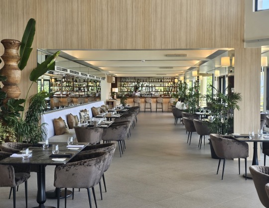
You can find tables for bigger groups as well as smaller tables for intimate dates and gatherings. This would probably look so romantic and colorful during sunset.

We were also welcomed by none other than Chef Ariel Manuel who runs the kitchen at Sintâ. The restaurant is owned by PYC Foods Corporation which also owns and operates One World Deli, Pardon My French, Tokugawa, and more. PYC is an importer and distributer of high-quality meats and seafood, so there will be no shortage of premium ingredients here at Tagaytay’s newest dining destination.
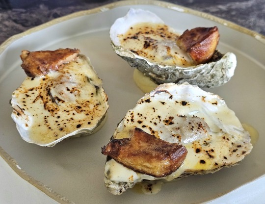
From farm-fresh ingredients to innovative culinary creations, every bite at Sinta is a testament to Chef Ariel's passion for excellence and dedication to his craft. The menu at Sintâ showcases some of the classic favorites found at Lolo Dads like the Baked Oyster with Foie Gras (P650/pc). This was our first time to try this popular starter and we were really amazed!

These are big Hokkaido oysters baked with chardonnay cream reduction served on its own shell and placed on top of soba noodles. To make it even more impressive, a piece of foie gras is added to complete the set. It's an astounding combination of flavors that really shows Chef Ariel's mastery of French and Asian cuisines.

The Tartare (P1,420) is also one of the specialties at Sintâ. This is prepared live tableside with its brunoise of Angus beef tenderloin plus condiments and apple kimchi.

It doesn't get fresher than this as the server will grind the raw beef and mix it together with the ingredients while you watch. Sintâ might be one of the very few restaurants in the country that does this tableside. We loved the tartare as it doesn't even taste like raw beef at all.

Another appetizer we enjoyed was this Fresh Ocean Trout Scallopine (P1,300) and ceviche with radish kraut, soy jelly, mustard leaf puree and salmon roe. Sintâ gets its fresh seafood from the best regions in the world so you know that the quality of their dishes are really excellent.

For our mains, my mother in law had the Chilean Seabass (P3,340) which is baked with a caramelized unagi crust, truffle, potato terrine, and doused with a ginger and green onion sauce. The combination of seabass and unagi is truly a match made in heaven.

We also ordered a few items From the Grill. You can't go wrong with the Maine Lobster (P4,280) which comes with two pieces of Maine lobster tail (450g) served with vegetables and sauces. The lobster was cooked perfectly and was absolutely one of the best I have ever had.

Another amazing main course you can order is the Eye of Rib Eye (P2,770). The juicy loin of rib eye steak is topped with its sisig-like rendered fat, making it doubly delicious. Onion jam and anticucho sauce are also added to complete this unique steak dish.

If you love eating lamb, then you must order their Rack of Lamb (P3,590). This comes with three huge pieces of Herb de Provence crusted rack of lamb with melted garlic and Roquefort cheese risotto. The lamb is very tender and doesn't taste gamey at all.

To end our fantastic lunch, Chef Ariel served his specialty French dessert, the Souffle (P550). Unlike other versions, this one is a coffee-flavored soufflé with baked cappuccino meringue and vanilla crème anglaise. Then there's also the Declension (P550), a declined mango and mascarpone cheese “mille feuille” with berry and balsamic ice cream.

Sintâ Restaurant by Chef Ariel Manuel is now our new favorite fine dining restaurant in Tagaytay. The elegant property fuses art and culinary creativity from the moment you step inside the lobby until the end of your gastronomic journey. Add the majestic view of Taal Lake to the story and you have the perfect dining destination that will make the drive to Tagaytay all worth it. Congratulations to PYC Foods Corporation for another great new venture.

Sintâ Restaurant
5385 Mystic Hills, Sitio Catmon, Ligaya Drive San Francisco, Tagaytay City
(0962) 453-3570
www.sintarestaurant.ph
www.facebook.com/sintarestaurantph
0 notes
Text
A Whole Lotta Shaking Going On: Earthquakes, Volcanic Eruptions and The Ring of Fire (Shared from Mu the Motherland) has been published on Elaine Webster - http://elainewebster.com/a-whole-lotta-shaking-going-on-earthquakes-volcanic-eruptions-and-the-ring-of-fire-shared-from-mu-the-motherland/
New Post has been published on http://elainewebster.com/a-whole-lotta-shaking-going-on-earthquakes-volcanic-eruptions-and-the-ring-of-fire-shared-from-mu-the-motherland/
A Whole Lotta Shaking Going On: Earthquakes, Volcanic Eruptions and The Ring of Fire (Shared from Mu the Motherland)
A Whole Lotta Shaking Going On: Earthquakes, Volcanic Eruptions and The Ring of Fire
The Taiwanese are no strangers to earthquakes and the latest on April 3rd 2024 was a doozy. Measuring 7.4 on the Richter scale, it took lives, leveled buildings, crumbled infrastructure, and wreaked havoc on the landscape. The area continues to rattle with sizeable quakes recorded in Japan and the Northern Mariana Islands—all of which sit on the infamous Pacific Ring of Fire where 90 percent of the world’s earthquakes occur. In addition, the Ring of Fire houses about 75 percent of the world’s active and dormant volcanoes.
The Ring of Fire encircles the Pacific Ocean, and is the result of tectonic plate movements. It covers an area from New Zealand, along the eastern edge of Asia, across the Aleutians, and down the coasts of North and South America.
The mythical continent of Mu, purportedly located in the Pacific Ocean, is said to have sunk into the sea thousands of years ago. Theories of Mu’s existence often draw parallels with Atlantis, suggesting it was a highly advanced civilization that met its demise through cataclysmic geological events. Some proponents of this theory have speculated that the Ring of Fire’s intense seismic and volcanic activity could be linked to the sinking of Mu, proposing that the continent’s disappearance was the result of massive earthquakes or volcanic eruptions.
For those of us that believe that Mu and Atlantis existed, the recent seismic events, seem like a repeat performance of pre-history. As mentioned in prior posts, we wonder if humanity is once again at the same crossroads as it was during the last destruction. Does the earth literally react to our energies? Are seismic and volcanic activities simply a matter of cause and effect? —mankind’s karma? Will wars, greed, misused technology, pollution, and the blatant disrespect of Mother Earth once again throw the world and its inhabitants into chaos and destruction? Unless we change our ways, we think so.
The Edgar Cayce readings contain evidence that we reincarnate through many lifetimes, and often with the same souls with which we have karmic ties. On the same note, many religions teach that there are cycles that repeat, and that God (or however you perceive the divine) because of love, continues to offer us the opportunity to spiritually progress. We appear to be at the same crucial point in our development as when the last cataclysmic events occurred. Just saying.
So, let’s get back to science. The study of the Ring of Fire involves a multidisciplinary approach, leveraging a variety of scientific methods and technologies to understand the complex geological processes at play. Key among these methods are:
Seismology: Seismologists use seismographs to detect and measure the vibrations caused by earthquakes, which are prevalent in the Ring of Fire. This data helps in mapping fault lines and understanding the dynamics of tectonic plate movements. Analysis of seismic waves allows scientists to pinpoint the location, depth, and magnitude of earthquakes, offering insights into the stress points within the Earth’s crust.
Volcanology: Researchers study volcanoes through direct observation, satellite imagery, and by monitoring volcanic gases, ash emissions, and lava flows. Instruments like spectrometers can measure gas compositions, while thermal cameras and satellites monitor heat signatures from erupting or potentially active volcanoes. This information is crucial for predicting volcanic eruptions and assessing their potential impact.
Plate Tectonics Study: Scientists use GPS and satellite data to track the movement of tectonic plates. This geodetic monitoring provides precise measurements of how plates are shifting, which is vital for understanding the mechanics of subduction zones and the forces driving volcanic and seismic activity.
Oceanography: In the marine regions of the Ring of Fire, oceanographers study the seafloor through sonar mapping and submersible vehicles. This exploration reveals underwater volcanoes, hydrothermal vents, and deep-sea trenches, contributing to our knowledge of the oceanic components of tectonic plate boundaries.
Geochronology and Paleomagnetism: To understand the historical activity within the Ring of Fire, scientists date rocks through radiometric dating techniques. Additionally, the study of paleomagnetism, which examines the magnetic properties of rocks, provides clues about past movements of the Earth’s plates and the history of magnetic field reversals.
Remote Sensing and Satellite Imagery: Satellites equipped with various sensors provide a broad, comprehensive view of the Earth’s surface, including the Ring of Fire. They offer valuable data on topography, thermal anomalies indicating volcanic activity, and changes in the landscape due to seismic events.
Computational Modeling: With the data gathered from these various methods, scientists use computer models to simulate geological processes, such as magma movement beneath volcanoes or the stress accumulation along fault lines. These models are invaluable for predicting future activity and for testing hypotheses about the underlying causes of volcanic and seismic events.
By integrating these diverse scientific methods, researchers can piece together a comprehensive picture of the Ring of Fire’s dynamic geological processes. This interdisciplinary approach is essential for advancing our understanding of Earth’s geology and for mitigating the risks associated with living in one of the planet’s most active geological zones.
The bottom line is that remnants of the lost continent of Mu can be found in the form of microcontinents or submerged landmasses in the Pacific. This leads to an intriguing question: Could the geological tumult within the Ring of Fire be responsible for the disappearance of Mu? This hypothesis hinges on the idea that the intense seismic and volcanic activity characteristic of the Ring of Fire could have led to the submergence or fragmentation of this ancient landmass. To explore this connection, we must evaluate geological evidence, such as the distribution of seismic activity, the history of volcanic eruptions, and the patterns of tectonic plate movement in the Pacific region. If Mu existed, its fate might have been sealed by the same dynamic earth processes that continue to shape the Ring of Fire today.
In conclusion, while the connection between the Ring of Fire and the lost continent of Mu remains speculative, it opens up fascinating avenues for exploration in both geology and mythology. The Ring of Fire’s role in our planet’s seismic activity is undeniable, and its study provides crucial insights into Earth’s geological processes. The legend of Mu, whether real or mythical, challenges us to consider the possibilities of Earth’s ancient past and the mysteries that may still lie beneath the ocean’s depths.
Further research and exploration are essential in unraveling these mysteries and understanding the complex dynamics of our planet. As we continue to study the Ring of Fire, we may find more clues about the lost continent of Mu, bridging the gap between science and myth, and expanding our knowledge of Earth’s fascinating history.
Meanwhile, let your love shine.
youtube
0 notes
Photo
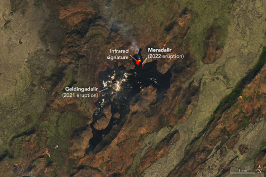
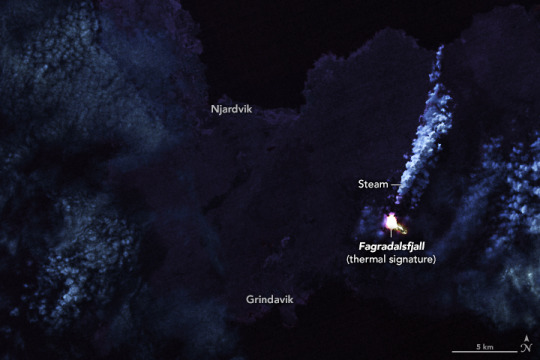
Eruption in Fagradalsfjall, Iceland On August 3, 2022, a new volcanic eruption began in the Fagradalsfjall fissure zone on Iceland’s Reykjanes Peninsula. The eruption site in the Meradalir Valley is about a kilometer northeast of last year’s eruption in the Geldingadalir Valley. The recent eruptions come after an 870-year quiet period in the Krýsuvík–Trölladyngja volcanic system. This volcanic system is composed of two groups of fissures or swarms, named Fagradalsfjall and Krýsuvík. The fissures trend northeast-southwest for 50 kilometers (30 miles) in the rift zone where the North American and Eurasian plates are moving apart, according to the Catalogue of Icelandic Volcanoes. Starting in early August, the Meradalir eruption emitted fountains of lava along a 300-meter (1,000-foot) segment of the fissure. The brilliant display attracted crowds of tourists, who hiked in to see the slow-moving basaltic lava as it flowed east-northeast. Iceland’s Institute of Earth Science estimated that 10.6 million cubic meters (14 million cubic yards) of lava had covered an area of 1.25 square kilometers (0.5 square miles) by August 15. Early on, the eruption was emitting lava at a rate of up to 32 cubic meters (40 cubic yards) per second, which soon slowed to an average of 11 cubic meters per second. By August 16, the Meradalir eruption rate had dropped to 2 cubic meters (3 cubic yards) per second. The March 2021 eruption in Geldingadalir issued 5 to 10 cubic meters (7 to 13 cubic yards) of lava per second during its first three weeks. The first image above was acquired on August 16, 2022, by the Operational Land Imager (OLI) on Landsat 8. The image includes a combination of visible and infrared light (bands 6-5-3), which helps distinguish the heat signature of the lava. The second image also shows the thermal infrared signature of the lava flow, but at night. It was acquired on August 7, 2022, with OLI and the Thermal Infrared Sensor (TIRS) on Landsat 8. According to the Catalogue of Icelandic Volcanoes, the Krýsuvík-Trölladyngja system has been "moderately active" during the Holocene Epoch. At least 10 eruptions, lasting from a few years to decades, have occurred over the past 8,000 years. This suggests an eruption interval of 400 to 1,000 years, with an average of more than 750 years. The last significant eruption before 2021 happened in the 12th century, when four lava flows ejected 220 million cubic meters (287 million cubic yards) of lava. The molten rock covered more than 36 square kilometers (14 square miles) and reached the north and south coasts of the Reykjanes Peninsula. NASA Earth Observatory images by Lauren Dauphin and Joshua Stevens, using Landsat data from the U.S. Geological Survey. Story by Sara E. Pratt.
7 notes
·
View notes
Text
Templates of Light
Each crystal has its own unique energy signature. These “templates of light” are encoded with all you need to activate your own power. The key is to find a crystal that is attuned to your own personal energy or that raises your energetic resonance to ensure well-being and expand your consciousness.
THE POWER OF GEMS
Not only flashy gemstones hold power. Since antiquity, crystals of all sorts have served as protective amulets. Humble stones such as Flint were magical carriers for the soul, for metaphysical workings, or for shamans to use on their otherworldly journeys. Many stones produced incandescent sparks or could be super-heated into streams of gold, silver, copper, and other precious metals. Just
as magical were the sky rocks that fell to Earth, bringing with them iron to forge tools and create weapons.
Egyptologist Wallis Budge explained, “Each stone possessed a sort of living personality, which could experience sickness and disease, and could become old and powerless and even die.” However,
in Egyptian medicine, stones could also heal. The Greek philosopher Plato believed stones were living beings, produced by a fermentation process induced by “a life-giving intelligence descending
from the stars.” According to many myths, crystals solidified from ice, a view reinforced by the bubbles of water sometimes found within a crystal.
HOW CRYSTALS FORM
Crystals are, for the most part, created by the Earth’s awesome power. Boiled, compressed, and excoriated, some were born of volcanoes, glaciers, earthquakes, and immense pressure; others
dripped into being through osmosis, gas bubbles, and nature’s gentler forces.
Some so-called crystals don’t actually have a crystalline structure. Amber, for instance, is fossilized tree resin, and volcanic Obsidian formed so fast it didn’t have time to crystallize. How a crystal forms affects how its power works. Those that grew slowly tend to emit their power gently; those that were on an accelerated path of growth blast their power out to the world. Paradoxically, some of the youngest geological stones have the highest vibrations and the greatest power to transform our world.
0 notes
Text
Researchers probe molten rock to crack Earth’s deepest secrets - Technology Org
New Post has been published on https://thedigitalinsider.com/researchers-probe-molten-rock-to-crack-earths-deepest-secrets-technology-org/
Researchers probe molten rock to crack Earth’s deepest secrets - Technology Org
New research focused on the quantum structure of elements under extreme conditions has implications for understanding Earth’s evolution, interpreting unusual seismic signals, and even studying exoplanets for insights into habitability.
Deep inside rocky planets like Earth, the behavior of iron can greatly affect the properties of molten rock materials: properties that influenced how Earth formed and evolved. Scientists used powerful lasers and ultrafast X-rays to recreate the extreme conditions in these molten rock materials, called silicate melts, and measure the properties of iron. Image credit: Greg Stewart/SLAC National Accelerator Laboratory
Deep inside rocky planets like Earth, the behavior of iron can greatly affect the properties of molten rock materials: properties that influenced how Earth formed and evolved.
In fact, the evolution of our entire planet may be driven by the microscopic quantum state of these iron atoms. One special feature of iron is its “spin state,” which is a quantum property of the electrons in each iron atom that drives their magnetic behavior and reactivity in chemical reactions. Changes in the spin state can influence whether iron prefers to be in the molten rock or in solid form and how well the molten rock conducts electricity.
Until now, it’s been challenging to recreate the extreme conditions in these molten rock materials, called silicate melts, to measure the spin state of iron. Using powerful lasers and ultrafast X-rays, an international team of researchers at the Department of Energy’s SLAC National Accelerator Laboratory, Stanford University, Universite ́ Grenoble Alpes, Laboratoire pour l’Utilisation des Lasers Intenses (LULI), and Arizona State University overcame this challenge. They showed that at extremely high pressures and temperatures, the iron in silicate melts mostly has a low-spin state, meaning its electrons stay closer to the center and pair up in their energy levels, making the iron less magnetic and more stable.
The results, published Friday in Science Advances, support the idea that certain types of molten rock might be stable deep inside Earth and other rocky planets, potentially lending a hand in the creation of magnetic fields. The research has potential implications for understanding Earth’s evolution, interpreting seismic signals, and even the study of exoplanets.
“In terms of exploring Earth’s history, we’re investigating processes that took place over 4 billion years ago,” said collaborator Dan Shim, a researcher at Arizona State. “The only way to study this is by using modern technology that operates in femtoseconds. The contrast between these immense time scales is both eloquent and startling: it’s akin to the idea of a time machine.”
Asteroid bombardment and magmatic oceans
About 4.3 to 4.5 billion years ago, early Earth underwent intense impacts, getting pummeled by asteroids as large as cities. These impacts produced so much heat that they could have completely melted the outer layers of the planet, creating a deep ocean of molten rock.
“It’s been theorized that under the immense pressure of these impacts, the molten rock may have became denser than the solid rock,” said collaborator and SLAC scientist Arianna Gleason. “This denser magma would have sunk towards the core, capturing the chemical signatures of that era. Some believe remnants of this magma layer may still exist today, holding clues from 4.5 billion years ago. Volcanoes like those in Hawaii could be releasing these ancient chemical signatures, providing us a glimpse into Earth’s distant past.”
At shallow depths, molten rock takes up more space than the same material when it’s solid. But as you go deeper and the pressure increases, this difference decreases. The inclusion of iron, especially its spin state, plays a big role in determining these properties. Prior research has shown mixed results about the spin state of iron in similar conditions: some studies found a rapid change in iron’s spin state under high pressures, while others saw a slower, more gradual change.
This new study provides the first direct look at iron’s behavior in real molten rock under extreme conditions.
“While we can glean a lot from studying rocks and fossils, some aspects of Earth’s early history are lost because few records from that time exist,” Shim said. “That’s what makes this study unique. Earth’s formation was a tumultuous process, involving intense impacts and resulting in a globally molten rock layer. The pressure in this layer was immense. We study this by simulating the conditions through laboratory experiments.”
At the Matter in Extreme Conditions (MEC) experimental hutch at SLAC’s Linac Coherent Light Source (LCLS), the team was able to recreate the extreme pressures that would have been found in early Earth’s magmatic ocean by blasting samples with powerful lasers that transform the solid material into a silicate melt in a matter of nanoseconds. Then, the scientists used femtosecond X-ray pulses from LCLS to study the electronic structure of elements like iron under these extreme conditions, providing insights into how electronic configurations change under different conditions and revealing that the molten magma did indeed become denser than a solid under specific conditions.
“By understanding Earth’s internal dynamics, we can refine models of tectonic movement and other geological phenomena,” Gleason said. “Moreover, as Earth’s layers are interconnected, these findings have implications for climate science.”
Understanding our planet
In this research the team concentrated on low iron content melts. But as material rains down towards the Earth’s center, it’s theorized to absorb more iron, making it denser. To follow up, the team plans to study melts with higher iron content. They also hope to experiment with melts containing some water, furthering our understanding of Earth’s water cycle and climate.
The research could also shed light on peculiar seismic velocities deep within Earth’s mantle. These anomalies have puzzled scientists for decades. Some theories suggest these zones could be remnants of magma from 4.5 billion years ago, while others believe they result from tectonic plates that have sunk into the Earth’s interior, spreading low melting point material. By comparing different hypotheses using seismic imaging, the team aims to determine the origins of these zones and distinguish between ancient and more recent materials.
“As technology advances, we’re at the forefront of addressing grand challenges that range from mineralogy to climate science, connecting various research areas,” said SLAC scientist and collaborator Roberto Alonso-Mori. “The sheer volume of information we can gather has transformed our capabilities. It’s a game-changer. It’s exhilarating to develop novel techniques and apply them to pressing questions with such a diverse team.”
Source: Stanford University
You can offer your link to a page which is relevant to the topic of this post.
#Asteroid#asteroids#Astronomy news#atom#atoms#Behavior#billion#challenge#chemical#chemical reactions#Chemistry & materials science news#cities#climate#climate science#deep ocean#dynamics#earth#electricity#electronic#electrons#energy#Evolution#Exoplanets#experimental#form#Fossils#Fundamental physics news#game#hand#History
0 notes
Text

Have you ever wondered about the source of gemstone colors and names? We all have a favorite gemstone based on a color that means something special to us (possibly a birthstone), but most of us never consider how that color came to be in the stones we love.
It turns out that nature is an amazing chemist, mixing minerals, and using temperature and light to produce unbelievable beauty in stones all around the world. And many you’ll learn about right here.Unlocking the Secrets Of Beautiful Gemstone Colors And Names
Obtained via methods such as open-pit, underground, and alluvial mining, gemstones come to us from all around the world, from volcanos, mines, and even rivers and streams, but the source of their captivating beauty goes much deeper.
And although the most sought-after gemstones usually possess a good degree of transparency and vibrancy, rarity, and a well-proportioned cut, the quality of each gemstone is based on more than meets the eye.
Dive deep into the complex world of mineralogy and crystallography to discover their splendid secrets.
Gemstone Colors And Names: Transition Metals
The color of several gemstones originates from the inclusion of what’s known as a transition metal in the stone’s structure. This takes the form of an impurity in what we would see as an otherwise colorless crystal lattice. These inclusions play a significant role in the stone’s makeup, including both its hardness and its color.
Various transition metals react differently in visible light. The result is the stone’s distinctive color. The responsible transition metal within the gemstone is often a chemical portion containing mineral impurities.
A tiny amount of the transition metal is all that’s needed to influence the necessary change in the stone’s complex internal anatomy to result in what we perceive as the beautiful intensity of their different colors.
What Makes Gemstones Original Colors?
Mesmerizing natural stones with their spectacular variation in color and transparency becomes less of a mystery when as we explore the basics of their mineral formation and what makes each gemstone’s distinctive characteristic.
Minerals, substances with a certain chemical structure, originate through processes like crystallization from molten magma, precipitation (rain from liquid), or the replacement of their original composition from other minerals or rocks.
In turn, the minerals’ physical and chemical composition, including color and transparency, are determined by their specific chemical arrangement.
Different minerals absorb different wavelengths of visible light, which possess different ranges of colors — isn’t that fascinating? This leads to the mineral’s unique color we all find so attractive.
Gemstones often demonstrate color variation because of impurities or trace elements within their crystalline structure. This is known as the allochromatic quality of the stone, which describes the influence of impurities within the stone.
Gemstones And Their Colors: The Value Of Impurities
Chromium and vanadium give emeralds their signature green hue; titanium and iron are responsible for the beautiful blue of the sapphire; chromium produces the compelling red flame in the ruby; and one of the most favorite gemstones, the coveted (and famously resilient) diamond, which can range from colorless to tinted, comes from impurities in its carbon framework.
The transparency of the crystal is determined by the crystal lattice arrangement of the tiny components seen with the help of science and powerful lenses. These, too, are the product of impurities in the crystal’s composition.
If the atoms are arranged in a regular pattern, it is likely to be transparent. Alternatively, an irregular arrangement or impurities and defects may lead to opaqueness, or cloudiness, in the crystal’s structure.

Heat can also play a role in a gemstone’s color. Heat-treated minerals can shift a stone’s color from one state to another. This is the case of a gemstone like zoisite, named tanzanite by Tiffany & Company.
Once heated, the mostly brownish stone undergoes a remarkably beautiful color change. Natural (metamorphic) heating, as well as deliberate, intentional heat treatment, can shift the stone’s color to the more alluring blue the stone is famous for.
Another means to revise a gemstone’s color involves manipulating the way the stone reflects light. Adding a mineral like iron can change the color by influencing the way the gemstone refracts the natural light we see reflected from the stone itself.Tanzanite Stone
Our beloved diamonds are the clearest of all and rank among the most popular gemstones. They consist only of pure carbon atoms that are aligned in a specific geometric pattern which allows visible white light to travel through without scattering or being blocked.
This is the amazing nature of the diamond’s unique sparkle. Likewise, quartz, beryl, and topaz get their translucent beauty from the same phenomenon, although the density of the diamond reflects it in a league all of its own.Diamond Crystal Gemstone
Opaque, or semi-transparent gemstones, get their coloration from the absorption of certain wavelengths of light or by having impurities or structural defects in their crystal lattice. These gems can have solid-colored appearances, patterns, or markings on the surface. Examples of opaque gemstones are turquoise, jasper, and agate.
Corundum, an aluminum oxide-based mineral, can display a range of hues and transparencies. These depend on the composition of the gemstone and any impurities it contains.
For example, corundum may be red (ruby), blue (sapphire), yellow, pink, or purple; this is due to the presence of chromium (red), titanium, and iron (blue) or iron (yellow) in its complex crystal structure.
Gemstones also exhibit a wonderful range of clarities, from transparent to translucent to opaque.
Translucent gemstones allow some visible light to pass through them but are not completely transparent.
These gemstones include the precious opal and moonstone, both of which exhibit what is often called a play of color in gemstones, during which white light waves travel through the stone’s interior which acts like a prism, producing a wonderful kaleidoscope of colors.
A Play Of Color
The otherwise pure light we see with our naked eyes is actually a series of waves or wavelengths.
In fact, the colors we see in light have their own distinct wavelengths that react differently depending on the nature of the object that the light interacts with (or that is absorbing visible light). As a result, the color of a gemstone is determined by the way light interacts with it.
This is why a red gemstone that absorbs the entire spectrum of visible light except red will appear red itself. By the same phenomenon, gemstones absorbing all visible light will appear black — another fascinating fact.
1 note
·
View note
Text
Introduction
An extensive proteomic analysis was performed on a set of 12 bones of human victims of the eruption that in AD 79 rapidly buried Pompeii and Herculaneum, allowing the detection of molecular signatures imprinted in the surviving protein components. Bone collagen survived the heat of the eruption, bearing a piece of individual biological history encoded in chemical modifications. Here we show that the human bone proteomes from Pompeii are more degraded than those from the inhabitants of Herculaneum, despite the latter were exposed to temperatures much higher than those experienced in Pompeii. The analysis of the specimens from Pompeii shows lower content of non-collagenous proteins, higher deamidation level and higher extent of collagen modification. In Pompeii, the slow decomposition of victims' soft tissues in the natural dry–wet hydrogeological soil cycles damaged their bone proteome more than what was experienced at Herculaneum by the rapid vanishing of body tissues from intense heat, under the environmental condition of a permanent waterlogged burial context. Results herein presented are the first proteomic analyses of bones exposed to eruptive conditions, but also delivered encouraging results for potential biomarkers that might also impact future development of forensic bone proteomics.
Ancient protein analysis provides clues about human and animal life and diseases from the past[ 1]. Bone, a mineralised extracellular matrix rich in connective tissue components, is one of the most abundant sources of ancient proteins and one of the most abundant biomaterials in the archaeological record. The diagenesis, in particular when heat-induced, of organic matter in archaeological bone, initially represented mostly by type I collagen (90%), lipoproteins and mucopolysaccharides[ 2], is a complex phenomenon[ 3],[ 4], linked to several factors in the depositional environment[ 2],[ 5],[ 6]. The analysis of how diagenesis acted to produce or prevent specific modifications led to coin the term "diagenetiforms" to describe the variety of distinct molecular species arising from chemical modifications by environmental conditions[ 7].
Mass spectrometry-based techniques have been widely applied to characterize collagen in ancient proteomes and its chemical modifications occurred pre- and post-mortem[ 8]. Apart from the extensive observation of deamidation of asparagines and glutamines Gln[ 9]–[12] and methionine oxidation[13], a few other diagenetically induced modifications were also detected, such as: (i) aminoadipic acid formation from lysine, (ii) tryptophan oxidation products[13], (iii) advanced glycation end-product (AGEs)[13]–[15], (vi) backbone cleavage[14],[15], while complex oxidative reactions occurring on prolines have been hypothesised but not characterised in details[13],[14]. So far though, no study characterized the modifications occurred post-mortem in the proteome of archaeological bones from individuals who died due to the nearly instantaneous exposure to extreme temperature.
Here we applied a bottom-up proteomic approach to investigate the proteome and the chemical modifications present in the bone proteins of humans from Herculaneum[16] and Pompeii (Italy)[17], who died during the eruption of the Vesuvius in 79 AD. The accurate description of the catastrophic event affecting Herculaneum and Pompeii provided by ancient sources, and the peculiar burial conditions[18]–[20], offer a unique opportunity to analyse lifeways in a relatively large cohort of perfectly coeval individuals who lived and died together[21], whose bone proteome experienced such extreme conditions.
Herculaneum, Pompeii and other Roman settlements up to 20 kms away from the volcano were suddenly hit by successive hot pyroclastic currents and buried by up to tens of meters thick volcanic ash deposits produced by the 79 AD eruption, that killed everyone who had not been evacuated or managed to flee[16]. The skeletal remains were in an excellent state of preservation as a result of the unusual death and burial conditions: instant death caused by hot pyroclastic surges at temperatures between approx. 300 (Pompeii) and 500 °C (Herculaneum)[16],[17],[22].
In Herculaneum, death was followed by vanishing of soft tissues and rapid replacement by volcanic ash[19]. Here, some evidence suggests that the volcanic environment was characterised by a drop in temperature of the first pyroclastic surge during its emplacement. Evidence of rapid cooling of the volcanic ash cloud may account for the preservation of organic tissue residues[22],[23] and organic compounds[24]. At Herculaneum the ash-bed deposit was permanently waterlogged by groundwater[25],[26], as revealed by the early 1980s archaeological investigations of the victims' skeletons on the ancient beach, thus a permanent system of hydraulic pumps was activated[20]. The burial environment of the victims was most likely able to inhibit microbial attack to bone and related diagenetic processes[27].
On the other hand, the temperature of approx. 300 °C experienced by the victims in Pompeii was sufficient as well to instantly kill people, but it was not hot enough to cause rapid soft tissue disappearing as in the case of Herculaneum. Therefore, in Pompeii the victims' corpses were preserved intact inside the ash deposit after its rapid cooling and hardening around them[16]. The cavity formed around the victim's body after the slow disappearance of the flesh would then be filled with plaster of Paris in order to obtain plaster casts, technique adopted for the first time on human victims in 1863 by Giuseppe Fiorelli[28]. Over the last century and up to the present, in the case of bodies of victims found in the ash surge deposit, this technique has been used to replicate the features of the body.
In this paper, our objective was a proteomic profiling of the bones of the eruption victims, using a bottom-up proteomic approach and an unbiased discovery of chemically modified peptides[29], in search for signatures of the high temperatures and environmental conditions the bodies were exposed to. Bone collagen survived even the harshest conditions of temperature imposed by the volcano eruption, bearing encoded in the chemical modifications a piece of individual biological history. For comparison, the bones from a coeval skeletal population from the Campanian region were considered (Baia, Scalandrone locality, II sec. AD, Roman Imperial Age, Puteoli, Naples, Italy). The necropolis site of Baia Scalandrone was chosen as a control because it is coeval with the sites of Herculaneum and Pompeii and, as with these, the burial ground is of volcanic origin. Furthermore, unlike the two Vesuvian sites, the bodies of these individuals were not exposed to heat.
Most interestingly, bones are frequently found in archaeological and forensic contexts, and their characterization for the study of past populations (e.g., age at death or details of funerary practices), or for victim characterization in forensic investigations is of unquestionable relevance[30]–[33]. Actually, burned bones might be the only remains found in forensic scenarios (e.g. from terrorist attacks, explosions or fires) from which identify victims or obtain information, and the present study is expected to contribute to a full molecular characterisation of bones that have been exposed to heat.
Results
Proteome of human bones
A shotgun proteomics approach by LC–MS/MS (Figure S2) was applied to skeletal samples excavated from the archaeological sites of Pompeii (7 samples) and Herculaneum (5 samples)[16],[19]. Samples collected from three individuals from Roman Imperial Age (II sec. AD) cemetery in Baia Scalandrone were also analysed to compare results from the AD 79 eruption victims with this coeval skeletal population. All specimens are illustrated in the Table S1 and Figure S1. Moreover, raw data from proteomic analyses of archaeological bones from a completely independent excavation site, from the Hitotsubashi site (AD 1657–1683) in Tokyo, Japan, were used for comparative evaluation of the results. Samples H-162, H-142 from[34] were selected because proteins have been extracted with almost the same protocol and analysed on the same instrument as herein samples.
Very stringent criteria for protein identification were used: only peptides with score higher than 70 were considered and proteins were considered as identified only when 2 or more peptides have been detected (Table S2).
The number of identified proteins in the samples from Herculaneum, Pompeii and Baia Scalandrone varied among individuals from 2 to 27 and includes collagenous and non-collagenous proteins (NCPs) (Table S3). The expected dominance of collagen in bone tissue is reflected by the result that the two chains of type 1 collagen, namely collagen alpha-1 (I) and collagen alpha-2 (I), were confidently identified in all the samples. Of the 15 samples, 5 contained only type 1 collagen chains (four samples out of seven from Pompeii, 2.8 proteins on average, ± 1.12, and one sample from Baia Scalandrone) while samples from Herculaneum exhibited a higher protein content (13 proteins on average, ± 7). Bone proteome complexity is affected by several factors, including burial age[35]. In this case, since bones from Pompeii and Herculaneum are exactly coeval, the volcanic environmental conditions during death and burial appear to have played a significant role in protein survival in the two different sets of bone samples[36]. Raw data of Control samples were processed with the same constrains as the samples herein analysed, and a large number of proteins, (38 ± 0) were identified, as already reported[34].
Despite a general large variability among the individuals within the different groups (Figure S3), samples can be grouped depending on the site of origin in respect to NCP content. We definitively observed that bone samples from Herculaneum exhibit more NCPs than those from Pompeii. Venn diagram using the ensembles of the proteins identified in each sample group shows that the proteins identified in Pompeii bones are common to all three groups, while several other proteins are shared exclusively by Herculaneum and Baia Scalandrone bones (Figure S4, Table S4).
The non-collagenous proteins identified in this study agree with those expected for archaeological bones[37],[38]. Most of the identified NCP proteins are small leucine-rich proteoglycans (SLRPs) from the extracellular matrix (namely chondroadherin, biglycan, decorin, lumican and osteomodulin), all involved in biomineralisation or interacting with fibrillar collagen, (such as vitronectin and pigment epithelium-derived factor). Moreover, alpha-2-HS-glycoprotein, also known as fetuin-A is a bone matrix protein, known to have a high affinity for apatite. It is worth mentioning that several of the NCPs are related to the coagulation pathway (namely, prothrombin and antithrombin III, and Protein Z-dependent protease inhibitor), and can be functionally connected (Figure S5).
Diagenetically induced modifications in bone proteins
Modifications of amino acids, such as oxidation of methionine, deamidation of asparagine and glutamine, as well as the backbone cleavage, are all degradation phenomena commonly observed and routinely searched for in ancient/aged proteins[13],[39].
Deamidation
To begin with, deamidation of asparagine (N) and glutamine (Q) residues, among the most common and most informative diagenetically derived modifications in proteins[11],[12],[40], was examined. While extensive deamidation increases heterogeneity of the samples, it is a general and relevant glance on the myriad changes that archaeological bone proteins undergo and is influenced by the age and more generally by the preservation state of the bone sample under consideration[11],[40]–[42]. Extensive protein deamidation (N,Q) has been consistently observed in ancient samples, and it has been routinely measured as part of the palaeoproteomics analysis of archaeological and paleontological specimens as a global indicator of sample preservation quality, since rates and levels of deamidation are affected by several chemical and environmental factors[40]. As expected, proteins in our samples are extensively deamidated and asparagine sites are much more deamidated than glutamines[40],[43], (Fig. 1). Moreover, we split the evaluation of the deamidation levels for collagenous (Fig. 1) and non-collagenous proteins (Figure S6) and, in agreement to what already reported by[15], peptides from non-collagenous proteins showed very high to complete deamidation in comparison to peptides derived from collagenous proteins. On average, peptides from Pompeii samples are the most deamidated (see bulk deamidation per archaeological site, Fig. 1).
Graph: Figure 1 Overall percentage of deamidation for asparagine and glutamine residues of collagenous bone proteins from Pompeii, Herculaneum, Baia Scalandrone and control (H-162, H-142 from[34]). Error bars represent standard deviation and numbers above each bar represent the number of deamidation sites the data is based on.
It is worth mentioning that Control samples are significantly more recent (1657–1683 AD)[34] than samples from the Vesuvius area, and they are definitively less deamidated than Pompeii and Herculaneum bone samples. Variations were observed from individual to individual in the three groups from the Vesuvius area (Figure S7–S8). As a general trend, we can confidently assert that the lower the number of surviving proteins the higher the deamidation level, and it is worth observing that the few NCPs identified in samples from Pompeii are almost completely deamidated. Apparently, other factors rather than temperature might have played the biggest role in deamidation. In fact, the skeletal remains from Baia Scalandrone, which were not exposed to hot pyroclastic flows, but had been buried in the volcanic soil of the Campi Flegrei area (Bay of Puteoli, Gulf of Naples, Italy), exhibited a level of deamidation only slightly lower than that measured for bones from Herculaneum and Pompeii.
We analysed the distribution of deamidation level along the sequence of collagen type I chains, to explore the possibility of hot spots for deamidation rather than an average distribution. Figure S9 illustrates the deamidation values at single deamidation sites along collagen alpha-1 (I) and alpha-2 (I) chains. The label size indicates the relative intensity of each position in each sample. The values for Control are always well below those calculated for samples from the volcanic areas, in agreement with the global deamidation level calculated in Fig. 1. This difference is even more evident in glutamines, conceivably because glutamine deamidates more slowly. There is a trend in the deamidation; there are some zones where deamidation is more pronounced than others. This trend is almost reproducible in the samples of Pompeii, Herculaneum and Baia Scalandrone suggesting that the deamidation profile is quite robust for samples similar as concerns age and burial soil and also that three-dimensional arrangement might affect the local deamidation level.
Oxidation of methionines
With the same approach the oxidation of methionines (M) was evaluated. Figures S10–S11 illustrate the global oxidation levels of collagenous and non-collagenous proteins in all the samples. Apart from the zero values of control samples, all the proteins in all groups are almost totally oxidized (100%), demonstrating that methionine oxidation follows another pattern than deamidation. Furthermore, we investigate the oxidation values at single oxidation sites along collagen alpha-1 (I) and alpha-2 (I) chains. Almost all oxidation values are either 1 or 0, meaning that methionines are either fully oxidized or not oxidized at all (Figure S12). However, it is worth saying that several methionines were not detected, despite the generally good protein sequence coverage.
Backbone cleavage
Backbone cleavage of the polypeptide chain is also expected as a degradation feature in ancient proteins[15],[44],[45], and can be evaluated since, upon trypsin hydrolysis, semi-tryptic peptides will be generated. Search for semi-tryptic peptides was carried out only on collagen type I chains for comparative purposes, since they are the only polypeptide chains shared among all the samples. The frequency of semitryptic peptides was evaluated as percentage of semitryptic peptides over the total number of identified peptides for each chain, on the basis of spectrum matches (PSMs).
Figure 2 shows the relative abundance of peptide-spectrum matches (PSMs) of semitryptic peptides over the total number of peptides of collagen alpha-1 (I) and alpha-2 (I) chains as a bulk per archaeological site (Fig. 2A) and in the single samples (Fig. 2B). The frequency of backbone cleavages is generally high. However, no clear-cut difference was observed among the samples from the volcanic areas or with the control sample.
Graph: Figure 2 Backbone cleavages in collagen alpha 1(I) and collagen alpha 2 (I) in bone samples from Pompeii, Herculaneum, Baia Scalandrone and control (H-162, H-142[34]). Overall occurrence per samples groups (A) and in the single samples (B), evaluated as percentage of peptide-spectrum matches (PSMs) of semitryptic peptides over the total number of peptide-spectrum matches (tryptic plus semitryptic peptides).
The peptides showed a clear pattern derived from extended terminal hydrolysis occurring in regions of the collagen chains rather than in specific peptide bonds (Figure S13). A manual alignment of all the semitryptic peptides in the four different groups (Pompeii, Herculaneum, Baia Scalandrone, Control) to COL1A1 and COL1A2 sequences, however, reveals that while in case of controls the number cleavages are spread along the sequences, in the samples of Pompeii, Herculaneum and Baia Scalandrone they are localized in some regions of the protein sequences. These hot spots are between 266–286, 481–511, 772–796 and 1051–1118 sites of COL1A1 with a window of ± 2 amino acids, and in COL1A2 between 154–167, 232–250, 320–340, 425–438, 499–517, 681–708, 965–1006 and 1042–1066 with a window of ± 2 amino acids (Figure S14).
The cleavage frequency was then re-evaluated considering the regions rather than the single peptide bonds, by calculating the number of PSMs with semitryptic cleavages identified in a region divided by the total PSMs in the same region, including both tryptic and semitryptic matches. As shown in figure S14, the regions listed above are more hydrolysed in the samples from Pompeii, Herculaneum and Baia Scalandrone. The higher frequency of observed backbone cleavage seems to suggest a different state of preservation of bones embedded in volcanic deposits from those from agricultural soil.
Other diagenetically induced chemical modifications
Data-depended peptide algorithm of MaxQuant[29],[46] was used for an blind search of chemical modifications (CMs) in the samples. The CMs were ranked by their occurrence within the dataset. The modifications were chosen after filtering with localization probabilities of ≥ 80% for modified peptides and occurrence of detection of DP Cluster Mass ≥ 5times for each sample (see Fig. 3). As expected, hydroxylation of prolines is fairly abundant, actually overwhelming most of the other modifications (and therefore omitted from the figure), as well as deamidation at asparagines and glutamines.
Graph: Figure 3 Peptide-spectrum matches (PSMs) of "dependent peptides" with mass shifts in the type I collagen chains in the sample groups of Pompeii, Herculaneum, Baia Scalandrone and control samples (H-142, H-162[34]). Mass shifts were selected after filtering with localization probabilities of ≥ 80% for modified peptides and occurrence of detection of DP Cluster Mass ≥ 5 times for each sample. Reported data only include mass shifts corresponding to known oxidative modifications with matching amino acid targets (Unimod, http://www.unimod.org/).
As a second step, the selected CMs were inserted as variable modifications in standard MaxQuant searches, by setting the modifications as variable in separate runs, for each group separately, as detailed in Table S2. To confirm peptide assignment, we manually inspected MS/MS spectra (and some examples are reported in the supplementary information, Figures S23–S28) thus allowing to confidently assess the site localization of the chemical modifications.
The frequency of modified residues in respect to the amino acid detection is reported in the tables S5 (A–E). Each position was considered only once in this calculation, even when the position was present in overlapping peptides. Furthermore, the frequency of chemical modifications at a specific primary structure position was semiquantitatively evaluated using the MaxQuant calculation of mod/base ratio as reported in[47] (Figures S19–S22).
Interestingly, a high occurrence of mass shifts on lysine (K) and arginine (R) (Figs. 4 and 5) was observed, all, as expected, in correspondence of trypsin missed cleavages, that were interpreted as glycation products, with a high incidence in the samples group of Pompeii. Protein glycation involves the binding of reducing sugar carbonyl groups to protein amino groups, or the reaction of α-dicarbonyls such as glyoxal or methylglyoxal, that are continuously formed during oxidative degradation of sugars, with lysine and arginine residues, leading to a series of molecular reactions collectively called Maillard reaction that generate a variety of complex compounds called advanced glycation end products (AGEs)[48]–[51]. Among lysine-derived AGEs, Nε-(carboxymethyl)lysine (CML) and Nε-(carboxyethyl)lysine (CEL) are the most studied representatives and were significatively observed in the samples from the eruptive area (Fig. 4). Formylation at lysine side chains, oxidative deamination of lysine to aminoadipic acid, another marker of protein carbonyl oxidation[52] that can be associated to decomposition after death[13], and carbamylation, that has been reported as a hallmark of protein aging[53], were all also observed in collagen from samples from Herculaneum and Pompeii. Among arginine-related AGEs we detected the hydroimidazolones MG-H1 and G-H1 formed by reaction of arginine side chain with the oxoaldehydes methylglyoxal and glyoxal[54], respectively, and a substantial formation of ornithine (Figure S15 and fragmentation spectra at Figure S27)[55], that was also recently identified in ancient dental enamel proteins[56].
Graph: Figure 4 Extent of modified lysine residues, reported as percentage of modified over detected (modified plus unmodified) ones.
Graph: Figure 5 Extent of modified arginine residues, reported as percentage of modified over detected (modified plus unmodified) ones.
These modifications are less frequent in the control sample and, within samples from the volcanic areas, such modifications are significantly higher in bone collagen from Pompeii (Figs. 4 and 5).
Histidine is one of the targets of oxidative modifications[39], generating 2-oxohistidine and dioxohistidine that can evolve further to break down to aspartic acid. An extensive oxidation of histidine residues in collagen chains from the bones from the eruptive area was observed (Fig. 6). In fact, more than 65% of collagenous histidine residues in Pompeii and Herculaneum bone samples have been found modified (Table S5D). Interestingly, extensive evolution to aspartic acid has been observed in all the samples coming from the volcanic sites, comprising those from Baia Scalandrone, but not in the control samples, suggesting an influence of the alkalinity of volcanic soil in the final degradation product[57].
Graph: Figure 6 Extent of modified histidine residues, reported as percentage of modified over detected (modified plus unmodified) ones.
Mass shifts that are consistent with the Cα-Cβ bond cleavage of the side chains of serine and threonine, which result in the formation of glycine (G) (− 30.011 Da and − 44.026 Da, respectively) were observed (Figs. 7, S16, S28). This modification resembles what recently reported on histidine residues[39] and generally postulated as a result of radical transfer to backbone following oxidation reactions[58]–[60], although it has never been reported so far for serine and threonine residues. However, this modification is not a prerogative of the bone samples here analysed, from volcanic sites, since it has been consistently observed also in the ancient bone control samples.
Graph: Figure 7 Extent of Cα-Cβ bond cleavage at serine and threonine reported as percentage of modified over detected (modified plus unmodified) residues.
Proline is a rather complex and often neglected target of chemical modification. The abundance of this residue in collagen, exceeding 20% of the total amino acids in human type I collagen, however, increases the rate of detection of modifications on this peculiar residue, although the abundant and variable incidence of hydroxylation makes detection of any other modification quite challenging (see Figure S17 for the occupancy of hydroxylation of proline along the sequences of COL1A1 and COL1A2). It has already been suggested that an increased level of hydroxylated prolines might result from a non-enzymatic oxidation[61]. The peculiar cyclic structure of proline results in an oxidative fate different from that of other aliphatic side chain[62]. Unfortunately, some oxidation products, such as glutamic semialdehyde are isobaric with hydroxylation[62],[63], impairing their unequivocal identification. Nevertheless, consistent formation of pyroglutamic acid from proline (ΔM + 13.980 Da) and di- and tri-oxidation products (ΔM + 31.989 Da and + 47.983 Da respectively), with di-oxidation that also matches formation of glutamic acid (Figure S18), are eventually suggestive of oxidative diagenetic modification (double hydroxylation is not reported as a physiological post-translational modification) (Figures S23–S26). Most interestingly, a mass shift of ΔM − 2.001 Da, consistent with the loss of 2 hydrogens, was repeatedly detected and only in the samples of Pompeii and Herculaneum (Fig. 8). We suggest (Figures S18 and S26) that this mass shift is attributed to 3,4 dehydro-proline, which is the only stable form of the five possible isomers of olefinic proline[64], and could arise from dehydration of 4-hydroxyproline or 3-hydroxyproline. From now it will be called Dhp, with a mass shift of − 18.001 Da from hydroxyproline and ΔM − 2.001 Da from proline.
Graph: Figure 8 Extent of modified proline residues, reported as percentage of modified over detected (modified plus unmodified) ones.
We also explored the occupancy of the non-enzymatic identified modifications along the sequence of COL1A1 and COL1A2. In general, the distribution of modifications is uneven, with residues with high modification occupancy and sites with low occupancy (Figures S19–S22). However, as far as the glycation products, that are the most striking peculiarity of Pompeii samples, rather interestingly, the G-H1 and MG-H1 modifications seem to be localized in some specific arginine positions, namely positions 564, 574, 1014, 1026 and 1034 of COL1A1, and positions 448, 474, 673 and 691 in COL1A2 (Figure S19). Conversely, in agreement with the observation of a higher average modification of lysines (according to Table S5A), glycation products on lysines seems more spread along the polypeptide chain in Pompeii samples (Figure S20). It is worth mentioning that almost all the lysine and arginine were actually covered.
More than 65% of the detected histidines in the samples from Pompeii and Herculaneum have been found modified (Table S5D). Figure S21 reports the occupancy of the identified modifications along the sequence of COL1A1 and COL1A2 in all the sample groups. Pompeii and Herculaneum samples behave quite similarly, and histidine 267 in COL1A1 seems to be a rather hot spot for oxidation.
Proline oxidation products are quite spread along the sequences (Figure S22) and follow the general trend of samples from Pompeii which are more modified than the Herculaneum ones. This is in agreement also with the observation that collectively 8% of prolines have been found to be modified (differently from hydroxylation) in samples from Pompeii and Herculaneum (Table S5C), which are in turn more modified than those from Baia Scalandrone and Control samples. Position 592 seems to be a hot spot in all the cases.
Discussion
Bones can be considered time capsules, and individual history can be imprinted on their organic content[30]. Lack of intracellular proteins, extensive deamidation, backbone cleavage, oxidative chemical modifications, are all taphonomic marks of the diagenesis of organic matter. All these signs characterize the proteins extracted from the bones of human victims from Pompeii and Herculaneum, as a molecular imprint of the effects of the 79 AD eruption.
A striking feature is the almost complete absence of NCPs in the bones from Pompeii compared with those from Herculaneum, thus suggesting an incomplete consumption of the organic matter for the bones from Herculaneum. The latter hypothesis is in agreement with the evidence of preservation of organic tissue residues[22],[23] and organic compounds[24].
Bodies in Pompeii experienced a different fate than those in Herculaneum, and the differences are also imprinted molecularly. The body flesh of Pompeii victims slowly disappeared, thus resulting in cavities between the skeleton and the volcanic ash[19]. In Herculaneum, instead, soft tissues underwent a rapid thermally-induced vanishing resulting in the complete body skeletonization and bones exceptionally well preserved[16]. The different proteomic content observed in Herculaneum bones in comparison with those from Pompeii is the result of the different environmental conditions due to exposure to different pyroclastic flows: the Pompeii victims were affected by the third and fourth pyroclastic surges, while at Herculaneum people were hit and buried by the first surge, which did not reach Pompeii[16]. Local environmental conditions during the eruption such as the peak of maximum temperature of the ash cloud and the time needed for the ash deposit to cool would have produced unique effects on the victims' corpses and their bones.
The pathway of chemical reactions that break down the proteins within the bioapatite cage appears still fairly mysterious, with proteins normally degrading principally via a combination of two parallel as well as interplaying mechanisms: digestion by microbes and chemical modification/degradation[38], with time, temperature and burial environment all contributing to influence the kinetics of protein decay. For instance, the presence of many copper minerals such as sulfates, oxides, carbonates, and phosphates in the volcanic soil may increase the solubility of hydroxyapatite thus leading possibly to partial bone loss[65]. Proteome complexity is generally considered a hallmark of bone degradation, inversely proportional to age, with most of the samples older than 20,000 years containing predominantly and almost exclusively collagen that benefits of the interaction with the bioapatite cage that protects them from degradation[35]. We can observe that in five of the seven Pompeii bones samples, collagen chains were the only proteins to be detected, and in the other two samples, beside collagen, only chondroadherin and biglycan were identified. Moreover, the lower NCPs content, the higher deamidation level and, in general, the higher extent of modification of collagen in the bones from Pompeii in respect to the bone samples from Herculaneum, demonstrate a more degraded state possibly as a result of the slower decomposition of soft tissue.
Despite the higher temperature that the bodies experienced at Herculaneum than at Pompeii, a good number of NCPS were identified in most of the bones. Only proteins stabilized by the binding to collagen or to the inorganic component of bones survived in Herculaneum, while all other proteins probably decayed rapidly due to the intense heat of the pyroclastic surge. The most common NCPs detected in the Herculaneum bones include Alpha-2-HS-glycoprotein, biglycan, chondroadherin, pigment epithelium derived factor (PEDF), lumican, and prothrombin, all proteins that are known to bind collagen or calcium ions. This evidence is in good agreement with proteins mostly identified in ancient bones[38],[66]. Moreover, it was recently observed that fetuin-A (herein reported as Alpha-2-HS-glycoprotein), a serum glycoprotein, is relatively stable after death[36]. Here we observe that this protein, that prevents mineral precipitation during mineralization process by stabilizing supersaturated mineral solutions by forming soluble colloidal nanospheres[66], is among the NCP survivors to the volcanic environmental conditions at Herculaneum.
Interestingly, in our samples, also Vitronectin survived quite well (it was identified in six of the seven samples from Herculaneum, as frequently as biglycan). This is an abundant multifunctional glycoprotein found in serum, extracellular matrix, and bone, involved in various physiological processes such as cell attachment, spreading, and migration, which interacts also with collagen type I[67].
It is worth mentioning that none of the NCPs recently detected by immunological methods in calcined bone tissue[31] has been identified herein by proteomic approach, while the set of proteins identified is in agreement with those recently identified by similar proteomic approach in rat model bones[68].
NCPs were absent in samples from Pompeii. It might be hypothesized that, in the case of bones from Pompeii, where the body soft tissue survived much longer than in Herculaneum, proteins underwent a massive degradation process, possibly speeded up by the hot burial environment, thus resulting in skeletal remains with the fewest and most modified proteins.
Oxidative modifications in the 79 AD bone samples are extensive, very close to what expected to occur in a cooking process, which is still a debated question on a molecular basis[58]. Diagenetic increase of AGEs correlates with oxidative conditions[69],[70] and extensive glycation products were observed in the samples from Herculaneum and Pompei, always more pronounced in those from Pompeii, likely originating from reactions with the sugars originally present in the extracellular matrix. Histidine was herein confirmed as oxidative target among the amino acids[58] and formation of radicals at C-α backbone can also eventually lead to backbone fragmentation[58], thus suggesting an oxidative origin at the basis of the extensive backbone cleavage observed rather than hydrolysis in an environment where water evaporation is expected.
Several oxidative processes have been postulated to occur on prolines and hydroxyprolines upon heating, according to chemical pathway depicted by Hellwig[58], who predicts hydroperoxides formation from addition of oxygen to radical at the aliphatic side-chain of prolines, as stable intermediates in protein oxidation.
The high incidence of prolines in collagen allowed to highlight the occurrence of oxidative modifications on this peculiar side chain, some of which possibly explained as modifications originating from hydroxyproline (such as that corresponding to a ΔM − 2.001 Da when considering proline as the unmodified amino acid).
It is interesting to observe that modifications (although identified throughout the sequence), appear to be more pronounced in specific regions. In Fig. 9, diagenetic modifications are collectively showed along the collagen sequences, highlighting a different behaviour of the samples from Herculaneum and Pompeii in respect to those of Baia Scalandrone and control samples, that appear clearly less modified, with modifications spread along the sequence. This suggests a strong three-dimensional effect in directing chemical modifications events, an aspect that will deserve further future investigation.
Graph: Figure 9 Comparative analysis of the global "damage signatures" in COL1A1 and COL1A2 from human bones of the different archaeological sites. The figure represents the sum of the average modified/unmodified values of K, R, S, T and P diagenetic modifications (except hydroxyproline and deamidation) at the specific primary structure positions of COL1A1 and COL1A2.
These data do not claim to be conclusive of differences that we have highlighted in the diagenetic processes when comparing skeletal remains from Herculaneum and Pompeii, but rather demonstrate that molecular differences exist and can be seen as a perspective on the chemist's approach to read through the processes that alter proteins in bone during burial. The history written in the molecules, a kind of "chemical life history tracer". Despite the intra-samples' variability observed, paleoproteomic analyses revealed that diagenetic processes generated by different environmental conditions are significantly reflected in the protein survival and modification. Why proteins survived better in the bones of the Herculaneum victims, whose body flesh rapidly disappeared, and why modifications were more evident in Pompeii bones are the main questions to be answered.
In this regard, it is important to highlight that bones from soils subjected to natural dry–wet hydrogeological cycles, as the case of Pompeii, show a low level of organic matter and high porosity[71]. The oxygen-rich environment during dry periods leads to a rapid degradation of the bone's organic matter, and favours as well microbial activity[72]. Water level fluctuation induces leaching out and degradation of collagen due to increased solubility, leading to rapid destruction of the skeleton[73]. In contrast, permanently waterlogged sediments, as is the case with Herculaneum[74], being anoxic, are able to inhibit microbial attack and related diagenetic processes[ 5],[27]. The main characteristic of bone buried in a reducing and waterlogged stable environment is a high level of preservation of the organic and mineral matter, with a consequent low level of porosity/breakdown of the osseous structure[71]. Some microbes were also reported to have a preservative effect over time on bones[73],[75].
Soil chemistry also has an influence on bone preservation. Acidic soils are found to induce bone mineral loss, since an acidic environment is a main condition for bioapatite dissolution[76]. This diagenetic process can be increased by events as repeated wetting/drying soil cycles[77], which in turn may accelerate the degradation of collagen[78]. In an acidic/corrosive soil, rapid bone mineral destruction and chemical alteration by microbial attack will occur. Vice versa, under alkaline to neutral conditions the organic and mineral bone components will be better preserved[76],[78]. This seems to fit the case of the 79 AD eruption, where the chemical composition of volcanic deposits is primarily basic (alkaline-potassium sediment)[79]. A correlation between high fluorine (F) concentrations and alkaline soils has been also highlighted[80]. At Herculaneum, the waterlogged ash bed deposit is characterized by a fluoride-rich environment[25]. Fluorine enrichment of the bone transform bioapatite into a more thermodynamically stable phase[81], thus giving the bone greater hardness, as also detected at Herculaneum[20],[26].
Overall, the volcanic soils from the Campanian region are characterized by a high alkalinity (alkaline-potassium magmatism)[82], with values even more marked for the Phlegraean Fields than those detectable for the Vesuvius area[83],[84]. In addition, the alkalinity of groundwater from the Campanian volcanic areas, which originates from the leaching of alkaline-potassium pyroclastic deposits[85], further supports the evidence of good preservation of organic and mineral matter in the bone[76],[78]. Therefore, contrary to the assumptions of some authors[86], the long-term good preservation of organic matter (i.e., collagen and other proteins) in the Herculaneum bones emerges as the result of the chemical-physical burial environment (ash bed deposit) rather than the effect of a not-so-high ash surge temperature. More in general, the extent of preservation of organic molecules in bones from Herculaneum and Pompeii, on the one hand, and Baia Scalandrone, on the other, regardless of whether or not they were exposed to heat, above all reflects the peculiarity of the interactions between the chemical-physical composition and the hydrogeological regime of the volcanic soils in which the bones were buried, characteristics that being different for each of the sites, produced different effects on the organic bone preservation (Fig. 10).
Graph: Figure 10 Sequence of biological and taphonomic events concerning the 79 AD human victims from Herculaneum and Pompeii, in comparison with the skeletons from the Baia Scalandrone graveyard.
Finally, it is also important to stress that, in an anoxic environment, the extent of bone preservation depends on the mechanism by which the body is buried in the soil over time[78]. In this regard, the sequence of biological and taphonomic events that affected the victims' corpses in Pompeii and Herculaneum, and the way the flesh of the body buried in the ash bed disappeared, appears to play a major role (Fig. 10). In Pompeii, the body tissues of the victims, killed by heat at 250–300 °C[17] and then buried intact, underwent slow decay. The slow decomposition of soft tissues in a cycle of periodic wetting/drying of the soil appears to be the cause of poor preservation of organic matter. At Herculaneum, instead, after the rapid vanishing of soft tissue by ≥ 500 °C exposure[22], the permanently waterlogged ash bed in which the skeleton was buried must have inhibited the microbial chemical modifications, allowing the long-term survival of organic matter. Such a type of environmental context seems to explain the reason of the highlighted good preservation of proteins, as well as the survival of collagen and DNA[24],[87].
Archaeological as well as forensic sciences will possibly benefit from the results herein obtained, since burned human skeletal remains are a common object of study for biological anthropologists, but they also represent a frequent type of evidence in the forensic scenario. Forensic proteomics is still in the early stages of development[88], and the characterization of bone exposed to heat could be useful as an auxiliary strategy[31]. So far, a few proteomic analyses of bones in forensic context have been explored to estimate biological age (age-at-death)[30],[36],[89] and post-mortem interval (PMI)[30],[32],[36] of skeletal tissue[88], or to distinguish individuals[90], looking mainly at residual proteome complexity or to protein deamidation. Our results suggest that additional information can be found by expanding the set of modifications of proteins to look for, unveiling more details about taphonomic agents that may affect bone death processes, leading to find potential biomarkers for medicolegal investigations that can provide information about environmental parameters at the time of death.
Methods
The skeletal elements of 15 individuals from the archaeological sites of Pompeii ( 7), Herculaneum ( 5) and Baia Scalandrone ( 3) were analysed. Table S1 describes each specimen and its related information and Figure S1 shows pictures of the samples and of the EDTA solubilised fraction. All necessary permits were obtained for the study of the human specimen from the Ethics Committee for Biomedical Activities, AOU Federico II, Naples, Italy, Protocol N. 101/17.
Protein extraction, digestion and analyses
Bone samples were prepared as described in[34] with slight modifications. Figure S2 represents the whole procedure, and a detailed description of the protocol is provided in the supplementary materials. Samples were processed as reported in[91], and detailed in the supplementaries. Samples were separated on a 15 cm column (75 μm inner diameter) in-house laser pulled and packed with 1.9 μm C18 beads (Dr. Maisch, Germany) on an EASY-Nlc 1000 (Proxeon, Odense, Denmark) connected to a Q-Exactive HF (Thermo Scientific, Bremen, Germany).
Data analysis
The resulting raw files (EvoG_sample name, in total 15 files) were searched and analysed using the MaxQuant (MQ) software[92] against a UniProt database (759,512 sequences, 37,179,137 residues) with Homo sapiens as taxonomic restriction (20,199 sequences, 928,813 residues). Details of the different runs for standard proteins identification and searches for diagenetically induced modifications are provided in supplementary information and schematised in Table S2.
Author contributions
L.B., G.N., designed research; G.N., I.R.P. and L.B. performed research; P.P. collected samples and cured the anthropological aspects of the conceptualization; G.N., I.R.P. and L.B. analysed data; F.S., F.D.P., G.M., E.C. and L.B. supervised data analysis; G.N., P.P, G.M., E.C. and L.B. wrote the paper.
Funding
G.N., E.C. and L.B were supported by the European Union's Horizon 2020 Research and Innovation Program under the Marie Sklodowska-Curie Grant Agreement No. 722606, TEMPERA (Teaching Emerging Methods in Palaeoproteomics for the European Research Area).
Data availability
LC–MS/MS data have been deposited to ProteomeXchange platform (http://proteomecentral.proteomexchange.org) with the dataset identifier PXD020462.
Competing interests
The authors declare no competing interests.
Supplementary Information
Graph: Supplementary Information.
Supplementary Information
The online version contains supplementary material available at https://doi.org/10.1038/s41598-022-12042-6.
Publisher's note
Springer Nature remains neutral with regard to jurisdictional claims in published maps and institutional affiliations.
References
1 Warren M. Ancient proteins tell their tales. Nature. 2019; 570: 433-436. 1:CAS:528:DC%2BC1MXht1yktbjK. 31243383. 10.1038/d41586-019-01986-x. 2019Natur.570.433W
2 Kendall C, Eriksen AMH, Kontopoulos I, Collins MJ, Turner-Walker G. Diagenesis of archaeological bone and tooth. Palaeogeogr. Palaeoclimatol. Palaeoecol. 2018; 491: 21-37. 10.1016/j.palaeo.2017.11.041
3 Mamede AP, Gonçalves D, Marques MPM, Batista de Carvalho LAE. Burned bones tell their own stories: a review of methodological approaches to assess heat-induced diagenesis. Appl. Spectrosc. Rev. 2018; 53: 603-635. 10.1080/05704928.2017.1400442. 2018ApSRv.53.603M
4 Marques MPM. Profiling of human burned bones: oxidising versus reducing conditions. Sci. Rep. 2021; 11: 1-13. 10.1038/s41598-020-79139-8. 2021NatSR.11.5M. 1:CAS:528:DC%2BB3MXnsFemsw%3D%3D
5 Hedges REM. Bone diagenesis: an overview of processes. Archaeometry. 2002; 44: 319-328. 1:CAS:528:DC%2BD38XotlOiu7g%3D. 10.1111/1475-4754.00064
6 Collins MJ. The survival of organic matter in bone: a review. Archaeometry. 2002; 3: 383-394. 10.1111/1475-4754.t01-1-00071
7 Cleland TP, Schroeter ER, Colleary C. Diagenetiforms: a new term to explain protein changes as a result of diagenesis in paleoproteomics. J. Proteomics. 2021; 230. 1:CAS:528:DC%2BB3cXitFahs7%2FF. 32992016. 10.1016/j.jprot.2020.103992
8 Van Huizen NA, Ijzermans JNM, Burgers PC, Luider TM. Collagen analysis with mass spectrometry. Mass Spectrom. Rev. 2019; 1: 1-27
9 Perez Hurtado P, O'Connor PB. Deamidation of collagen. Anal. Chem. 2012; 84: 3017-3025. 1:CAS:528:DC%2BC38XhsVSqt7c%3D. 10.1021/ac202980z
Orlando L. Recalibrating equus evolution using the genome sequence of an early Middle Pleistocene horse. Nature. 2013; 499: 74-78. 1:CAS:528:DC%2BC3sXhtVWgt7zI. 23803765. 10.1038/nature12323. 2013Natur.499.74O
Van Doorn NL, Wilson J, Hollund H, Soressi M, Collins MJ. Site-specific deamidation of glutamine: a new marker of bone collagen deterioration. Rapid Commun. Mass Spectrom. 2012; 26: 2319-2327. 22956324. 10.1002/rcm.6351. 2012RCMS.26.2319V. 1:CAS:528:DC%2BC38XhtlWqt7bK
Wilson J, Van Doorn NL, Collins MJ. Assessing the extent of bone degradation using glutamine deamidation in collagen. Anal. Chem. 2012; 84: 9041-9048. 1:CAS:528:DC%2BC38XhsVGisbbF. 23030643. 10.1021/ac301333t
Cappellini E. Proteomic analysis of a pleistocene mammoth femur reveals more than one hundred ancient bone proteins. J. Proteome Res. 2012; 11: 917-926. 1:CAS:528:DC%2BC3MXhsV2nu77M. 22103443. 10.1021/pr200721u
Cleland TP, Schroeter ER, Schweitzer MH. Biologically and diagenetically derived peptide modifications in moa collagens. Proc. R. Soc. B Biol. Sci. 2015; 282: 1-6. 1:CAS:528:DC%2BC2MXhs1eqtbrO
Hill RC. Preserved proteins from extinct Bison latifrons identified by tandem mass spectrometry; hydroxylysine glycosides are a common feature of ancient collagen. Mol. Cell. Proteomics. 2015; 14: 1946-1958. 1:CAS:528:DC%2BC2MXhtFWntrzP. 25948757. 4587324. 10.1074/mcp.M114.047787
Mastrolorenzo G. Archaeology: herculaneum victims of Vesuvius in AD 79. Nature. 2001; 410: 769-770. 1:CAS:528:DC%2BD3MXjtVemt74%3D. 11298433. 10.1038/35071167. 2001Natur.410.769M
Mastrolorenzo G, Petrone P, Pappalardo L, Guarino FM. Lethal thermal impact at periphery of pyroclastic surges: evidences at pompeii. PLoS ONE. 2010; 5. 20559555. 2886100. 10.1371/journal.pone.0011127. 2010PLoSO.511127M. 1:CAS:528:DC%2BC3cXotVyqsb8%3D
Petrone P. Human corpses as time capsules: new perspectives in the study of past mass disasters. J. Anthropol. Sci. 2011; 89: 3-6. 21730365
Petrone P. A hypothesis of sudden body fluid vaporization in the 79 AD victims of Vesuvius. PLoS ONE. 2018; 13. 30256793. 6157861. 10.1371/journal.pone.0203210. 1:CAS:528:DC%2BC1cXitlCisL%2FL
Petrone P. The herculaneum victims of the 79 AD vesuvius eruption: A review. J. Anthropol. Sci. 2019; 97: 1-22
Soncin S. High-resolution dietary reconstruction of victims of the 79 CE Vesuvius eruption at Herculaneum by compound-specific isotope analysis. Sci. Adv. 2021; 7: eabg5791. 1:CAS:528:DC%2BB3MXitVKku7rO. 34433561. 8386925. 10.1126/sciadv.abg5791. 2021SciA.7.5791S
Petrone P. Heat-induced brain vitrification from the Vesuvius eruption in c.e. 79. N. Engl. J. Med. 2020; 382: 383-384. 31971686. 10.1056/NEJMc1909867
Petrone P. Preservation of neurons in an AD 79 vitrified human brain. PLoS ONE. 2020; 15. 1:CAS:528:DC%2BB3cXitVOlurnP. 33022024. 7537897. 10.1371/journal.pone.0240017
Martyn REV. Capturing Roman dietary variability in the catastrophic death assemblage at Herculaneum. J. Archaeol. Sci. Rep. 2018; 19: 1023-1029
Petrone P, Giordano M, Giustino S, Guarino FM. Enduring fluoride health hazard for the vesuvius area population: the case of AD 79 herculaneum. PLoS ONE. 2011; 6. 1:CAS:528:DC%2BC3MXotVWisL4%3D. 21698155. 3116870. 10.1371/journal.pone.0021085. 2011PLoSO.621085P
Petrone P, Guarino FM, Giustino S, Gombos F. Ancient and recent evidence of endemic fluorosis in the Naples area. J. Geochem. Explor. 2013; 131: 14-27. 1:CAS:528:DC%2BC38XhvVyrurrN. 10.1016/j.gexplo.2012.11.012
Tressaud, A. Fluorine and the environment. Fluorine and the environment, agrochemicals, archaeology, green chemistry and water vol. 2 (Elsevier, 2006).
Stefani Grete. Uno alla volta. I calchi. (Mondadori Electa S.p.A, 2010).
Savitski MM, Nielsen ML, Zubarev RA. ModifiComb, a new proteomic tool for mapping substoichiometric post-translational modifications, finding novel types of modifications, and fingerprinting complex protein mixtures. Mol. Cell. Proteomics. 2006; 5: 935-948. 1:CAS:528:DC%2BD28XkslCls7o%3D. 16439352. 10.1074/mcp.T500034-MCP200
Mickleburgh HL. Human bone proteomes before and after decomposition: investigating the effects of biological variation and taphonomic alteration on bone protein profiles and the implications for forensic proteomics. J. Proteome Res. 2021; 20: 2533-2546. 1:CAS:528:DC%2BB3MXlslGmt70%3D. 33683123. 8155572. 10.1021/acs.jproteome.0c00992
Díaz-Martín RD, Ambrosio JR, Flores RM, Gonzáles-Pozos S, Valencia-Caballero L. Cytoskeletal and extracellular matrix proteins resist the burning of bones. Forensic Sci. Int. 2019; 305. 31704515. 10.1016/j.forsciint.2019.110027. 1:CAS:528:DC%2BC1MXitFemsbvP
Prieto-Bonete G, Pérez-Cárceles MD, Maurandi-López A, Pérez-Martínez C, Luna A. Association between protein profile and postmortem interval in human bone remains. J. Proteomics. 2019; 192: 54-63. 1:CAS:528:DC%2BC1cXhs1Cgs7jE. 30145274. 10.1016/j.jprot.2018.08.008
Solari A. Cooked bones? method and practice for identifying bones treated at low temperature. Int. J. Osteoarchaeol. 2015; 25: 426-440. 10.1002/oa.2311
Sawafuji R. Proteomic profiling of archaeological human bone. R. Soc. open Sci. 2017; 4. 28680659. 5493901. 10.1098/rsos.161004. 2017RSOS.461004S. 1:CAS:528:DC%2BC1cXisV2jt7bI
Wadsworth C, Buckley M. Proteome degradation in fossils: Investigating the longevity of protein survival in ancient bone. Rapid Commun. Mass Spectrom. 2014; 28: 605-615. 1:CAS:528:DC%2BC2cXisFSjsbk%3D. 24519823. 4282581. 10.1002/rcm.6821. 2014RCMS.28.605W
Procopio N, Williams A, Chamberlain AT, Buckley M. Forensic proteomics for the evaluation of the post-mortem decay in bones. J. Proteomics. 2018; 177: 21-30. 1:CAS:528:DC%2BC1cXivVSmurs%3D. 29407476. 10.1016/j.jprot.2018.01.016
Jiang X. Method development of efficient protein extraction in bone tissue for proteome analysis. J. Proteome Res. 2007; 6: 2287-2294. 1:CAS:528:DC%2BD2sXkvFKlsb4%3D. 17488005. 10.1021/pr070056t
Buckley M, Wadsworth C. Proteome degradation in ancient bone: Diagenesis and phylogenetic potential. Palaeogeogr. Palaeoclimatol. Palaeoecol. 2014; 416: 69-79. 10.1016/j.palaeo.2014.06.026
Mackie M. Palaeoproteomic profiling of conservation layers on a 14th century italian wall painting. Angew. Chemie Int. Ed. 2018; 57: 7369-7374. 1:CAS:528:DC%2BC1cXosFSqtL0%3D. 10.1002/anie.201713020
Schroeter ER, Cleland TP. Glutamine deamidation: an indicator of antiquity, or preservational quality?. Rapid Commun. Mass Spectrom. 2016; 30: 251-255. 1:CAS:528:DC%2BC2MXitVygtbfI. 26689157. 10.1002/rcm.7445. 2016RCMS.30.251S
Pal Chowdhury M. Collagen deamidation in archaeological bone as an assessment for relative decay rates. Archaeometry. 2019; 61: 1382-1398. 1:CAS:528:DC%2BC1MXitVOhtrnF. 10.1111/arcm.12492
Welker F. Variations in glutamine deamidation for a Châtelperronian bone assemblage as measured by peptide mass fingerprinting of collagen. Sci. Technol. Archaeol. Res. 2017; 3: 15-27
Presslee S. Palaeoproteomics resolves sloth relationships. Nat. Ecol. Evol. 2019; 3: 1121-1130. 31171860. 10.1038/s41559-019-0909-z
Orsini S, Yadav A, Dilillo M, McDonnell LA, Bonaduce I. Characterization of degraded proteins in paintings using bottom-up proteomic approaches: new strategies for protein digestion and analysis of data. Anal. Chem. 2018; 90: 6403-6408. 1:CAS:528:DC%2BC1cXptVCitbk%3D. 29733588. 10.1021/acs.analchem.8b00281
Vinciguerra R, De Chiaro A, Pucci P, Marino G, Birolo L. Proteomic strategies for cultural heritage: form bones to paintings. Microchem. J. 2016; 126: 341-348. 1:CAS:528:DC%2BC28XislShsQ%3D%3D. 10.1016/j.microc.2015.12.024
Tyanova S, Temu T, Cox J. The MaxQuant computational platform for mass spectrometry-based shotgun proteomics. Nat. Protoc. 2016; 11: 2301-2319. 1:CAS:528:DC%2BC28XhslynsL7O. 27809316. 10.1038/nprot.2016.136
Martin DR, Dutta P, Mahajan S, Varma S, Stevens SM. Structural and activity characterization of human PHPT1 after oxidative modification. Sci. Rep. 2016; 6: 1-12. 10.1038/s41598-016-0001-8. 1:CAS:528:DC%2BC2sXhslGmur3N
Perrone A, Giovino A, Benny J, Martinelli F. Advanced glycation end products (AGEs): biochemistry, signaling, analytical methods, and epigenetic effects. Oxid. Med. Cell. Longev. 2020; 2020: 3818196. 32256950. 7104326. 10.1155/2020/3818196. 1:CAS:528:DC%2BB3cXit1Glsb7I
Rabbani N, Thornalley PJ. Glycation research in amino acids: a place to call home. Amino Acids. 2012; 42: 1087-1096. 1:CAS:528:DC%2BC38XjsVWhs7w%3D. 20981459. 10.1007/s00726-010-0782-1
Soboleva A, Schmidt R, Vikhnina M, Grishina T, Frolov A. Maillard proteomics: opening new pages. Int. J. Mol. Sci. 2017; 18: 2677. 5751279. 10.3390/ijms18122677. 1:CAS:528:DC%2BC1cXitFCqtbrI
Bachi A, Dalle-Donne I, Scaloni A. Redox proteomics: Chemical principles, methodological approaches and biological/biomedical promises. Chem. Rev. 2013; 113: 596-698. 1:CAS:528:DC%2BC38XhslalurzN. 23181411. 10.1021/cr300073p
Sell DR, Strauch CM, Shen W, Monnier VM. 2-Aminoadipic acid is a marker of protein carbonyl oxidation in the aging human skin: effects of diabetes, renal failure and sepsis. Biochem. J. 2007; 404: 269-277. 1:CAS:528:DC%2BD2sXlt1ejs7w%3D. 17313367. 1868793. 10.1042/BJ20061645
Gorisse L. Protein carbamylation is a hallmark of aging. Proc. Natl. Acad. Sci. U. S. A. 2016; 113: 1191-1196. 1:CAS:528:DC%2BC2MXitV2isL%2FJ. 26712018. 10.1073/pnas.1517096113. 2016PNAS.113.1191G
Chetyrkin S. Glucose autoxidation induces functional damage to proteins via modification of critical arginine residues. Biochemistry. 2011; 50: 6102-6112. 1:CAS:528:DC%2BC3MXns1Knu7w%3D. 21661747. 10.1021/bi200757d
Sell DR, Monnier VM. Conversion of arginine into ornithine by advanced glycation in senescent human collagen and lens crystallins. J. Biol. Chem. 2004; 279: 54173-54184. 1:CAS:528:DC%2BD2cXhtVyltb7E. 15489230. 10.1074/jbc.M408946200
Cappellini E. Early Pleistocene enamel proteome from Dmanisi resolves Stephanorhinus phylogeny. Nature. 2019; 574: 103-107. 1:CAS:528:DC%2BC1MXhslKhsr3O. 31511700. 6894936. 10.1038/s41586-019-1555-y. 2019Natur.574.103C
Hawkins CL, Davies MJ. Detection, identification, and quantification of oxidative protein modifications. J. Biol. Chem. 2019; 294: 19683-19708. 1:CAS:528:DC%2BB3cXjt1ans7Y%3D. 31672919. 6926449. 10.1074/jbc.REV119.006217
Hellwig M. The chemistry of protein oxidation in food. Angew. Chemie Int. Ed. 2019; 58: 16742-16763. 1:CAS:528:DC%2BC1MXhsFSrtLjJ. 10.1002/anie.201814144
Headlam HA, Mortimer A, Easton CJ, Davies MJ. β-Scission of C-3 (β-Carbon) alkoxyl radicals on peptides and proteins: a novel pathway which results in the formation of α-carbon radicals and the loss of amino acid side chains. Chem. Res. Toxicol. 2000; 13: 1087-1095. 1:CAS:528:DC%2BD3cXntFWjtbs%3D. 11087430. 10.1021/tx0001171
Headlam HA, Davies MJ. Markers of protein oxidation: Different oxidants give rise to variable yields of bound and released carbonyl products. Free Radic. Biol. Med. 2004; 36: 1175-1184. 1:CAS:528:DC%2BD2cXjtVemtL4%3D. 15082071. 10.1016/j.freeradbiomed.2004.02.017
Mikšík I, Sedláková P, Pataridis S, Bortolotti F, Gottardo R. Proteins and their modifications in a medieval mummy. Protein Sci. 2016; 25: 2037-2044. 27543755. 5079257. 10.1002/pro.3024. 1:CAS:528:DC%2BC28XhtlOmur3J
Xu G, Chance MR. Hydroxyl radical-mediated modification of proteins as probes for structural proteomics. Chem. Rev. 2007; 107: 3514-3543. 1:CAS:528:DC%2BD2sXos1egs7s%3D. 17683160. 10.1021/cr0682047
Amici A, Levine RL, Tsai L, Stadtman ER. Conversion of amino acid residues in proteins and amino acid homopolymers to carbonyl derivatives by metal-catalyzed oxidation reactions. J. Biol. Chem. 1989; 264: 3341-3346. 1:CAS:528:DyaL1MXhvVygsbk%3D. 2563380. 10.1016/S0021-9258(18)94071-8
Mauger AB, Witkop B. Analogs and homologs of proline and hydroxyproline. Chem. Rev. 1966; 66: 47-86. 1:CAS:528:DyaF28XksFaisw%3D%3D. 10.1021/cr60239a003
Memoli V. Total and fraction content of elements in volcanic soil: Natural or anthropogenic derivation. Sci. Total Environ. 2018; 625: 16-26. 1:CAS:528:DC%2BC2sXitVCktrrJ. 29287209. 10.1016/j.scitotenv.2017.12.223. 2018ScTEn.625.16M
Jahnen-Dechent W, Heiss A, Schäfer C, Ketteler M. Fetuin-A regulation of calcified matrix metabolism. Circ. Res. 2011; 108: 1494-1509. 1:CAS:528:DC%2BC3MXntF2htrk%3D. 21659653. 10.1161/CIRCRESAHA.110.234260
Leavesley DI. Vitronectin—Master controller or micromanager?. IUBMB Life. 2013; 65: 807-818. 1:CAS:528:DC%2BC3sXhsVertbjO. 24030926
Johnston E, Buckley M. Relative protein abundances and biological ageing in whole skeletal elements. J. Proteome Res. 2021; 20: 538-548. 1:CAS:528:DC%2BB3cXitFSltLbF. 33089684. 10.1021/acs.jproteome.0c00555
Wiemann J. Fossilization transforms vertebrate hard tissue proteins into N-heterocyclic polymers. Nat. Commun. 2018; 9: 4741. 30413693. 6226439. 10.1038/s41467-018-07013-3. 2018NatCo.9.4741W. 1:CAS:528:DC%2BC1cXitFKmsLvL
Vistoli G. Advanced glycoxidation and lipoxidation end products (AGEs and ALEs): an overview of their mechanisms of formation. Free Radic. Res. 2013; 47: 3-27. 1:CAS:528:DC%2BC3sXht1Witr%2FP. 23767955. 10.3109/10715762.2013.815348
Reiche I. A multi-analytical study of bone diagenesis: the Neolithic site of Berey (Paris, France). Meas. Sci. Technol. 2003; 14: 1608-1619. 1:CAS:528:DC%2BD3sXnsVKjs7g%3D. 10.1088/0957-0233/14/9/312
Bocherens H. Diagenetic evolution of mammal bones in two French Neolithic sites. Bull. Soc. Géol. France. 1997; 168: 555-564
Kendall C. Diagenesis of archaeological bone and tooth. Palaeogeogr. Palaeoclimatol. Palaeoecol. 2018; 491: 21-37. 10.1016/j.palaeo.2017.11.041
Capasso, L. I fuggiaschi di Ercolano. Paleobiologia delle vittime dell'eruzione vesuviana del 79 d. C. (Roma, 2001).
Tjelldén AKE, Kristiansen SM, Birkendal H, Jans MME. The pattern of human bone dissolution-A histological study of Iron Age warriors from a Danish wetland site. Int. J. Osteoarchaeol. 2018; 28: 407-418. 10.1002/oa.2666
Berna F, Matthews A, Weiner S. Solubilities of bone mineral from archaeological sites: the recrystallization window. J. Archaeol. Sci. 2004; 31: 867-882. 10.1016/j.jas.2003.12.003
Pike, A. W. G, Nielsen-Marsh, C. M, & Hedges R. E. M. Bone Dissolution & Hydrology. Proceedings of Archaeological Sciences 1997, British Archaeological Reports (Oxford, 2001).
Nielsen-Marsh CM. Bone diagenesis in the European Holocene II: taphonomic and environmental considerations. J. Archaeol. Sci. 2007; 34: 1523-1531. 10.1016/j.jas.2006.11.012
Civetta L, Galati R, Santacroce R. Magma mixing and convective compositional layering within the Vesuvius magma chamber. Bull. Volcanol. 1991; 53: 287-300. 10.1007/BF00414525. 1991BVol.53.287C
Saxena S, Rani A. Fluoride ion leaching kinetics for alkaline soils of Indian origin. J. Sci. Res. Rep. 2012; 1: 29-40
Straulino L. Approach to the knowledge of preservation of pleistocenic bone. Rev. Mex. de Cienc. Geol. 2019; 36: 170-182. 10.22201/cgeo.20072902e.2019.2.1036
Piochi M, Bruno PP, De Astis G. Relative roles of rifting tectonics and magma ascent processes: Inferences from geophysical, structural, volcanological, and geochemical data for the Neapolitan volcanic region (southern Italy). Geochem. Geophys. Geosyst. 2005; 6: Q07005. 1:CAS:528:DC%2BD2MXpvFOmsbo%3D. 10.1029/2004GC000885. 2005GGG..6.7005P
Pappalardo L. Chemical and isotopical evolution of the Phlegrean magmatic system before the Campanian Ignimbrite (37 ka) and the Neapolitan yellow tuff (12 ka) eruptions. J. Volcanol. Geotherm. Res. 1999; 91: 141-166. 1:CAS:528:DyaK1MXntVaitL8%3D. 10.1016/S0377-0273(99)00033-5. 1999JVGR.91.141P
Isaia, R, Iannuzzi, E, Sbrana, A, Marianelli, P, Donadio, C, Conforti, A, D'Argenio, B. (eds). Note illustrative della Carta Geologica d'Italia alla scala 1:50.000, foglio 446–447, Napoli, ISPRA.
Conticelli, S. et al. Leucite-bearing (kamafugitic/leucititic) and –free (lamproitic) ultrapotassic rocks and associated shoshonites from Italy: constraints on petrogenesis and geodynamics. J. Virt. Explor.36, paper 20, https://doi.org/10.3809/jvirtex.2010.00251 (2010).
Martin R. A re-evaluation of manner of death at Roman Herculaneum following the AD 79 eruption of Vesuvius. Antiquity. 2020. 10.15184/aqy.2019.215
Guarino FM. Recovery and amplification of ancient DNA from Herculaneum victims killed by the 79 AD Vesuvius hot surges. Turk. J. Biol. 2017; 41: 640-648. 1:CAS:528:DC%2BC1cXitV2htr%2FN. 10.3906/biy-1702-48
Duong VA, Park JM, Lim HJ, Lee H. Proteomics in forensic analysis: applications for human samples. Appl. Sci. 2021; 11: 3393. 1:CAS:528:DC%2BB3MXhs1ymsLrM. 10.3390/app11083393
Procopio N, Chamberlain AT, Buckley M. Intra- and interskeletal proteome variations in fresh and buried bones. J. Proteome Res. 2017; 16: 2016-2029. 1:CAS:528:DC%2BC2sXmtlyhu7w%3D. 28436665. 10.1021/acs.jproteome.6b01070
Mason KE, Anex D, Grey T, Hart B, Parker G. Protein-based forensic identification using genetically variant peptides in human bone. Forensic Sci. Int. 2018; 288: 89-96. 1:CAS:528:DC%2BC1cXotlGjsb4%3D. 29738994. 10.1016/j.forsciint.2018.04.016
Lanigan LT. Multi-protease analysis of Pleistocene bone proteomes. J. Proteomics. 2020; 228. 1:CAS:528:DC%2BB3cXhs1GjsbjF. 32652221. 10.1016/j.jprot.2020.103889
Cox J, Mann M. MaxQuant enables high peptide identification rates, individualized p.p.b.-range mass accuracies and proteome-wide protein quantification. Nat. Biotechnol. 2008; 26: 1367-1372. 1:CAS:528:DC%2BD1cXhsVWjtLzJ. 19029910. 10.1038/nbt.1511
0 notes
Last Seen Blogs

mo-ros-world-blog
7Oda

anotiliii36-blog
Antolini

theali-wings
thealiwings

phantastragoria
Celestial Emissary