ihearthisto
A histology blog (with a twist) 🔬
A blog all about histology! See your life through a different objective lens
This blog is a collection of images that will help you identify patterns in tissues & learn more about how your body works at the microscopic level
1537 posts
Don't wanna be here? Send us removal request.
Last Seen Blogs

nekodream
Untitled

zan9a
Sans titre

esyok-az
題名未設定

i-miss-cedar-point
this is just a sideblog to dump dsmp stuff

rosedd3
Untitled
Text

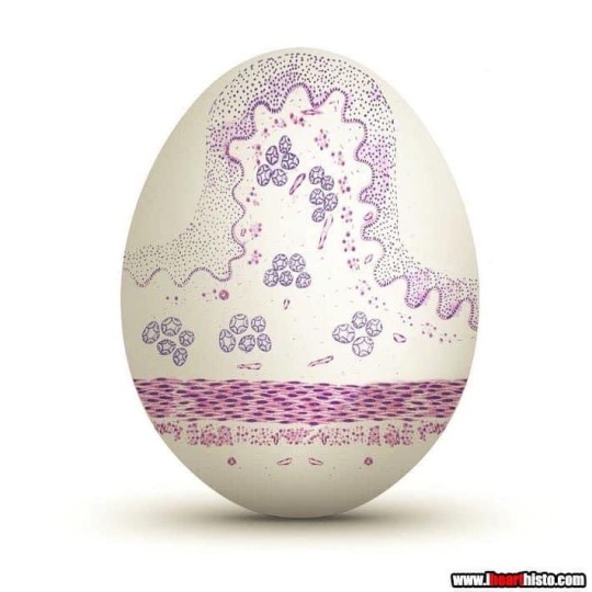
🥚 Esoph-egg-us 🥚
✅Non-keratinized stratified squamous epithelium
✅Circumscribing muscularis mucosae
✅Submucosal glands
✅Dual layered muscularis externa
It just isn’t Spring until you have painted your very own Esoph-egg-us
So get cracking and I don’t want to hear any eggscuses
i♡histo
#histology#science#pathology#med school#med student#ihearthisto#vet science#vet school#anatomy#premed#happy easter#easter#egg#spring#esophagus#digestive system#dentistry#dental school#dental student
69 notes
·
View notes
Photo

🎬🤖 💦 C3-PEE-O 💦🤖🎬
MAY THE FOURTH BE WITH YOU!
Enjoy this protocol droid in a renal tubule.
i♡histo
This familiar protocol droid is in the renal tubules of the kidney. His head and surrounding tubules are different regions of the long tubes known as nephrons that travel through the kidney. These structures convey the glomerular filtrate (fluid filtered from the blood), The different components of the nephron modify this fluid by secreting and reabsorbing water and ions from the filtrate in order to produce the concentrated urine that we excrete when we go pee.
C-3PO’s eyes and mouth are actually chunks of mineral that have formed inside a tubule. These crystals can sometimes form when the urine is being concentrated. Concentrating the urine is an essential process that allows your body to reabsorb water and stop you from dehydrating. The crystals can form larger renal calculi (kidney stones) that can travel along the tubes that convey the urine out of your body: ureters, bladder or urethra. they can become lodged along these tubes at any point causing a blockage.
The image was submitted by veterinarian Dr Allison Watson via the ihearthisto Facebook page - it is actually the kidney of a chicken!
#histology#anatomy#science#pathology#kidney#renal#c3po#r2d2#nurse#star wars#urine#dentistry#medlab#autopsy#biology#premed#meded#nursing#med school#med student#medicine#vet science#vet school#vet student#histotech#histo#ihearthisto\
18 notes
·
View notes
Photo
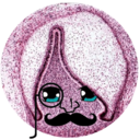
🏈 Super ‘Bowel’ Sunday LVII💩
This football-shaped egg is the spawn of the female whip worm Trichuris trichiura.
They can be found in water contaminated by feces or in delicious half-time hotdogs 🌭 handled by unsanitary fingers. Once ingested it makes its way into your intestines and after about 3 months will hatch into a worm.
Once mature, the worm burrows her head and mouth parts into the delicate and comfortable mucosa of your intestines to feed. It is here that she will trigger an inflammatory response and causes bouts of abdominal pain with plenty of (sometimes bloody) diarrhea. A disease called trichuriasis.
While she is feeding she dangles her vulva into the lumen of your intestine and has sex with as many nearby male worms as possible.
Our little parasite now becomes a momma and starts firing out somewhere between 2,000-10,000 of these little eggs…
Per day!
This life of feeding and egg-laying, if left untreated, can last for the lifespan of a roundworm which can be around 5 years… that’s a very busy worm and lots of eggs.
All those eggs get flushed out of your body with your bloody diarrhea and, depending on where you live, ‘touchdown’ in the local water supply or back on that hotdog ready to be ingested once again and the whole process starts over.
i♡histo
#histology#science#pathology#pathologists#anatomy#autopsy#parasite#superbowl#worms#parasitology#intestines#patrickmahomes#kansascitychiefs#premed#meded#nursing#medschool#medstudent#medicine#education#vetscience#vetschool#histotechnology#histotech#pathArt#sciArt#ihearthisto
27 notes
·
View notes
Text
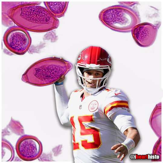
🍕Happy Pizza Day 🍕
Sometimes cells remind me of food.
These are epithelial cells from an adenocarcinoma of the lung.
i♡histo
An adenocarcinoma is a tumor that is derived from the epithelial cells of glandular tissues. The pizza was obtained from a nodule on the lung. An endoscope with an ultrasound attached to it was used to guide a needle directly into the nodule and some cells were then sucked out for observation. The technique is called endoscopic ultrasound fine needle aspiration or EUS/FNA for short. Aspirates like this allow pathologists to determine whether the cells in the nodule looked normal or cancerous so they can recommend appropriate treatment and/or surgery to remove the tumor.
#histology#science#pathology#med school#med student#ihearthisto#vet science#vet school#anatomy#premed#pizza#pizzaday#cells#dentistry#dental school#medlab#histopathology#biomedical science
56 notes
·
View notes
Text

🎅 Have a Howelly-Jolly Christmas 🎄
A festive finding in the blood of an asplenic patient 💉
i❤️histo
A Howell–Jolly body is a cytopathological finding whereby small remnants of nuclear DNA are present in normally anuclear circulating erythrocytes.
During development in the bone marrow, late orthochromatophilic erythroblast normally expel their nuclei. However, in some cases, a small portion of DNA remains (the purple dots in the erythrocytes wearing the Santa hats).
Under normal circumstances if these irregular erythrocytes make it into the blood, they are removed from circulation by the spleen.
As a result, the presence of erythrocytes with Howell-Jolly bodies in peripheral blood smears like this usually signifies a damaged or absent spleen - because a healthy spleen would normally filter this type of red blood cell.
📷 by exlibrisadpugno via reddit
#histology#science#pathology#med school#med student#ihearthisto#vet science#vet school#anatomy#premed#nurse#nursing#cytology#christmas#dentistry#dental school#histotechnology#biomedical#medblr
97 notes
·
View notes
Photo

🏆⚽🐛 The Worm Cup 🐛⚽🏆
The scolex (head) of the intestinal tapeworm looks like the soccer/football World Cup trophy!
i♡histo
www.ihearthisto.com
This image shows the pork tapeworm Taenia solium, an intestinal parasite found in countries where pork is eaten. This is just the head of the worm (the scolex) with its terrifying looking, hooked rostellum which is used to attach itself onto your intestinal wall so that you don’t poop it out easily.
Full grown worms can grow to around two or three meters in length and are composed of numerous segments each of which form their own reproductive unit.
The life cycle of the parasite begins when pigs ingest the tenia solium eggs (from fecal contamination) and these eggs develop into larvae in the pigs which migrate through the intestinal wall and form cysts in the muscle and tissues. Once slaughtered for meat, the larvae can be ingested by humans if the pork is eaten uncooked or undercooked. Once in the human small intestine the larvae grow into the large adult worms where they persist often unnoticed due to the lack of symptoms associated with their existence. The worms release eggs into the intestines which are released during defecation thus completing the cycle.
A major complication of infection with T. solium is Cysticercosis. This is a parasitic tissue infection caused by the larval cysts of the tapeworm. These larval cysts infect brain, muscle, or other tissue, and are a major cause of adult onset seizures. A person only gets cysticercosis by swallowing the eggs found in the feces of a person who has an intestinal tapeworm NOT from contracting a tapeworm by eating undercooked pork itself. So people living in the same household with someone who has a tapeworm have a much higher risk of getting cysticercosis than people who don’t because they can be exposed to the eggs released in the stool.
#histology#anatomy#pathology#science#med school#med student#medblr#microbiology#tapeworm#nurse#nursing#vet school#vet student#vet science#biomedical#soccer#football#premed#biology#histologia#histo#ihearthisto#worldcup#julesrimet
58 notes
·
View notes
Photo

🥚🐣🩸 Golem Blood 🩸🐣🥚
A clump of erythrocytes surrounding a monocyte in a blood smear.
i♡histo
This rock Pokémon has evolved from a clump of erythrocytes/red blood cells (Golem's boulder shell) that are partially surrounding a monocyte (Golem's head).
Erythrocytes are the main type of cell found in your blood. They are born in the red marrow inside your bones. Before they are released into the circulation, they eject their nucleus - you can see that the cells do not contain a purple nucleus like the neighboring monocyte. Ejecting their nucleus means that they have more room inside them to store the important protein hemoglobin. Hemoglobin transports oxygen & carbon dioxide around the body - it's the erythrocytes that carry the oxygen you breathe from the lungs to all the cells in your body! It makes sense to maximize the carrying capacity of each of these cells. However, not having a nucleus comes at a price. The nucleus contains the DNA info required for cells to make new proteins and divide. No nucleus means erythrocytes cannot divide or grow/repair easily like other cells. As a result they have a short lifespan (approx 120days) after which they are removed from circulation by macrophages in the liver or spleen. Your bone marrow will continue to make new erythrocytes to replace the old ones for your entire life.
The monocyte is a type of leukocyte (white blood cell). It's a large cell capable of leaving the circulation and entering surrounding connective tissues where it becomes known as a macrophage! Macrophages eat foreign invaders, cellular debris or old cells (like those old erythrocytes!). They also form part of our immune system because they act as antigen-presenting cells that breakdown invaders into harmless component parts and show these parts to other leukocytes (like lymphocytes) in the hope of eliciting a controlled immune response to the invading pathogen.
#histology#science#pathology#autopsy#pathologists#anatomy#pokemon#golem#blood#hematology#dental school#premed#med ed#nurse#nursing#med school#med student#med lab#vet school#vet student#vet science#histotechnology#histo#pathArt#cells#ihearthisto
38 notes
·
View notes
Text

🌸🌸 Rectal Daisies 🌸🌸
A bouquet with a unique fragrance.
Here's the 🔬 story behind these pretty flowers...
i♡histo
Microscopically, the mucosal lining of the rectum & colon exhibits many intestinal glands or crypts of Lieberkuhn (the flowers) that are lined with mucus secreting goblet cells (the petals).
Mucus collects in the lumen of the gland (center of flower) and is squeezed out when the muscular wall contracts to push the fecal material out.
As the feces move through the colon, water is reabsorbed making them drier to conserve the water. As a result, the mucus secreted from these crypts is super important in this region because it ensures that the tube is well lubricated to allow the dry feces to be passed along the colon easily without causing any damage (the mucus decreases friction).
If you slice up the colon or rectum in a way that ensures you cut through the mucosa parallel with its surface then your rectum looks much more like a field full of daisies with pretty goblet cell petals than a chute for passing along poop 💩
#histology#science#pathology#med school#med student#ihearthisto#vet science#vet school#anatomy#premed#nurse#nursing#medlab#histotech#colon#GI#digestive system#rectum#sciart
214 notes
·
View notes
Text
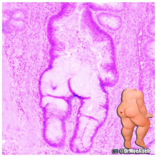
🍑 Bubble Butt Adenoma 🍑
A posterior view of a tubulovillous adenoma in the colon.
i♡histo
If you have ever had a colonoscopy then your doctor may have informed you that they found a number of polyps.
Polyps are slow growing lumps of tissue that grow on the inside wall of the colon and protrude, often from stalks, into the lumen of the colon like tiny mushrooms.
They are most often benign but can be an early warning sign for colorectal cancer.
To the naked eye, most polyps look similar but they can be classified based upon their microscopic appearance which means that it is important to biopsy a polyp to determine exactly what it is.
Some polyps may be described as tubular adenomas. These types of polyps look like that are composed of the many numerous tubular glands that line the rest of the colon. Except they look more irregular and contain few of the mucous secreting goblet cells you would expect to find in a regular intestinal gland (crypt of Lieberkuhn). The legs, arm and butt in the image left are the tubular gland part of this adenoma.
Some polyps may be described as being a villous adenoma. These look more shaggy/lumpy grossly. They look like this because under the microscope you will find that they are composed of many long, branching finger-like projections called villi. Villi are not a typical finding in the colon and imply a faster rate of polyp growth.
A polyp that has features of both types is often referred to by doctors as a 'tubulovillous' adenoma.
It is important to know the type of adenoma causing the polyp because villous adenoma have a higher chance of becoming cancer quicker than tubular adenomas.
Original 📷: @DrMoeAtieh [Twitter]
#histology#science#pathology#med school#med student#ihearthisto#vet science#vet school#anatomy#premed#nurse#nursing#buttstuff#colon#digestive system#cancer#polyp#med lab#histotechnology
56 notes
·
View notes
Text

😀 Bob Lobule 😀
A happy looking hepatic artery, bile ductule (eyes) and portal vein (mouth) bordering a number of hepatic lobules.
His name is Bob 😀
i♡histo
This is a hematoxylin and eosin stained slice through a liver.
The liver is composed of numerous roughly hexagonal shaped lobules. Each lobule has a venule at its center (you can see one bottom middle of this image) and a series of hepatic triads at its periphery (top left, bottom left).
A triad is a collection of three structures:: tiny branches of the major tubes that enter/leave the liver at its porta hepatis. The hepatic artery, the hepatic portal vein and the hepatic ducts that contain bile.
Once in the liver these tubes of the porta hepatis branch massively to form the triads.
Bob's face is formed from some of these branches prior to them forming the smaller triads. His left eye is an arteriole, his right eye is a bile ductule and his mouth is a hepatic portal venule.
Blood in the vessels of the triads flows into the fine sinusoids that pass between adjacent hepatocytes. The hepatocytes process the nutrient rich blood before it flows into a central vein.
The central veins of each lobule converge to form tributaries that ultimately unite to form the hepatic veins that drain into the inferior vena cava.
In this way all blood, rich in raw nutrients and toxins absorbed from the GI tract is processed by the liver before it enters the systemic circulation!
#histology#science#pathology#med school#med student#ihearthisto#vet science#vet school#anatomy#premed#nurse#nursing#liver#digestive system#dentistry#biomedical#medlab#medicine#emoji#faces in things
53 notes
·
View notes
Photo

🌐✨ Happy Star Wars Day ✨🌐
May the Fourth be with you!
i♡histo
Star Wars histology from top left, clockwise:
1. The Graafian follicle Death Star
In a galaxy far, far away an intergalactic superweapon is halted in metaphase II of meiosis amid a surge in Luteinizing Hormone.
2. Jabba the Corpus Albicans
“makingsa lee ka bok pateesa… beeska chata wnow kong bantha poodoo”
(translation) “you may have been a good friend..but now you are bantha fodder”
The corpus albicans is a structure in the ovary that is formed when the corpus luteum regresses.
3. Tusken Raider in the Liver
Despite what you see here, Tusken Raiders are not native to the human liver. If you think that, then you are making a wookie mistake.
The image is actually a portal triad and demonstrates the major structures that enter and leave the liver: hepatic artery, hepatic portal vein and bile duct.
4. The Empire Strikes Back (at the Liver)
Liver histology is definitely where it’s At-At!
A region of connective tissue among the hepatocytes in the liver.
---
Images by @ihearthisto, @drjohnrajala, @zenonich and @hopkins_gi_path respectively
#histology#anatomy#science#pathology#med school#med student#med lab#star wars#May 4th#may the 4th be with you#nurse#nursing#dental school#dental student#dentistry#vet science#vet school#veterinary#pre med#sci art#ihearthisto
95 notes
·
View notes
Text
🥚 Esoph-egg-us 🥚
✅Non-keratinized stratified squamous epithelium
✅Circumscribing muscularis mucosae
✅Submucosal glands
✅Dual layered muscularis externa
It’s just not Spring until you have painted your very own Esoph-egg-us
So get cracking and I don’t want to hear any eggscuses
i♡histo
#histology #science #pathology #pathologists #anatomy #autopsy #eggs #easter #spring #biology #esophagus #digestive #premed #meded #nurse #nursing #medschool #medstudent #medicine #education #vetscience #vetschool #dentistry #histotechnology #histologica #histotech #histo #pathArt #sciArt #ihearthisto
#histology#science#pathology#med school#med student#ihearthisto#vet science#vet school#anatomy#premed#nurse#nursing#dentistry#dental school#esophagus#spring#easter#egg#digestive system#medlab
45 notes
·
View notes
Text

😾 Grumpy Cord 😾
A transverse slice through a spinal cord that looks like Grumpy Cat.
i♡histo
Being the center of the nervous system, transmitting neural signals between the brain and the entire body and controlling independent neural reflexes…is just so awful 😾.
The pale central face is the 'grey matter' and is home to the many neuron cell bodies that run through the spinal cord. The surrounding darker region is composed of the axons of these neurons as the exit, enter, ascend and descend through the spinal cord on their way to innervate muscles, or returning information about pain back from the skin, or relaying information about body position back to the brain.
#histology#science#pathology#med school#med student#ihearthisto#vet science#vet school#anatomy#premed#nurse#nursing#dentistry#dental school#spinal cord#cns#nervous system#neuroscience#neuroanatomy#grumpy cat#cat
134 notes
·
View notes
Text
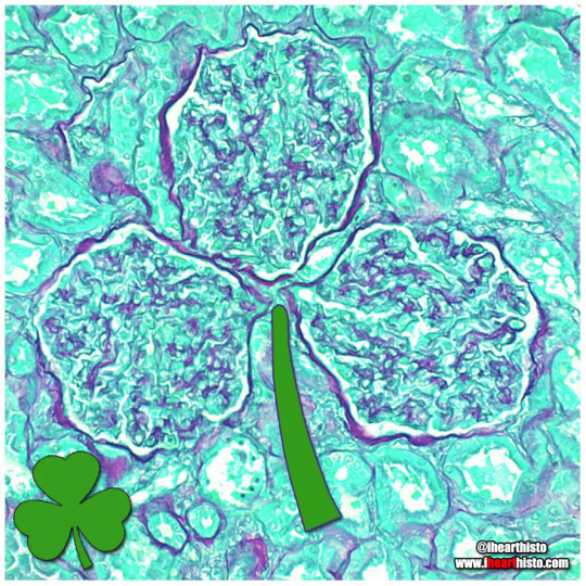
☘️ Shamrock Gloms ☘️
For each petal on the shamrock,
This brings a wish your way.
Good health. Good luck. Happiness.
For today and every day.
Happy St. Patrick’s Day!
i♡histo
Three renal corpuscles (glomerulus + their surrounding Bowman's capsules) floating in a sea of distal and proximal convoluted tubules within the cortex of the kidney.
These three small structures are knotted balls of capillaries (glomeruli) surrounded by a specialized epithelium (Bowman's capsule) that is composed of cells called podocytes. These cells have tiny interlocking legs that form a small slit between them.
This structural organization is responsible for filtering your blood to produce a fluid that then travels within tubes continuous with the Bowman's capsule called nephrons. In these nephrons the tubular fluid is modified by reabsorbing and secreting ions and conserving water to produce urine for excretion.
#histology#science#pathology#med school#med student#ihearthisto#vet science#vet school#anatomy#premed#nurse#nursing#shamrock#st. paddy's day#st. patrick's day#st patricks day#green#kidney#renal#medlab#biomedicine#dental#dental school#dental student#pathart
99 notes
·
View notes
Text

⚡ Lord Voldermis ⚡
A biopsy of a region of skin-that-shall-not-be-named (dermis/hypodermis junction shhh), complete with nerves, vessels, sweat glands and hair follicles.
i♡histo
📷 by @nejiby
#histology#science#pathology#med school#med student#ihearthisto#vet science#vet school#anatomy#premed#nurse#nursing#skin#dermatology#harry potter#voldemort#biomedical#medical#med#medlab#dentistry#dental school
30 notes
·
View notes
Text
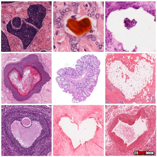
💗 You can find LOVE in the strangest of places (2022 edition) 💗
By row starting top left:
1. 💗 in a skin cylindroma
2. 💗 in an hepatic ductule
3. 💗 in a pancreas
4. 💗 in a warty penile growth
5. 💗 in a mucus-y colon
6. 💗 in a region of hypodermis
7. 💗 in a secondary oocyte
8. 💗 in a chondrosarcoma
9. 💗 in a small artery
💗 Happy Valentine's Day 💗
Tag a friend with the histo heart you want to share with them and spread the love!
i♡histo
Images by:
@ihearthisto [1-4, 5-9]
@donna.horncastle [5]
#histology#science#pathology#med school#med student#ihearthisto#vet science#vet school#anatomy#premed#valentine's day#valentine#heart#hearts#nurse#nursing#dental school#dental student#dentistry#sciart#histotechnology#medicine#med lab
968 notes
·
View notes
Photo
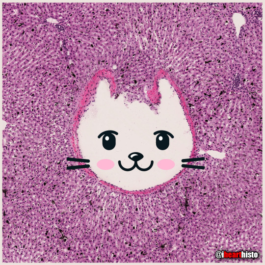
🐱 Kitty Liver 🐱
Check meowt!
I'm a purrfect hepatic vein surrounded by hepatic lobules!
i♡histo
This is a hematoxylin and eosin stained slice through a liver that was injected with carbon/ink prior to fixation.
The liver is compose of numerous roughly hexagonal shaped lobules. Each lobule has a venule at its center (the central vein; top right) and a series of hepatic triads at its periphery. A triad is a collection of three structures - in this case branches of the hepatic artery, hepatic portal vein and hepatic duct.
Blood in the hepatic artery (oxygenated) and hepatic portal vein (rich in nutrients absorbed from the intestines) travels in sinusoids (the many narrow white spaces in this image) towards the central vein of each lobule. On its way through the sinusoids the blood is processed by the the many hepatocytes that line this region (the pink cells).
Within the sinusoids reside many liver macrophages (Kupffer cells) that phagocytose debris traveling through sinusoids. Normally these cells are invisible in standard H&E preparations but recall that this tissue was injected with ink! The ink was phagocytosed by the macrophages filling their cytoplasm with carbon so that they are now visible as black cells in the sinusoids.
Once the blood enters the central vein of each hepatic lobule they drain into the hepatic veins (kitty!) until the blood reaches the inferior venal cava.
In this way all blood, rich in raw nutrients and toxins absorbed from the GI tract is processed by the liver before it enters the systemic circulation!
Pawsitively unbeliverable!
#histology#science#patholgoy#anatomy#pathologists#autopsy#kitty#cat#liver#hepatic#digestive system#GImed#medical#nursing#nurse#kitten#med school#med student#medicine#education#vet science#vet school#histotechnology#histotech#histo#pathart#sciArtt#ihearthisto
22 notes
·
View notes