
Last Seen Blogs
sanabibi666
😇SanaBibi😈666

mobileesports
Mobile eSports Games
wobblypanda-blog1
WobblyPanda

eeostre
;
rguedes02
Sobre coisas
Photo

⏳History of headache 🎯Meningiomas are extra-axial tumors and represent the most common tumor of the meninges. They are a non-glial neoplasm that originates from the meningocytes or arachnoid cap cells of the meninges and are located anywhere that meninges are found, and in some places where only rest cells are presumed to be located. Imaging :Typical meningiomas appear as dural-based masses isointense to grey matter on both T1 and T2 weighted imaging enhancing vividly on both MRI and CT #ultrasound #fetal #heart #anatomy #sonography #radiology #radiologist #theradiologypage #radiologie #radiologi #radiologia #radtech #radiologylove #radiographer #radiologista #xray #xrays #mri #imaging #surgery #medicine #medschool #medstudent #futuredoctor #futureradiolgist #radiologyresident #medstudent #medicalschool #medicalstudent #xraytech #theradiologyinsider Case courtesy of Frank Gaillard, <a href="https://radiopaedia.org/?lang=us">Radiopaedia.org</a>. From the case <a href="https://radiopaedia.org/cases/9935?lang=us">rID: 9935</a> https://www.instagram.com/p/CoVD1byvY99/?igshid=NGJjMDIxMWI=
#ultrasound#fetal#heart#anatomy#sonography#radiology#radiologist#theradiologypage#radiologie#radiologi#radiologia#radtech#radiologylove#radiographer#radiologista#xray#xrays#mri#imaging#surgery#medicine#medschool#medstudent#futuredoctor#futureradiolgist#radiologyresident#medicalschool#medicalstudent#xraytech#theradiologyinsider
0 notes
Photo
Subdural hemorrhage (SDH) is a collection of blood accumulating in the subdural space, the potential space between the dura and arachnoid mater of the meninges around the brain. Subdural hemorrhage can happen in any age group, is mainly due to head trauma and CT scans are usually sufficient to make the diagnosis. #ultrasound #fetal #heart #anatomy #sonography #radiology #radiologist #theradiologypage #radiologie #radiologi #radiologia #radtech #radiologylove #radiographer #radiologista #xray #xrays #mri #imaging #surgery #medicine #medschool #medstudent #futuredoctor #futureradiolgist #radiologyresident #medstudent #medicalschool #medicalstudent #xraytech #theradiologyinsider Case courtesy of Frank Gaillard, <a href="https://radiopaedia.org/?lang=us">Radiopaedia.org</a>. From the case <a href="https://radiopaedia.org/cases/35891?lang=us">rID: 35891</a> https://www.instagram.com/p/CoVGeJlvI-A/?igshid=NGJjMDIxMWI=
#ultrasound#fetal#heart#anatomy#sonography#radiology#radiologist#theradiologypage#radiologie#radiologi#radiologia#radtech#radiologylove#radiographer#radiologista#xray#xrays#mri#imaging#surgery#medicine#medschool#medstudent#futuredoctor#futureradiolgist#radiologyresident#medicalschool#medicalstudent#xraytech#theradiologyinsider
0 notes

Photo

Chaos in the medicine Casualty . . . #tvm #medicalcollege #trvm #radiology #radiologymemes #radiologist #mbbs #internship #housesurgeons #medicine #casualty #surgery https://www.instagram.com/p/Cn43hFjvO6t/?igshid=NGJjMDIxMWI=
#tvm#medicalcollege#trvm#radiology#radiologymemes#radiologist#mbbs#internship#housesurgeons#medicine#casualty#surgery
0 notes
Photo
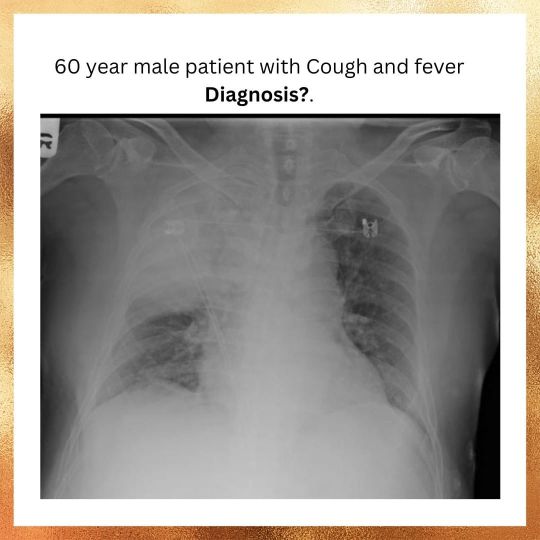
60 year old male patient with cough and fever . . . Diagnosis? #ultrasound #radiographer #mbbs #radiography #residency #doctors #anatomy #anatomia #medschool #medicalschool #imaging #medicine #radiologia #residency #pg #Physician #radiology #medstudent #nurse #nursingschool #nursingstudent #radtech #mri #ct ##chest #overlay #mbbs #medicalstudent #anatomy #bacteriology #bacteria https://www.instagram.com/p/Cn4hH93pzGb/?igshid=NGJjMDIxMWI=
#ultrasound#radiographer#mbbs#radiography#residency#doctors#anatomy#anatomia#medschool#medicalschool#imaging#medicine#radiologia#pg#physician#radiology#medstudent#nurse#nursingschool#nursingstudent#radtech#mri#ct#chest#overlay#medicalstudent#bacteriology#bacteria
2 notes
·
View notes
Photo
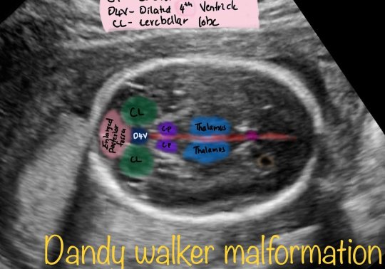
Dandy walker malformation #fetal #ultrasound #sonography #neonates #malformation #radiology #radiopaedia #radiologist #art https://www.instagram.com/p/CncmMulhZZl/?igshid=NGJjMDIxMWI=
1 note
·
View note
Photo

Findings?? #radiologist #xray #chest #radiology Presentation:🩺 Central lines should end at the junction of the vena cava and the right atrium. This is true for all central lines, including femoral,jugular, and subclavian lines, peripherally inserted central catheters (PICC lines), and tunneled catheters. X-Ray Findings:🩻 The tip of the central line will not be in the correct position. It might be coiled, be too long or two short, or might track down an incorrect vein. Management:⚕️🏥 The central line needs to be replaced. It cannot simply be repositioned. https://www.instagram.com/p/Cne-FxgviK3/?igshid=NGJjMDIxMWI=
1 note
·
View note
Photo
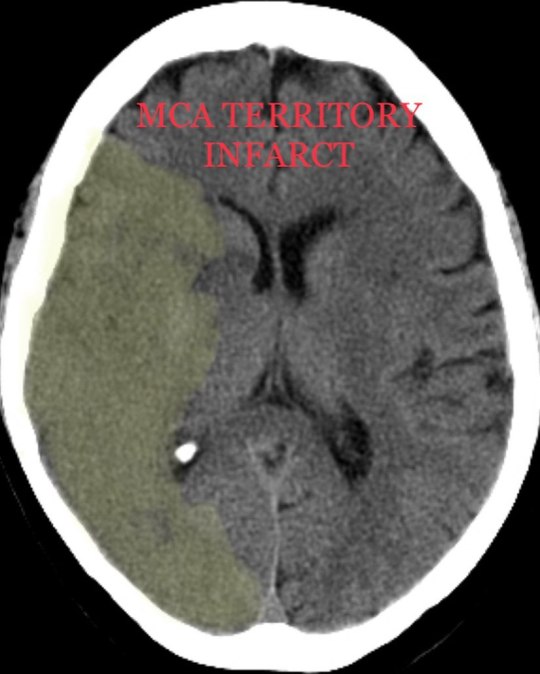
There is diffuse loss of the grey - white matter differentiation in the right MCA territory.Notice how on the left you can see the differentiation between the brighter grey matter and darker subcortical white matter. This distinction is lost on the right. Edema that blurs the grey-white matter differentiation is called cyto-toxic edema and is indicative of ischemic injury to the brain. #radiology #mca #radiologist #emergency #emergencymedicine #stroke #infarct #radiologyinsider https://www.instagram.com/p/CnxGW_Sv8GN/?igshid=NGJjMDIxMWI=
1 note
·
View note
Photo

The left MCA is hyperdense compared to the right MCA. This is highly suggestive of MCA thrombosis. See if you can follow this bright vessel into the sylvian fissure. It is important to look for signs of MCA distribution ischemia. Always look at the insula first. This is one of the first areas to show changes of infarction. #radiologist #radiology #stroke #emergencymedicine #emergency #ct #mri #ultrasound #sonologist #mca #ctscan #anatomy #signs https://www.instagram.com/p/CnxHr0Hvijn/?igshid=NGJjMDIxMWI=
#radiologist#radiology#stroke#emergencymedicine#emergency#ct#mri#ultrasound#sonologist#mca#ctscan#anatomy#signs
1 note
·
View note
Photo

Giant splenic abscess with suspected reactive adenopathy. There is a large septated fluid collection in the spleen with peri-splenic stranding and thickening of the adjacent anterior para-renal and lateral conal fascia. No internal air. There are scattered borderline enlarged mesenteric and retroperitoneal lymph nodes. No inflammatory change involving the bowel. #radiology #ultrasound #radiography #radiographer #mbbs #mbbsstudent #mbbsdiaries #residency #doctor #doctors #anatomy #anatomia #medschool #medicalstudent #imaging #nurse #nursingstudent #nursingschool #physician #radtech #mri #ct #overlay https://www.instagram.com/p/CnzmTlWv87w/?igshid=NGJjMDIxMWI=
#radiology#ultrasound#radiography#radiographer#mbbs#mbbsstudent#mbbsdiaries#residency#doctor#doctors#anatomy#anatomia#medschool#medicalstudent#imaging#nurse#nursingstudent#nursingschool#physician#radtech#mri#ct#overlay
1 note
·
View note
Photo

Large hepatic contusions and active bleeding There is hemoperitoneum around the liver, spleen, in the gall bladder fossa, and in the pelvis. There are large contusions in the superior right lobe of the liver (Segment VII) with evidence of active bleeding .There is an additional linear laceration in the posterior-inferior right lobe. Two more rounded, hypoattenuating lesions in the liver probably represent cysts and not trauma. Lastly, there is a right adrenal hemorrhage. Take home points: • Active bleeding looks like extravascular contrast (as bright as the contrast in the arteries) surrounded by hematoma which diffuses into the hematoma on delayed imaging. • Appearance of adrenal hematoma which can look like a hyperdense adrenal mass. • Trauma patients can have hepatic cysts or hemangiomas which should not be confused with acute injury. #ultrasound #radiology #radiopedia #sonography #trauma #emergency #emergencymedicine #accident #medicalstudent #mbbs #residency #surgery #orthopaedic #medicine #internalmedicine #nurse #nursingstudent #ct #mri #knowledge https://www.instagram.com/p/Cn1muhcP0YK/?igshid=NGJjMDIxMWI=
#ultrasound#radiology#radiopedia#sonography#trauma#emergency#emergencymedicine#accident#medicalstudent#mbbs#residency#surgery#orthopaedic#medicine#internalmedicine#nurse#nursingstudent#ct#mri#knowledge
1 note
·
View note
Photo

Long history of respiratory wheeze and chronic cough. Diagnosis??✌ Case courtesy of Frank Gaillard, Radiopaedia.org, rID: 15388. . #radiology #radiologist #theradiologyinsider #radiologie #radiologi #radiologia #radtech #radiologylove #radiographer #radiologista #xray #xrays #mri #imaging #surgery #medicine #medschool #medstudent #futuredoctor #futureradiolgist #radiologyresident #medstudent #medicalschool #medicalstudent #xraytech #mbbs #mbbsstudent #basic https://www.instagram.com/p/Cn3MIQQJNJ_/?igshid=NGJjMDIxMWI=
#radiology#radiologist#theradiologyinsider#radiologie#radiologi#radiologia#radtech#radiologylove#radiographer#radiologista#xray#xrays#mri#imaging#surgery#medicine#medschool#medstudent#futuredoctor#futureradiolgist#radiologyresident#medicalschool#medicalstudent#xraytech#mbbs#mbbsstudent#basic
3 notes
·
View notes
Photo

Long history of respiratory wheeze and chronic cough. Diagnosis??✌ Case courtesy of Frank Gaillard, Radiopaedia.org, rID: 15388. . #radiology #radiologist #theradiologyinsider #radiologie #radiologi #radiologia #radtech #radiologylove #radiographer #radiologista #xray #xrays #mri #imaging #surgery #medicine #medschool #medstudent #futuredoctor #futureradiolgist #radiologyresident #medstudent #medicalschool #medicalstudent #xraytech #mbbs #mbbsstudent #basic https://www.instagram.com/p/Cn3MIQQJNJ_/?igshid=NGJjMDIxMWI=
#radiology#radiologist#theradiologyinsider#radiologie#radiologi#radiologia#radtech#radiologylove#radiographer#radiologista#xray#xrays#mri#imaging#surgery#medicine#medschool#medstudent#futuredoctor#futureradiolgist#radiologyresident#medicalschool#medicalstudent#xraytech#mbbs#mbbsstudent#basic
0 notes
Text
Take home points
• CT criteria for appendicitis include: non filling with air or contrast, > 6mm thick, hyperemic mucosa, surrounding stranding, and thickening of the cecum at the origin.
• Trace the colon from anus to cecum. Find the appendix along the cecum on the opposite side of the ileocecal valve from the ascending colon.
#radiologist #radiology #surgery #pathology #biochemistry #physiology #mbbs #medicalstudent #medico #medicine #ortho #ct #mri #study #anatomy #radiologyinsider
1 note
·
View note
Text
#radiology #foreignbody #emergency #nurse #nursingstudent #residency #mbbs #mbbsstudent #radiologist
1 note
·
View note
Photo
Large hepatic contusions and active bleeding There is hemoperitoneum around the liver, spleen, in the gall bladder fossa, and in the pelvis. There are large contusions in the superior right lobe of the liver (Segment VII) with evidence of active bleeding .There is an additional linear laceration in the posterior-inferior right lobe. Two more rounded, hypoattenuating lesions in the liver probably represent cysts and not trauma. Lastly, there is a right adrenal hemorrhage. Take home points: • Active bleeding looks like extravascular contrast (as bright as the contrast in the arteries) surrounded by hematoma which diffuses into the hematoma on delayed imaging. • Appearance of adrenal hematoma which can look like a hyperdense adrenal mass. • Trauma patients can have hepatic cysts or hemangiomas which should not be confused with acute injury. #ultrasound #radiology #radiopedia #sonography #trauma #emergency #emergencymedicine #accident #medicalstudent #mbbs #residency #surgery #orthopaedic #medicine #internalmedicine #nurse #nursingstudent #ct #mri #knowledge https://www.instagram.com/p/Cn1muhcP0YK/?igshid=NGJjMDIxMWI=
#ultrasound#radiology#radiopedia#sonography#trauma#emergency#emergencymedicine#accident#medicalstudent#mbbs#residency#surgery#orthopaedic#medicine#internalmedicine#nurse#nursingstudent#ct#mri#knowledge
1 note
·
View note
Photo

Large hepatic contusions and active bleeding There is hemoperitoneum around the liver, spleen, in the gall bladder fossa, and in the pelvis. There are large contusions in the superior right lobe of the liver (Segment VII) with evidence of active bleeding .There is an additional linear laceration in the posterior-inferior right lobe. Two more rounded, hypoattenuating lesions in the liver probably represent cysts and not trauma. Lastly, there is a right adrenal hemorrhage. Take home points: • Active bleeding looks like extravascular contrast (as bright as the contrast in the arteries) surrounded by hematoma which diffuses into the hematoma on delayed imaging. • Appearance of adrenal hematoma which can look like a hyperdense adrenal mass. • Trauma patients can have hepatic cysts or hemangiomas which should not be confused with acute injury. #ultrasound #radiology #radiopedia #sonography #trauma #emergency #emergencymedicine #accident #medicalstudent #mbbs #residency #surgery #orthopaedic #medicine #internalmedicine #nurse #nursingstudent #ct #mri #knowledge https://www.instagram.com/p/Cn1muhcP0YK/?igshid=NGJjMDIxMWI=
#ultrasound#radiology#radiopedia#sonography#trauma#emergency#emergencymedicine#accident#medicalstudent#mbbs#residency#surgery#orthopaedic#medicine#internalmedicine#nurse#nursingstudent#ct#mri#knowledge
0 notes