#phenol red indicator
Text
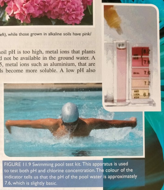
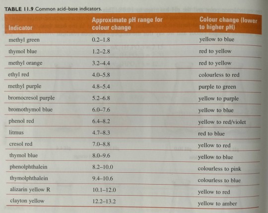
Kits for testing the pH of pool water (figure 11.9) use phenol red indicator, which changes colour over the pH range of 6.4-8.2.
"Chemistry" 2e - Blackman, A., Bottle, S., Schmid, S., Mocerino, M., Wille, U.
1 note
·
View note
Text

1 note
·
View note
Text


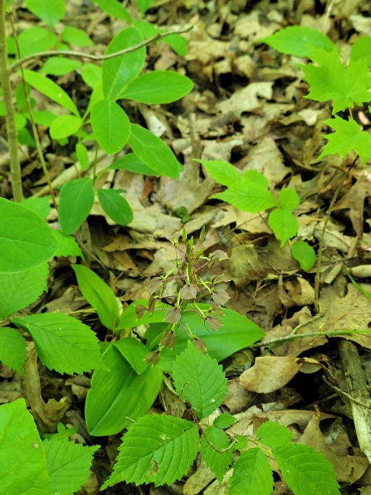


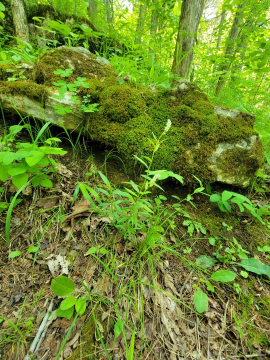
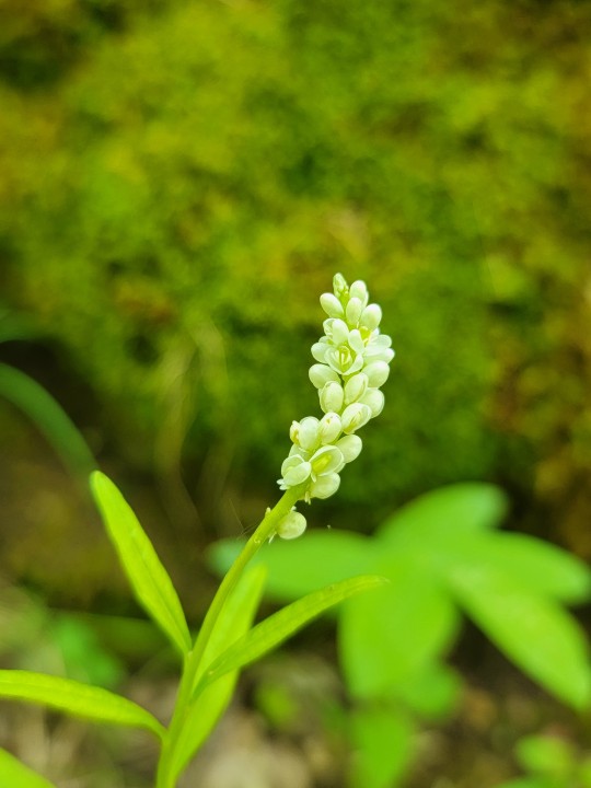
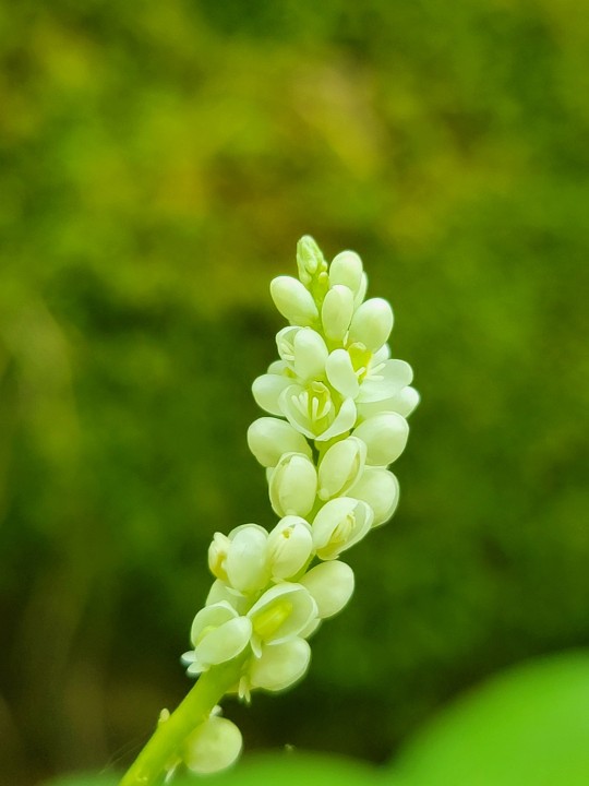

Two fun finds from Davis Memorial, Adams co. Both Seneca snake root, and lily leaved tway-blade were in bloom. Ive actually never seen Lily leaf tway blade in full anthesis.
Lily Leaf Tway Blade
Liparis liliifolia
a species of tway blade orchid known to have a fairly decent range and is an indicator species of fungal diverse hillsides with alkaline soil aggregate structure. The species it's self is considered to have unique nectar spur morphology and shows signs of insect mimicry in it's shape; with this said, it can be pollinated fairly easily by many generalists and mainly a long bodied small fly in the genera Pegoplata, aka short horned flys. which makes that spur, a mystery since we don't know if the fly is praying on moths or if it is praying on other long tongued insects looking for nectar, or if it's just somehow attracted to the flower proper.
Seneca Snake Root, senegal milkwort
Polygala senega

a rocky mesic hillside species that should be more common but is easily poached for its roots much like goldenseal, goldenroot, or ginseng and is usually locally abundant in spots and increasingly rare out of monitored preserves. The root can be boiled in tea form for mucus expectorants. High doses of powdered senega root or tincture are emetic and irritating to the GI tract, can cause reduced inflammation but also nerve transport/communication issues. The name is derived from it's anti inflamatory properties and nerve disrupting properties alone and was used but first nation tribes like the seneca/senagal and manatoba in aid for rattlesnake bites; this would need to be used with nervains( specifically Verbena spp. and blood clotter plants to work fully like Rattlesnake master, Eryngium yuccifolium.
Rattle Snake Master research is on going for usages:
https://pubmed.ncbi.nlm.nih.gov/18499203/
lipophilic chemicals that are associated are pretty interesting too.
when you add a lipid breaking and a protein breaking stew of chemicals, as well as phenolic bioactive compounds that are readily digestible and useful in a tea you can see why the plants were all used in conjunction to fight venom.
haemotoxic venom of adders can cause latent hemoraging at pressure points where platelet stacking and fat can cause massive stacking events. It mainly causes the opposite though in the fact that it disrupts the clotting cascade causing leaking veins and bleeding to not stop.
#ohio#botany#wildflowers#plantblr#ecology#cottagecore#Eryngium#polygala#polygala senega#liapris#liapris liliifolia#liparis#liparis liliifolia
13 notes
·
View notes
Text
Get the Best Quality of XLD Agar at TM Media
XLD Agar (Xylose Lysine Deoxycholate Agar) is a selective and differential medium used primarily for isolating and identifying enteric pathogens, particularly Salmonella and Shigella species. It contains xylose, lysine, lactose, sucrose, and the pH indicator phenol red. Salmonella colonies typically appear red with black centers due to hydrogen sulfide production, while Shigella colonies remain red. The medium's selectivity is enhanced by deoxycholate, which inhibits Gram-positive bacteria and some Gram-negative coliforms. XLD Agar is widely used in clinical and food microbiology.
Product link: https://www.tmmedia.in/product/xld-agar-2/
Website link: https://www.tmmedia.in/
Company Name: TM Media
Phone: +91-9999-1687-70
Address: 905, 9th Floor, Big joes Tower, Netaji Subhash Place, New Delhi
Mail:[email protected]

0 notes
Text
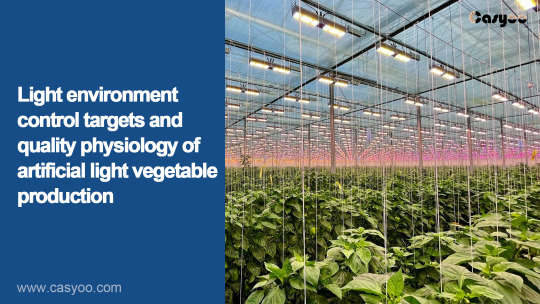
Light environment control targets and quality physiology of artificial light vegetable production
Quality and functional substance accumulation levels are one of the important indicators of concern in facility vegetable production. For vegetables, the main nutritional quality indicators include primary metabolites (sugar, protein, minerals, vitamins, cellulose, etc.) and secondary metabolites (anthocyanins, phenolics, flavonoids, lycopene, etc.). These indicators are controlled by light conditions. In other words, they are nutritional quality indicators that can be controlled by light conditions, which have prospects in the applications of LED lighting.
Hu et al.'s (2014) study showed that consuming 200g of fruits (equivalent to two apples) per day can reduce the risk of stroke by 32%; consuming 200g of vegetables per day can reduce the risk of stroke by 11%. The appearance quality and nutritional quality of vegetables are very important.
Zheng Xiaolei et al. (2011) studied the effects of different light qualities (white fluorescent light, red LED, blue LED, red/blue LED) on the growth and edge burning of loose leaf lettuce under plant factory conditions, providing a certain theoretical basis for the high-quality and efficient production of loose leaf lettuce in plant factories.
The results showed that red/blue LED can significantly increase the fresh weight, leaf number and leaf area of loose leaf lettuce, and reduce the nitrate content of loose leaf lettuce, but does not reduce the edge burn index; red LED can promote stem elongation, significantly reduces the burnt edge disease index and nitrate content, but is not conducive to dry matter, vitamin C accumulation and leaf area increase; blue LED inhibits the growth of loose leaf lettuce and increases nitrate content, but can significantly reduce burnt edge disease index. It showed that red LED is beneficial to the growth of loose leaf lettuce in the plant factory and reduces the occurrence of burnt edges.
The plants had longer internodes and thinner stems under red light. Under blue light, the internode is shorter and the stem is thicker, and the elongation and growth are inhibited to some extent (Jao and Fang, 2003).
Blue LED significantly inhibits the growth of loose leaf lettuce stems. Red light promotes stem elongation (Li and Kubota, 2009); while under blue LED, the stem length was 16% shorter than the control, indicating that blue light inhibited stem elongation. and the results were consistent with those of previous studies.
Blue light increased the activity of indoleacetic acid oxidase, decreased the level of auxin, and inhibited plant growth. Red and blue LED light can increase the vitamin C content of loose leaf lettuce compared with fluorescent light, and red LED can significantly reduce the vitamin C content of loose leaf lettuce. Chen Qiang et al. (2009) found the same result on tomatoes.
The effect of light quality on vitamin C content was related to the activity of its synthesis and decomposition enzyme activity. Galactolactone dehydrogenase (GalLDH) directly catalyzed the synthesis of vitamin C from galactolactone. Ascorbate oxidase and ascorbate peroxidase (AAP) are key enzymes in the oxidation of vitamin C in plants (An Huaming et al., 2005), and these three enzymes are sensitive to light. The mechanism of the different responses of the above three enzymes to different light properties needs to be further studied.
Vitamin C is an important nutrient component and an important index to evaluate the quality of loose leaf lettuce. Leafy vegetables are very easy to enrich nitrate, which is converted into nitrite during use, which is harmful to human health. Red LED can reduce the nitrate content of loose leaf lettuce, while blue LED can increase the nitrate content of loose leaf lettuce (Zheng Xiaolei et al., 2011; Deng Jiangming et al., 2000).
Blue LEDs increase nitrate content mainly because the process of absorbing nitrate nitrogen requires energy consumption, and blue light is likely to promote nitrate nitrogen absorption by stimulating ATP formation. The cogroups of nitrate reductase (NR) are flavin adenine dinucleotide and purine, and the chromophore of blue light receptor contains flavin and purine, so blue light is likely to directly stimulate NR, so that nitrate in loose leaf lettuce leaves can be reduced quickly, thus promoting the absorption and accumulation of nitrate.
The anthocyanin synthesis and accumulation of red leaf lettuce will decrease under the facility cultivation condition. Studies have shown that supplementing blue light and UV-B light at night can significantly improve anthocyanin synthesis. Shoji et al. (2010) studied the effect of increasing blue light intensity in red-blue composite light on anthocyanin synthesis. The accumulation of anthocyanins is the largest under blue light of 100umol/m2·s, the smallest under red light, and the red and blue composite light was in the middle. That is to say, the ratio of red to blue is very important in the synthesis of anthocyanins in red leaf lettuce.
0 notes
Text
Get the Best Quality of XLD Agar Composition (TM 492) at TM Media
If you are looking for Xylose Lysine Deoxycholate (XLD) agar (TM 492) manufactured by TM Medium, a distinctive culture medium, boasts a specialized blend of ingredients carefully selected to facilitate the isolation and differentiation of enteric pathogens. Featuring xylose and lactose as fermentable carbohydrates, lysine and thiosulfate as vital sulfur sources, and deoxycholate acting as a potent selective agent against gram-positive organisms, xld agar composition stands out for its precision. Incorporated phenol red serves as a pH indicator, transitioning the medium to a discernible yellow hue upon acidification from carbohydrate fermentation. Additionally, the inclusion of ferric ammonium citrate and sodium thiosulfate facilitates hydrogen sulfide production, distinguished by the formation of characteristic black colonies.
Visit the Website - https://www.tmmedia.in/

0 notes
Text
Phenolphthalein, for example, changes a solution from colourless to pink as the pH of the solution changes over a range of 8.2 to 10.0 (see table 11.9).
"Chemistry" 2e - Blackman, A., Bottle, S., Schmid, S., Mocerino, M., Wille, U.
#book quotes#chemistry#nonfiction#textbook#acid#base#indicator#color change#titration#methyl green#thymol blue#methyl orange#ethyl red#methyl purple#bromocresol purple#bromothymol blue#phenol red#litmus#cresol red#phenolphthalein#thymolphthalein#alizarin yellow r#clayton yellow
0 notes
Text
Exploring the Benefits of Basement Membrane Matrix HC (Phenol Red) in Cell Culture
Introduction
In the realm of cell culture techniques, researchers are constantly seeking improved tools and materials to mimic the physiological environment for better experimental outcomes. One such innovation is the Basement Membrane Matrix HC (Phenol Red), a cutting-edge product that offers numerous advantages for cell culture applications. This article will delve into the features, benefits, and potential applications of Basement Membrane Matrix HC (Phenol Red) to shed light on its significance in modern cell research.
Understanding Basement Membrane Matrix HC (Phenol Red)
Basement Membrane Matrix HC (Phenol Red) refers to a specialized extracellular matrix derived from natural sources like Engelbreth-Holm-Swarm (EHS) mouse sarcoma cells. It provides a biologically relevant scaffold that mimics the physiological conditions found in vivo. The addition of phenol red, a pH indicator, enables real-time monitoring of pH changes in cell culture, ensuring optimal conditions for cellular growth and functionality.
Advantages and Features
Biocompatibility: Basement Membrane Matrix HC (Phenol Red) possesses exceptional biocompatibility, allowing it to support the growth, proliferation, and differentiation of various cell types.
Structural Support: With its natural composition resembling the extracellular matrix, this matrix offers structural support to cells, facilitating their organization and development.
Cell Signaling: Basement Membrane Matrix HC (Phenol Red) contains essential proteins and growth factors that can actively influence cell behavior, promoting cell survival and enhancing cell-to-cell communication.
Versatility: This matrix supports a wide range of applications, including 3D cell culture, organoid culture, stem cell research, tissue engineering, and drug discovery, making it a valuable tool for diverse scientific investigations.
Ease of Use: Basement Membrane Matrix HC (Phenol Red)is available as a ready-to-use gel or solution, eliminating the need for complex preparation procedures and allowing researchers to save time and resources.
Applications in Cell Culture Research
Cancer Research: The use of Basement Membrane Matrix HC (Phenol Red) has facilitated the development of more accurate cancer cell models, enabling researchers to study tumor biology, metastasis, and drug responses in a physiologically relevant microenvironment.
Stem Cell Differentiation: Stem cells require a supportive niche for their differentiation into specific cell lineages. This matrix provides an ideal platform for inducing stem cell differentiation and studying tissue-specific functions and regenerative medicine.
Drug Screening and Toxicology: By culturing cells on Basement Membrane Matrix HC (Phenol Red), researchers can assess drug efficacy, toxicity, and metabolism accurately. This allows for the development of safer and more effective pharmaceutical interventions.
Tissue Engineering: The combination of Basement Membrane Matrix HC (Phenol Red) with appropriate scaffolds has opened new avenues for tissue engineering. It promotes cell adhesion, migration, and tissue formation, contributing to the development of functional 3D tissues and organs.
Neuroscience Studies: Basement Membrane Matrix HC (Phenol Red) has been successfully implemented in neurobiology research, offering a suitable microenvironment for studying neuron-astrocyte interactions, synaptic plasticity, and other aspects of brain function.
Conclusion
Basement Membrane Matrix HC (Phenol Red) represents a significant breakthrough in the field of cell culture research. Its biocompatibility, structural support, and ability to mimic the physiological conditions make it an invaluable tool for various applications. Researchers across numerous disciplines are increasingly relying on this matrix to advance their studies and gain deeper insights into cellular behavior. As the demand for more accurate cell models continues to grow, Basement Membrane Matrix HC (Phenol Red) is poised to play a pivotal role in shaping future advancements in cell culture research. Please visit MedChemExpress
0 notes
Text
Understand the Spiderman of the Microverse: XLD Agar
Learning XLD’s X, Y, and Z can be challenging, but not with us. In the previous article, we discussed the various needs and applications of XLD (click on the link if you haven’t read it). In this, we’ll talk more about its composition, principle, observations, and modifications.
What is the composition of XLD Agar?
It was developed by Welton Taylor in 1965. XLD stands for Xylose Lysine Deoxycholate Agar and is a bright pink or red-colored solid opalescent gel medium. Knowing its key contents will help you better understand its principles and differentiating abilities.
Ingredients g/l
Yeast Extract 3 g
L-lysine 5 g
Xylose 3.75 g
Lactose 7.5 g
Sucrose 7.5 g
Sodium Deoxycholate 1 g
Sodium Chloride 5 g
Sodium Thiosulfate 6.8 g
Ferric Ammonium Citrate 0.8 g
Phenol red 0.08 g
Agar 12.5 g
Distilled water 1 Litre
Yeast extract provides the medium with nutrients, vitamins, peptides, and other essential growth factors. Xylose, lactose, and sucrose are rich sources of fermentable carbohydrates. For the detection of fermentation of these carbs in the medium, a pH indicator, phenol red, is added. The addition of xylose to the medium accounts for the differentiation of various fermenting enteric bacteria from species of Shigella that do not ferment it. Most enteric pathogens, including Salmonella,can ferment xylose, which results in the formation of acidic byproducts and a change in the color of the medium from pink to yellow, whereas Shigella colonies remain red. In some cases, Salmonella imitates Shigella colonies and gives red colonies, which are then differentiated by the production of Hydrogen sulfide gas by the metabolism of thiosulfate and give black centers. Enterobacteria such as E. coli can ferment lactose in the medium.
XLD Agar’s composition can be adjusted according to one’s needs and choices and is hence available in many different variations, but the basic principle remains the same.
Observation and Inferences on XLD Agar:
Observation Inferences
Red colonies, some with black centers Salmonella spp.
Red colonies Shigella spp.
Yellow to orange colonies Coliforms
Pink, flat, and rough colonies Pseudomonas aeruginosa

While learning about XLD Agar, a question that must have run through your thoughts would’ve been, “Where is XLD Agar available?” Let’s look into it.
Where is XLD Agar available?
XLD Agar is available on the market as Dehydrated Culture Media in a number of variations according to one’s requirements, including plant-based and animal-based options.
These Dehydrated Culture Media can be dissolved in distilled water according to the concentration mentioned on the package, autoclaved, poured, and used.
TM Media, the microbiology division of Titan Biotech Ltd., offers a wide range of XLD Agar as Dehydrated Culture Media in a number of variations suitable for all your needs, operations, and specific requirements.

The product range includes the following:
XLD AGAR MODIFIED (as per ISO) TM 1621: XLD Agar, modified, is a selective and differential medium for the isolation of gram-negative enteric pathogens from clinical specimens or food products. It is a modification of the original formulation of Taylor that allows selective isolation of Salmonella typhi, E. coli, Salmonella enteritidis, Salmonella typhimurium, and Shigella flexneri. It is recommended by the ISO committee, and the composition and performance criteria of this medium are as per the specifications laid down in ISO 6579-1:2017.
XLD AGAR (VEG.) TMV 492: Xylose Lysine Deoxycholate Agar (Veg) is prepared by replacing Sodium Deoxycholate with synthetic detergent No.III, which makes the medium free of BSE/TSE risks. Xylose Lysine Deoxycholate Agar (Veg) is suitable for the isolation and identification of enteric pathogens from stool samples.
XLD AGAR (as per USP/EP/JP/BP) TMH 112: This medium is employed for pharmaceutical testing and non-sterile product testing for the detection of Salmonella after enrichment in Rappaport VassialidiasSalmonella Enrichment Broth in accordance with the harmonized method of USP/EP/BP/JP/IP.
XLD AGAR TM 1448: It is used for pharmaceutical testing and nonsterile product testing for the detection (or absence) of Salmonella after enrichment in Rappaport Vassialidias Salmonella Enrichment Broth in accordance with IP.
XLD AGAR TM 492: XLD Agar exhibits increased selectivity and sensitivity as compared to other plating media, e.g., SS Agar, EMB Agar, and Bismuth Sulphite Agar. The media formulation does not allow the overgrowth of other organisms over Salmonella and Shigella. Samples suspected of containing enteric pathogens, along with other mixed flora, are initially enriched in Modified Semisolid RV Medium Base.
Why choose TM Media’s XLD Agar?
TM Media’s superior-quality and diverse Microbiological Culture Media are certified by ISO, CE, GMP, FSSAI, and FSSC.
TM Media provides unrivaled convenience, dependability, and efficacy, as well as versatility and flexibility, by manufacturing a contamination-free medium with a longer shelf life and reproducible outcomes.
Conclusion:
XLD Agar is a selective and differential agar medium primarily used for the isolation and differentiation of different enteropathogenic organisms that are well-known to cause diseases like Food poisoning, Gastroenteritis, and other Digestive illnesses.
By choosing TM Media, consumers invest in precision, quality, and excellence. TM Media's product portfolio includes over 2000 Dehydrated Culture Media, along with Ready-to-Use Culture Media, Biological Media Bases, Media Supplements, Lab Chemicals, and many more.
#XLD Agar#dehydrated culture media#food microbiology#clinical microbiology#Microbiology#Xylose Lysine Deoxycholate Agar
0 notes
Text
Pomegranate Peel Powder benefits
Pomegranate peels have always been used as folk medicines, owing to their numerous beneficial compounds. Malaivel herbals offer you 100% Natural Pomegranate peel powder with no added substances and chemicals. It is also worth noting that complex bioactive compounds in pomegranate peel often exist in the form of a mixture, so the synergistic effect of different compounds can produce a variety of physiological activities. The extraction yield, antioxidant activity (DPPH and ABTS inhibition) and total phenolic contents were evaluated. Malaivel Pomegranate Peel Powder can be consumed as tea also, which help ease stomach upsets, hot flashes, haemorrhoids, conjunctivitis and several other ailments. Pomegranate peels are typically discarded and thought of as inedible, but they’re used regularly for various health and beauty benefits in Ayurvedic medicine, an alternative practice with roots in Indian culture. Lets Grab the brand new Malaivel Herbal Pomegranate Powder to experience numerous health benefits and healthy Glow of skin !
Product description :
Introduction :
The pomegranate peel is considered as an agro-waste but it can be a potential source of antioxidants, phenols and also possesses antibacterial and antifungal activity. Malaivel herbals offer you 100% Natural Pomegranate peel powder with no added substances and chemicals. The peels of pomegranate fruits are the major by-products produced during food processing of pomegranate enriched in antioxidants and broad-spectrum antimicrobial agents and can prevent food deterioration.
Ingredients :
Sun-dried Pomegranate skin or Punica granatum (Botanical name of Pomegranate).
Appearance :
Malaivel Herbal Pomegranate Peel powder looks like pale red or yellow in color.
Benefits of Malaivel Pomegranate Peel Powder:
* It will boosts skin cell regeneration.
* Malaivel Herbal Pomegranate Peel Powder is loaded with vitamin C, which helps to neutralize free radicals and repair damaged skin cells.
* It Hydrates skin and give a fresh look.
* It helps to reduce the signs of premature ageing.
* It helps to treat skin infections and heal
* This Natural Peel Powder helps to shrink pores.
How to use it:
* Take 1 tablespoon of Malaivel pomegranate peel Powder, make a paste of this powder with lemon juice or rose water and apply it all over your face, especially on your pimples or acne.
* Leave it off on the face, neck and hands. Let it dry and wash it off after 20 - 30 mins.
Highlights:
Malaivel Pomegranate peel Powder can be steeped in hot water and consumed as tea also. It has numerous health benefits for the body.
Fine prints:
Some studies and researches indicated pomegranate peel extract induces weight loss in mice, due to its effect on reducing serum TG and plasma glucose concentration. Malaivel Herbal Pomegranate Powder is very safe to consume as it is 100% natural.
Conclusion :
Malaivel Pomegranate Peel Powder is a good source of bioactive compounds, including phenolic acids, flavonoids and hydrolyzable tannins, which have beneficial health effects. Pomegranate peel powder will reduce the gastric ulcer area and ulcer index, gastric juice volume, and acidity. Pomegranate powder recovers gastric mucus content and gastric tissue at the histological level also.
0 notes
Text
Internal influencing factors when cultivating cells in cell factories
When we use cell factories to culture cells, the growth status of the cells is affected by various factors. These factors may seem inconspicuous, but if not paid attention to, they will cause heavy damage to the cells. So, how do small details when using cell factories affect cell growth, and how can we avoid pitfalls when using cell factories to culture cells?
1. Degree of contact between cells
Normal cells (diploids) have attachment contact inhibition, and cell growth stops after overgrowth. However, abnormal cells such as tumor cells have weak contact inhibition and can grow to higher cell densities, but some cells overgrow. The damage is difficult to reverse, so it is not recommended to have too high a cell density when culturing cells.
Important points: Generally, cells cultured in a cell factory can be passaged when the cell confluence reaches 70-80%. Do not overgrow the cells.

Cell Factory Systems
2. Cell growth attachments
Adherent cells will adhere to the wall under normal circumstances, but some cells themselves do not adhere firmly or have poor adhesion. At this time, coating attachments can promote cell adhesion. Therefore, when cultivating some special cells, the cell factory needs to be coated. Used after. Commonly used matrices include polylysine (D type, L type), gelatin, collagen, and extracellular matrix mixture, which can be selected according to needs during culture.
Key point: When the cell adhesion in the cell factory is poor, the matrix can be used to coat the cell factory.
3. pH value of culture medium
Most cell lines grow best when cultured in cell factories at around pH 7.4, and the indicator of conventional media is phenol red, so in most cases, the media we see are red, but different media There will be differences in color, for example DMEM is a little darker than 1640. When cultivating cells, you can also judge the cell status based on the color of the culture medium in the cell factory. For example, as the cells grow, the color of the culture medium will become lighter and yellower.
Key points: Pay attention to the color change of the culture medium when culturing cells, and deal with any abnormalities in a timely manner.
0 notes
Text
Keep Your Pet Safe The Ultimate Guide to Pet-Friendly Cleaning Products

Luckily, plenty of pet-friendly cleaning products are available today that can be used safely around your furry friend. To help you out, we’ve compiled this ultimate guide to selecting pet-safe cleaning items that won't cause any harm to your pet.
What are the most dangerous chemicals for dogs and cats?
There are a few key ingredients, such as chlorine bleach, ammonia and “phenols” (in some glass cleaners), that can be hazardous for your pet. Although these products may boast of being natural or organic, they still contain dangerous elements when ingested by pets. Some everyday household items like laundry detergent, fabric softener, oven cleaner and toilet bowl cleansers should also be avoided to ensure the health and safety of your pet.
What are safe cleaning products for pets?
These products are made with natural and non-toxic ingredients that are safe for your pets’ health. They are usually labelled as pet safe or animal-friendly, and you can also check for an EPA Safer Choice label, which indicates all ingredients have been reviewed for safety so that they won't cause harm to your pets.
What are some natural cleaners I can use at home?
To make your own pet-safe cleaning products, you can use common household items like baking soda, vinegar and lemon juice. These ingredients are non-toxic and provide a natural disinfectant that still effectively kills germs while being gentle on your pet’s sensitive skin. You can also opt for all-natural cleaning solutions such as castile soap and essential oils, which are safe to use around pets but should always be used in small amounts and diluted with water.
How Do I know if my dog is allergic to cleaning products?
Generally, pets can develop contact allergies with certain ingredients such as fragrances, dyes, and preservatives used in cleaning products. Signs of an allergic reaction include itching, redness, inflamed skin, hives, and excessive scratching. If you suspect your pet has an allergic reaction, avoid the product that caused it, and take your pet to the vet for a checkup. If your pet is having very mild reactions you could discontinue the use of the product and monitor them at home too.
What to do if your dog eats cleaning products?
It is not uncommon for pets to accidentally ingest cleaning products. If you suspect that your pet accidentally ingested something toxic, it is essential to seek veterinary assistance right away. Some common symptoms of ingestion include drooling, vomiting, diarrhea, lethargy, and seizures. Try to note the name of the product ingested and any other relevant information that can help the vet identify what has happened to your pet. We would not suggest waiting to see if the symptoms pass as this could lead to severe health complications for your pet.
Where can I find a pet-friendly floor cleaner?
Another thing to consider is that pets often spend a lot of time on the floor, so investing in pet-safe floor cleaners is a must. These pet-friendly floor cleaners are designed to disinfect floors without the use of harsh chemicals, and they are safe for pets to walk across. Plus, using a pet-safe floor cleaner means you won’t have to worry about your pet accidentally licking it off their paws! To obtain safe cleaners, check out pet stores nearby or purchase them from reputable online vendors. Carefully examine the labels to avoid purchasing products that contain toxic chemicals like ammonia, chlorine, bleach, and formaldehyde. Properly identifying harmful ingredients will help safeguard your pet's well-being.
What happens if my pet licks a cleaning product?
As previously stated, please contact your vet immediately if you suspect your pet has been poisoned by household cleaners. Symptoms can vary from mild to severe and may include diarrhea, excessive salivation, abdominal pain, and vomiting. Some poisons act quickly, so prompt veterinary attention is crucial for the best possible outcome.
In essence, our pets hold great significance in our households. As cherished members of our families, it is our responsibility to keep them secure and healthy. Without our beloved furry friends, life would not be the same. Therefore, picking cleaning items that are not harmful to pets is crucial and we must steer clear of toxic alternatives. It's important only to use pet-friendly cleaning products to ensure the safety of our furry companions. If your pet ingests a toxic substance, seek professional help and be on the lookout for signs of allergic reactions. Luckily, many excellent cleaning products are safe for pets, allowing us to keep them clean, healthy, and happy while managing our busy homes.
Genesis Supplies is the perfect destination for top-quality cleaning supplies that are pet-friendly and environmentally friendly. We are a well-known brand in North America and have partnered with several green cleaning product organizations in the US and Canada. Our wide selection of cleaning supplies caters to all your cleaning needs, be it for a restaurant, hotel, or office. So, if you are looking for sustainable cleaning supplies, visit us now and experience the best!
0 notes
Text
Antiproliferative Effect of Green Papaya on Lymphocytic Leukemic Cells

Abstract
Numerous edible plants have been reported to interfere with the carcinogenic process, and therefore, the regular consumption of these plant products may reduce the risk of developing cancer. We investigated the effect of papaya fruit and leaves on the cell proliferation of Jurkat T-lymphocytic and Daudi B-lymphocytic leukemia cells. Cells were treated with aqueous or methanolic extracts from leaves, skin, pulp, and seeds from green papaya. The papaya fractions were tested for total phenolic content, total flavonoids content, and anti-oxidation activity using chemical assays. Cell proliferation was measured using a WST-1 assay. Our data indicate that methanol and water extracts of seeds and leaves contained higher concentrations of total phenolic compounds and higher anti-oxidation activity than that of extracts from skin and pulp.
Both methanol and water extracts from leaves and skin potently inhibited the proliferation of leukemic Jurkat T-cells and Daudi B-cells. However, the effect was more potent on Jurkat T-cells, and the leaf extracts were more effective than that of skin extracts. None of the pulp or seed extracts showed inhibitory activity on leukemic cell proliferation. Although papaya leaves are not consumed as a food, leaf extracts have been used for the treatment of various conditions, including dengue and malaria fevers, gastric ulcers, low platelet counts, and cancers of the breast, lung, and cervix. Our data suggest that the consumption of papaya leaf extracts may also be beneficial in preventing and/or treating lymphocytic cancer. Isolation of active compounds from papaya leaves will also help in developing new drugs for cancer treatment.
Keywords: Polyphenols; Antioxidants; Anticancer; Jurkat cells; Daudi cells; Leukemia
Introduction
Leukemia is a cancer of the blood or bone marrow, as described by an anomalous multiplication of immature clonal hematopoietic cells. Leukemia is one of the cancers that can affect all races in the United States. The American Cancer Society estimates that there will be around 60,530 new cases of leukemia in the United States in 2020, resulting in 23,100 deaths [1]. Leukemia is classified based on its onset (acute or chronic), the affected blood cell type (lymphoblastic/lymphocytic or myeloid/myelogenous), the maturity stage of the blood cell, and phenotypic expression of the disease [2]. Leukemia, like other forms of cancer, can be treated by using a number of methods, including chemotherapy, radiation therapy, targeted therapy, or stem cell therapy, though it is easier to treat acute leukemia than chronic leukemia [3]. Although leukemia remains a common medical issue, there has been a decrease in incidence due to preventive measures, and death rates have also declined since 1991 due to recent advances in treatment strategies [4]. The 5-year survival rate for children with acute lymphocytic leukemia has greatly increased over time to about 90% [5]. Although there are improved treatment strategies and survival rates, the cost of such therapies remains an issue that hinders access for all patients [6]. There is a need for less expensive alternative strategies to increase treatment access and survival outcomes for all patients.
Recent advances in alternative and complementary medicine have shown that various herbs, fruits, and vegetables have compounds that could act as inhibitors of cancer formation, blockers of carcinogen interaction, and suppressors of tumor progression. For example, Korean red ginseng extract treatment inhibited the growth of U937 leukemia cells by downregulating the expression of human telomerase reverse transcriptase [7]. Another study confirmed that Kenaf seed oil causes the death of human leukemia cells HL60 and K562 by induction of apoptosis [8]. Studies demonstrated that a hot water extract of Euphorbia formosana (EFW) restrained the growth of THP-1 human leukemic cells by inducing apoptosis through caspase-dependent cell death via Fas and mitochondrial pathways [9]. Water, ethanol, and methanol extracts of garlic have exhibited lethal cytotoxic effects of 50-75% on cells harvested from 3 patients with acute lymphoblastic leukemia (ALL) and three patients with acute myeloid leukemia (AML) [10]. Typhonium flagelliforme (TF), a herbal plant in Malaysia, exhibited cytotoxic effects in P388 [11] and WEHI-3 [12] murine leukemia cells. Further, in vivo studies showed a significant decline in the cell count of immature monocytes and immature granulocytes, when the TF extract was orally administered for 28 days in a BALB/c leukemic mouse model [12]. Studies with grape seed extracts reported induction of apoptosis in Jurkat leukemic cells through a process that involves sustained JNK activation and Cip1/p21 up-regulation, culminating in caspase activation [13].
Papaya fruit has also been studied for its anticancer activities for colorectal [14], prostate [15,16], cervical [17], breast [18- 20], and skin cancers [21]. Papaya also effectively inhibits cancers of hematopoietic cell lines, including T cell lymphoma (Jurkat), plasma cell leukemia (ARH77), Burkitt’s lymphoma (Raji), anaplastic large cell lymphoma (Karpas-299), and human promyelocytic leukemia (HL-60) [22-23]. A study demonstrated that Benzyl isothiocyanate (BITC), a promising chemo preventive agent found in papaya fruits, when given followed by cisplatin (BITC/cisplatin) caused significant death in HL-60 cells [24]. Furthermore, a recent study reported that papaya leaf extract improved bone marrow blast counts and stabilized the hematological parameters of a 76-year-old male with untreated chronic myelomonocytic leukemia [25].
It is well Known that fruits, vegetables, and other medicinal plants contain polyphenols and flavonoids that exhibit various biological and pharmacological properties [26], which are often mediated by their anti-oxidation activity [27]. Based on these earlier studies, we evaluated water and methanol extracts from leaves, skin, pulp, and seeds of green papaya on T-lymphocytic leukemia and B-cell lymphocytic leukemia using Jurakt and Daudi cell lines, respectively, and hypothesized that the effect of papaya fractions on cell proliferation of leukemic cells is correlated to their phenolic contents and/or anti-oxidation activity.
Materials and Methods
Materials
The Jurkat T-cell line and Daudi B-cell line were purchased from ATCC (Manassas, VA). RPMI 1640 medium was purchased from Gibco (Grand Island, NY). Fetal Bovine Serum (FBS) was purchased from RAMBIO (Missoula, MT). Penicillin and streptomycin antibiotics (BP2959) and Phosphate Buffered Saline (PBS; BP399-550) were purchased from Fisher Scientific (Fair Ln, NJ). Folin Ciocalteu, sodium carbonate, aluminum chloride, Diphenyl-1-picrylhydrazyl (DPPH), quercetin, gallic acid, and trolox were purchased from Sigma Chemical Co. (St Louis, MO). WST-1 (MK400) was purchased from Takara (Kusatsu, Shiga, Japan). Green papaya was obtained from Randolph Farm at Virginia State University, Petersburg, VA.
Isolation of papaya leaves, skin, pulp, and seeds
The papaya samples were isolated as described previously [28]. Briefly, the papaya was washed with distilled water and blotted dry with a paper towel. The skin was peeled off using a kitchen peeler. An unskinned papaya was cut into half to remove seeds, and then the pulp was cut into small pieces. The leaves and seeds were washed with distilled water. All fractions (leaves, skin, pulp, and seeds) were freeze-dried. The dried leaves, skin, pulp, and seeds were ground to a fine powder using a mortar and pestle with liquid nitrogen added to keep the powder frozen. The dried powder was flash-frozen with liquid nitrogen and stored at -80 ℃ until used.
Preparation of extracts from papaya leaves, skin, pulp and seeds
The methanol and water extracts of papaya fractions were prepared as described previously [28]. Briefly, a known quantity (5g) of different fractions of freeze-dried papaya powder was homogenized with 200ml of 80% methanol or distilled water. The mixtures were placed on a shaker at room temperature overnight. The next day, the mixture was centrifuged at 2000 x g for 20 minutes using a Thermo Scientific centrifuge (Waltham, MA). The supernatants were collected, and the residues were washed two times by resuspending in the respective solutions, mixing, and placing on a shaker overnight. The collected supernatants were pooled, and the insoluble residues were discarded. The methanol extracts were dried in a nitrogen evaporator (Organomation Associates, Inc, Berlin, MA), whereas the water extract was freezedried. The dried extracts were stored in a -20℃ freezer. The dried extract was suspended into DMSO at a concentration of 250mg/ ml and was further diluted into media for cellular studies.
Characterization of anti-oxidation activity of papaya extracts
The oxidative properties of papaya were determined by assaying their total polyphenolic content (TPC), total flavonoid content (TFC), and anti-oxidation capacity (DPPH) as previously described [29].
Cell culture
Jurkat T-cells and Daudi B-cells were maintained in RPMI 1640 media supplemented with penicillin (100 units/ml), streptomycin (100μg/ml) and 10% FBS. All cell cultures were incubated in a humidified incubator at 37 °C and 5% CO2. Media was changed every three days, and cells were subcultured when they became confluent.
Cell proliferation assay
The effects of papaya leaf, skin, pulp, and seed extracts on cell proliferation were determined using a WST-1 assay as per manufacturer instructions and described previously [28]. Briefly, cells were isolated from a confluent flask, and about 10,000 cells were placed in each well of a 96 well plate and incubated in a CO2 incubator for 24 hours. The next day, media was removed, and cells were incubated in serum-free media containing varying concentrations of papaya leaf, skin, pulp, or seed extract for 24 hours. After treatment with papaya extract, 10 μl of WST-1 was added in each well, and the plate was read at 420 nm in a 96 well plate reader for the background reading. The plate was then incubated for 3 hours at 37 ℃ in a CO2 incubator, and the reduction of WST-1 dye due to mitochondrial oxidation was recorded at 420 nm. The change in absorbance was calculated by subtracting the initial reading from the final reading. The results are expressed as % change from control (untreated cells).
Data analysis
The data are expressed as mean ± SD for at least three replicates. All comparisons were made by one-way ANOVA with Tukey’s -HSD-post-hoc test using SPSS Statistics 20 software (IBM, Armonk, NY). All significant differences are reported as P < 0.05 and indicated by “*” whereas non-significant differences are not marked.
Results
Characterization of papaya extract for anti-oxidation activity
The anti-oxidation potentials of papaya extracts were determined by assessing their total polyphenol content, total flavonoid content, and by assaying their anti-oxidation capacity. The data showing the total phenolic contents are presented in Table 1 and reported previously [28]. The highest amounts of polyphenols were found in the seed extracts that ranged 14 -16 mg/g dry weight. There was no significant difference in total phenolic contents between water and methanol extracts from papaya seeds. The leaves were second highest in total phenolic content but had a significantly lower amount of total phenolic content than that of seeds (P < 0.05). The leaves contained 1- 4 mg/g dry weight total phenolic content. Water extracts of leaves contained less total phenolic content (~ 1 mg/g dry weight) compared to that of methanol extracts. The skin extracts contained total phenolic content ~ 0.5 - 1 mg/g dry weight, whereas the pulp extracts have very small amounts of total phenolic content (0.05 - 0.5 mg/g dry weight).
The data for the total flavonoid contents in various papaya fractions are presented in (Table 2) and reported previously [28]. The water extract of seeds contained the highest amounts of total flavonoid content (5 mg/g dry weight), whereas the methanol extracts contained lower amounts (2-2.5 mg/g dry weight, P < 0.05). In leaves, the methanol extract contained more total flavonoid content (~ 5mg/g dry weight) than that of water (~2 mg/g dry weight). The total flavonoid contents in pulp and skin were less than those of seeds and leaves. In the skin, higher total flavonoid contents were present in the methanol extracts (~ 3.2 - 3.5 mg/g dry weight) than in water extracts (1 mg/g dry weight). The pulp contained the lowest total flavonoid contents compared to other fractions. The total flavonoid content in pulp ranged from 1 - 1.5 mg/g dry weight.
The anti-oxidation capacities of papaya fractions were measured by assaying the inhibition of DPPH oxidation and are shown in (Table 3) and reported previously [28]. The seeds and leaves contained significantly more (P < 0.05) anti-oxidation capacity than that of the skin and pulp. The methanol fractions of seeds and leaves contained more anti-oxidation activity than water extracts. The methanol fractions of seeds and leaves inhibited DPPH oxidation by 75 - 85%, whereas the water extracts of these fractions inhibited DPPH oxidation by 50 - 70%. The skin and pulp inhibited DPPH oxidation from 25 - 35%. There were no significant differences between water or methanol extracts of skin and pulp.
Effect of papaya water extracts on T- cell leukemia
The effects of papaya water extracts from different fractions are presented in Figure 1. The treatment with leaf extract caused a dose-dependent decrease in cell viability. A concentration of the extract as low as 50 μg/ml caused significant cell death by 30% (P < 0.05). Upon increasing the extract concentration more cells died, reaching a plateau of almost 70% cell death (P < 0.05) at 150 μg/ml. Additional increases to 250 μg/ml concentration resulted in a very small change in cell viability (Figure 1).
The treatment with water extract from papaya seeds caused a dose-dependent decrease in cell viability. Extract concentrations as low as 150 μg/ml caused a significant 20% increase in cell death (P < 0.05). On further increasing the extract concentration, no further decrease in cell viability occurred, which reached a plateau at 250 μg/ml with 25% cell death (P < 0.05) (Figure 1)
The water extract of papaya skin started to significantly (P < 0.05) inhibit leukemic cell proliferation at 25 μg/ml, causing about 30% cell death. However, the leukemic cells progressively died in a dose-dependent manner reaching to about 80% cell death at 250 μg/ml of the dried papaya skin extract.
The water extract from papaya pulp was unable to cause significant cell death of Jurkat leukemic T-cells.
Effect of papaya methanol extracts on T- cell leukemia
The effects of papaya methanol extracts from different fractions are presented in Figure 2. The methanolic extract of papaya leaves appeared to be less effective than water extract. At 25 μg/ml a significant (P < 0.05) 30% decrease in cell viability resulted; however, the cell viability was decreased by only 60% (P < 0.05) on further increasing the methanolic extract concentration to 250 μg/ml. Similarly, the methanolic extract of papaya seeds appeared to be less effective than water extract. At 200 μg/ml, a significant P < 0.05) decrease in cell viability resulted (10%); however, the further decrease in cell viability was not significant at 250 μg/ml. The methanolic extract of skin also caused significant cell death by 25% at 25 μg/ml (P < 0.05). Further increase of the extract concentration resulted in a dose-dependent response reaching 80% cell death at 250 μg/ml .
The methanol extract from papaya pulp caused a modest significant effect by decreasing cell viability by 20% at 50 μg/ml ; however, on increasing the concentration of pulp extract to 250 μg/ml , there was no further increase in cell death.
Effect of papaya water extracts on B- cell leukemia
The effects of water extract from different papaya fractions on Daudi leukemia B-cells are presented in Figure 3. Treatment with leaf extract caused a dose-dependent decrease in cell viability. Concentrations as low as 25 μg/ml caused a significant 20% cell death (P < 0.05). On further increasing the extract concentration, significantly more cells (P < 0.05) died in a dosedependent manner, with approximately 50% cells dead at the highest concentration of 250 μg/ml. The water extracts from seeds and pulp have no significant effect on cell viability at any concentration. The extract of papaya skin started to significantly (P < 0.05) inhibit leukemic cell proliferation at 25 μg/ml causing about 25% cell death. However, on further increasing the extract concentration, no change in cell death was observed
Effect of papaya methanol extracts on B- cell leukemia
The effects of methanol extracts from different papaya fractions on Daudi leukemia B-cells are presented in Figure 4. The methanolic extract of papaya leaves appeared to affect Daudi B-cells, similar to water extracts. At 50 μg/ml, a significant 30% (P < 0.05) of cells died and the cell death was further increased in a dose-dependent manner to 80% (P < 0.05) at 250 μg/ml. Consistent with the water extract, the methanol extract of papaya pulp has no effect on Daudi B-cells. The methanolic extract of skin had a similar effect on Daudi B-cells as the water extract. It caused significant cell death by 25% at 25 μg/ml (P < 0.05). Increasing the concentration of extract resulted in a dose-dependent effect, reaching 50% cell death at 250 μg/ml.
Discussion
The present study was carried out to investigate the effect of water and methanol extracts of papaya leaves, skin, pulp, and seeds on Jurkat leukemia T- cells and Daudi leukemia B-cells. Water and methanol were used because phenolic compounds have different chemical characteristics and polarities, and because of these properties, compounds in different fractions of papaya may or may not be soluble [30]. We initially tested these fractions for their phenolic contents and their anti-oxidation activity. Most of the anti-oxidation activity in fruits and vegetables is due to their total polyphenolic contents [31]. Our data indicate that papaya seeds have higher amounts of polyphenols than that of other fractions (Table 1). Papaya leaves also contained substantial amounts of total phenolic contents but were 1/3 that of seeds. Other fractions have very little total phenolic content. In seeds, the amount of total phenolic content was similar in both extracts, but the leaf water extract has less total phenolic content than that of methanol. However, the water extract of skin contained almost twice the polyphenols than that of the methanol extract. Organic solvents are generally used to extract bioactive agents from fruits and vegetables. The most suitable solvents for the extraction of polyphenols are aqueous mixtures containing ethanol, methanol, acetone, and ethyl acetate. Ethanol has been known as a good solvent for polyphenol extraction and is safe for human consumption. Methanol has been generally found to be more efficient in the extraction of lower molecular weight polyphenols, whereas aqueous acetone is good for the extraction of higher molecular weight flavonoids [32]. Currently, over 8000 phenolic structures have been identified in fruits and vegetables [33], and flavonoids are one of the major phenolic classes comprising almost 4000 compounds present in different edible plants [33]. We also determined the total flavonoids in the papaya extracts. Our data show that water extracts of seeds and methanol extracts of leaves contained the highest amount of total flavonoid content, which represented about 30- 40% of total phenolic compounds in seeds and leaves (Table 2). The data suggest that most of the polyphenols in seeds are water-soluble flavonoids, whereas a higher proportion of polyphenols in leaves are less water-soluble flavonoids.
Next, the anti-oxidation activities were determined in these fractions. The leaves and seeds possess a higher anti-oxidation activity compared to that of other fractions (Table 3). Although leaves and seeds have different phenolic and flavonoid contents, they have similar profiles for their anti-oxidation activity. It was interesting to note that methanol extracts exhibited higher antioxidation activity than the water extracts. This difference could be due to the different natures of polyphenols.
Next, we analyzed the anticancer properties of papaya fractions on Jurkat cells and Daudi cells. The Jurkat cells are an immortalized line of human T lymphocyte cells that are often used to study acute T cell leukemia. The Jurkat cell line was established in the late 1970s from the peripheral blood of a 14-year-old boy with T cell leukemia [34] whereas the Daudi cell line is a wellcharacterized B lymphoblast cell line which was derived from a 16-year-old black male with Burkitt’s lymphoma [35].
A large number of phytochemicals from vegetables, fruits, and grains have been reported to possess anticancer properties and are promoted for cancer prevention and treatment [36-40]. Recently, multiple scientific reports have emerged about the medicinal benefits of papaya [16,41-43].
Our data indicated that none of the extracts from seeds or pulp possess significant inhibitory activity for cell proliferation in Jurkat leukemic T-cells or Daudi leukemic B-cells. The extracts from leaves and skin, however, showed significant inhibition of both Jurkat leukemic T-cells and Daudi B-cell growth. This effect was stronger on Jurkat T-cells and modest on Daudi B-cells. During the present study, we found that papaya seeds contained the highest amounts of phenolic compounds; however, none of the extracts from papaya seeds have any inhibitory activity for Jurkat leukemic cells. Our data clearly indicated that the inhibitory activity of these extracts was neither dependent on their phenolic contents, nor was it dependent on their anti-oxidation activity. It appears from this observation that the inhibitory compounds in papaya leaves and skin extracts acted on cellular mechanisms linked to cell growth. For example, phytochemicals have been shown to interfere with the stabilization of the microtubule structure, thereby inhibiting mitosis and cancer cell propagation [44]. Vincristine and vinblastine, isolated from the leaves of Madagascar periwinkle, were the first phytochemicals to be used clinically in combination with other anticancer agents in lymphomas, leukemias, and breast, lung, and ovarian cancers [45,46].
Epigallocatechin-3-gallate (EGCG), a polyphenol from the leaves of Camellia sinensis (processed to green tea), possesses anti-oxidation activity but it limited cancer cell proliferation by reducing DNA methylation through the inhibition of DNA methyltransferase together with reactivation of the silenced tumor suppressor genes [47]. Curcumin (diferuloylmethane), a polyphenol isolated from the rhizome of the turmeric plant, has also shown therapeutic efficacy on numerous disorders, including cancer [48]. Curcumin is reported to inhibit NF-κB signaling that triggers the intracellular inflammatory response as well as cellcycle- associated genes. By arresting the cell cycle and inducing apoptosis through the relaying pathways, curcumin interferes with angiogenesis and reduces tumor invasion.
An additional group of anticancer phytochemicals functions as inhibitors of topoisomerase I or II, which are the nuclear enzymes that control DNA supercoiling, eliminate tangles in the chromatin structure, and allow DNA to be replicated and transcribed. Thus, topoisomerase inhibitors can act as anticancer agents by inducing a delay of the cell cycle, followed by cell death [36]. β-Lapachone from the bark of the Lapacho (Handroanthus impetiginosus) plant, camptothecin from the bark/stem of Camptotheca acuminata (the Chinese happy tree), and podophyllotoxin from the root of the Mayapple (Podophyllum peltatum) plant are examples of phytochemicals inhibiting topoisomerases in cancer cells [49- 51]. We have not yet characterized the chemical composition of aqueous and methanolic extracts of leaves and skin or explored the molecular mechanisms that may be involved in inhibition of Jurkat leukemic cell growth. Clearly, additional experiments are required for isolation and identification of active compounds in papaya leaves and skin to determine the cellular mechanisms of their anticancer activity.
Our data suggest that consumption of papaya leaves or skin may be beneficial for preventing and/or treating leukemia, particularly T-cell leukemia. Historically, papaya leaves have been used in different cultures around the world, which suggests that the consumption of leaves extract is not toxic [52]. It is possible that anticancer compounds from leaves can be extracted by boiling them in water. However, the skin is not typically consumed because of its bitter taste. The skin can be dried and made into a powder that can be consumed in the form of pills. Alternatively, the active compounds from leaves and skin can be extracted and isolated in pure form, which can be the basis for new therapeutic leukemia drugs. In conclusion, this study suggests that leaves and skin of papaya contained anticancer activity for leukemic cells.
To Know More About Nutrition and Food Science International Journal
Please click on: https://juniperpublishers.com/nfsij/index.php
For more Open Access Journals in Juniper Publishers
please click on: https://juniperpublishers.com/index.php
#molecular gastronomy#food physics#food packaging#food physical chemistry#Juniper Publishers#juniper publishers open access
0 notes
Text
Two forms of Disposable Virus Sampling Tube Preservation
There are two main forms of disposable virus sampling tube preservation solutions. There are two main types of preservation solutions for virus blood collection tubes widely used in the market, one is a virus maintenance solution improved based on the transport medium, and the other is a preservation solution improved by nucleic acid extraction and lysate.
The main component of the former is Eagle's basal medium (MEM) or Hank's balanced salt, adding salts, amino acids, vitamins, glucose, and protein required for virus survival. This preservation solution uses phenol red sodium salt as an indicator, and the solution is pink when the pH value is 6.6-8.0. Necessary glucose, L-glutamine, and protein are added to the preservation solution. The protein is provided in the form of a fetal bovine serum or bovine serum albumin, which can stabilize the protein coat of the virus. Because the preservation solution is rich in nutrients, it is conducive to the survival of viruses but also to the growth of bacteria. If bacteria are contaminated in the preservation solution, they will multiply in large numbers, and the carbon dioxide in their metabolites will reduce the pH value of the preservation solution, from pink to pink. turns yellow. Therefore, most manufacturers have added antibacterial ingredients to the formula. The recommended antibacterial agents are penicillin, streptomycin, gentamicin, and polymyxin B. Sodium azide, and 2-formal are not recommended. Inhibitors such as methyl-4-isothiazolinone-3-one (MCI) and 5-chloro-2-methyl-4-isothiazolin-3-one (CMCI), because these components have an impact on the PCR reaction. Since the sample provided by this preservation solution is basically a live virus, the originality of the sample can be kept to the greatest extent, and it can be used not only for nucleic acid extraction and detection of the virus but also for the cultivation and isolation of the virus. However, when used for detection, it should be noted that nucleic acid extraction and purification must be performed after inactivation.
Another kind of preservation solution based on nucleic acid extraction lysate, the main components are balanced salts, EDTA chelating agent, guanidine salt (such as guanidine isothiocyanate, guanidine hydrochloride, etc.), anionic surfactants (such as dodecane Sodium sulfate), cationic surfactant (such as tetradecyltrimethylammonium oxalate), phenol, 8-hydroxyquinoline, dithiothreitol (DTT), proteinase K and other several or more components, This preservation solution directly lyses the virus to release nucleic acid and eliminates nucleolytic enzyme (RNase). If it is only used for RT-PCR, it is more suitable, but the lysate can inactivate the virus, and this kind of sample cannot be used for virus culture and isolation.
It is recommended to use EDTA salts (such as dipotassium ethylenediaminetetraacetic acid, disodium ethylenediaminetetraacetic acid, etc.) as the metal ion chelating agent used in the preservation solution of disposable protective virus blood collection tubes. It is not recommended to use heparins (such as sodium heparin, lithium heparin, etc.) ), so as not to affect the PCR detection.
1 note
·
View note
Text
Downregulation of miR-135b-5p Depresses Progression of Esophageal Cancer malignancy along with Plays a part in the Effect of Carfilzomib
avenaceum was essentially the most pathogenic varieties, followed by F ree p. graminearum. The particular pathogenicity regarding F ree p. oxysporum in soy bean cultivars has not been significantly not the same as that relating to P oker. tricinctum, yet has been substantially less than that of F ree p. graminearum. Within cloned studies, six, nine, 11 and seven cultivars ended up persistently ranked because the most resistant against P oker. avenaceum, Y. graminearum. F ree p. oxysporum along with Y. tricinctum, correspondingly. Cultivar Cherry Amber has been resistant to all 4 Fusarium varieties according to underlying decay seriousness, even though cultivar Altona has been proof against F ree p. avenaceum, P oker. oxysporum as well as At the tricinctum. A number of cultivars, 9004, Alternating current Harmony, Lanark and also Maple Pointer, every single confirmed effectiveness against 2 different Fusarium varieties.During berries growth, the particular power cyanidin-3-galactoside lowered #link# inside the remove of unbagged 'Golden Delicious' apples but elevated because of 'Red Delicious' oatmeal. Fruit getting entirely inhibited the actual combination of cyanidin-3-galactoside inside berries peel from the lime. Right after c-Met inhibitor -exposure to be able to natural light regarding Being unfaithful days and nights, 'Golden Delicious' synthesized an elevated quantity of anthocyanin in comparison to #link# previously unbagged berry, while 'Red Delicious' created lower than manage. Proteasome inhibitor -3-glycoside have also been diminished through getting remedy in the early developmental stage, nevertheless has been improved following getting rid of luggage delayed within berries growth. Even so, the particular combination in the half a dozen varieties of flavonols, quercetin-3-galactoside, quercetin-3-glucoside, quercetin-3-rhamnoside, quercetin-3-rutinoside, quercetin-3-xyloside and also quercetin-3-arabinoside, may well get various , etc . for you to bagging therapy, for their concentrations of mit greater from various amounts when compared with management. Chlorogenic chemical p in 'Red Delicious' apples as well as procyanidin B2 throughout 'Golden Delicious' didn't have any response to getting therapy even though the former chemical substance inside 'Golden Delicious' along with the second item throughout 'Red Delicious' does react, advising how the regulation of your functionality of such materials simply by sunshine irradiation may be genotype dependent. At the same time, your result of other phenolic fatty acids as well as flavanols and also phloridzin to the bagging therapy has been similar; their levels reduced together with berry getting and then elevated following handbag removing, indicating their combination regulation may be genotype self-sufficient. (Chemical) This year Elsevier N.Sixth is v. All privileges earmarked.A software involving stochastic modelling through diffusion strategies to the predicting involving berries dimensions and grade is presented in the present cardstock. Within a very first cycle, a new diffusion procedure that properly matches the accessible files for the period of berry expansion is constructed as well as projected. Next #link# , an offer is perfect for an activity using the chance changeover withdrawals from the fitted procedure for your part regarding quality to each and every fresh fruit. Proto-oncogene tyrosine-protein kinase Src are created from the values that discriminate involving calibers before crop, that allow us to create a conjecture via every single previous moment instantaneous.
1 note
·
View note
Text
Manufacturing of virus sampling tubes - automatic production line of virus sampling tubes
The virus sampling tube produced by GST nucleic acid detection virus sampling tube filling and capping packagingautomatic production line equipmentbelongs to medical device products. Most domestic manufacturers are registered as first-class products, and few companies are registered as second-class products. For the urgent needs of Wuhan and other places, many enterprises are applying for the first-class filing license through the "emergency channel". The virus sampling tube is composed of sampling swab, virus preservation solution and outer packaging. Since there is no unified national standard or industry standard, the products of various manufacturers vary greatly.

1. Sampling swab: The sampling swab directly contacts the sampling site, and the material of the sampling head is closely related to the subsequent detection. The sampling swab head should be made of Polyester (PE) synthetic fiber or Rayon (man-made fiber). Calcium alginate sponge or wooden stick swabs (including bamboo sticks) cannot be used, and the material of the swab head cannot be cotton. Because the fiber of cotton has strong adsorption of protein, it is not easy to be eluted into the subsequent preservation solution; and the wooden stick or bamboo stick containing calcium alginate and wooden components will also adsorb protein after being broken and soaked in the preservation solution, and even will It has an inhibitory effect on the subsequent PCR reaction. It is recommended to use synthetic fibers such as PE fiber, polyester fiber and polypropylene fiber for the material of the swab head. Natural fibers such as cotton are not recommended, nor nylon-based fibers, because nylon-based fibers (similar to toothbrush heads) are absorbent. Poor, resulting in insufficient sampling, affecting the detection rate. Calcium alginate sponge is prohibited for sampling swab materials! There are two types of swab handles: broken type and built-in type. The broken swab is placed in the storage tube after sampling, and then the tube cover is broken after being broken off near the sampling head; for the built-in swab, the sampling swab is directly placed into the storage tube after sampling, and the built-in cap of the storage tube is Align the small hole with the top of the handle and tighten the cap. Comparing the two methods, the latter is relatively safe. When a broken swab is used with a smaller storage tube, it may cause the liquid in the tube to splash out when broken, and full attention should be paid to the possible contamination risk caused by improper use of the product. The material for making the swab handle is recommended to use hollow polystyrene (PS) extrusion tube or polypropylene (PP) injection indentation tube, no matter what material is used, calcium alginate additives cannot be added; wooden sticks or Bamboo sticks. In short, the sampling swab should ensure the sampling amount and release amount, and the selected material must not contain substances that affect the subsequent detection.
2. Virus preservation solution: There are mainly two kinds of virus preservation solutions widely used in the market, one is a virus maintenance solution improved on the basis of transport medium, and the other is an improved preservation solution of nucleic acid extraction lysate.

The main component of the former is Eagle's basal medium (MEM) or Hank's balanced salt, adding salts, amino acids, vitamins, glucose and proteins required for virus survival. This preservation solution uses phenol red sodium salt as an indicator. The solution is pink at pH 6.6-8.0. The necessary glucose, L-glutamine and protein are added to the preservation solution, and the protein is provided in the form of fetal bovine serum or bovine serum albumin, which can stabilize the protein coat of the virus. Since the preservation solution is rich in nutrients, it is conducive to the survival of viruses but also to the growth of bacteria. If the preservation solution is contaminated with bacteria, it will multiply, and the carbon dioxide in its metabolites will reduce the pH of the preservation solution. turns yellow. Therefore, most manufacturers have added bacteriostatic ingredients in the formula. The recommended bacteriostatic agents are penicillin, streptomycin, gentamicin and polymyxin B, etc. Sodium azide, 2-methyl methacrylate are not recommended. Inhibitors such as yl-4-isothiazolin-3-one (MCI) and 5-chloro-2-methyl-4-isothiazolin-3-one (CMCI), as these components have an effect on the PCR reaction. Since the samples provided by this preservation solution are basically live viruses, the originality of the samples can be maintained to the greatest extent, and it can be used not only for the nucleic acid extraction and detection of viruses, but also for the cultivation and isolation of viruses. However, when it is used for detection, it should be noted that nucleic acid extraction and purification must be carried out after inactivation.
Another preservation solution based on nucleic acid extraction lysis solution, the main components are balanced salts, EDTA chelating agent, guanidine salts (such as guanidine isothiocyanate, guanidine hydrochloride, etc.), anionic surfactants (such as dodecane) Sodium sulfate), cationic surfactants (such as tetradecyl trimethyl ammonium oxalate), phenol, 8-hydroxyquinoline, dithiothreitol (DTT), proteinase K and other several or more components, This preservation solution is to directly lyse the virus to release nucleic acid and eliminate RNase. If it is only used for RT-PCR, it is more suitable, but the lysate can inactivate the virus, and this sample cannot be used for virus culture and isolation.
EDTA salts (such as dipotassium ethylenediaminetetraacetate, disodium ethylenediaminetetraacetate, etc.) are recommended for metal ion chelators used in virus preservation solutions, and heparins (such as sodium heparin, lithium heparin) are not recommended to avoid affecting PCR detection.

3. Preservation tube: The material of the preservation tube should be selected carefully. Some data suggest that polypropylene (Polypropylene) is related to the adsorption of nucleic acids, especially in the case of high tension ion concentration, polyethylene plastic (Polyethylene) is more than polypropylene (Polypropylene). Easy to grab DNA/RNA. Polyethylene-propylene polymer (Polyallomer) plastic and some specially treated polypropylene (Polypropylene) plastic containers are more suitable for DNA/RNA storage. In addition, when using breakable swabs, the storage tube should try to choose a container with a height of more than 8 cm to prevent the contents from splashing and contaminating when the swab is broken.
4. Water for the production of preservation solution: Ultrapure water used for the production of preservation solution should be filtered through an ultrafiltration membrane with a molecular weight of 13,000 to ensure the removal of biologically derived polymer impurities, such as RNase, DNase and endotoxin, and ordinary purification is not recommended. water or distilled water.
0 notes
Last Seen Blogs

ghostlygothdreamexpert
Untitled
ghostlygothdreamexpert
Untitled
ghostlygothdreamexpert
Untitled

planetariumhub
Untitled

congtybaove24h
Công Ty Dịch Vụ Bảo Vệ Mạnh Dũng