#chemosis
Text
Anime Sex Haven Milf Girls Fucked in Beach
Lolas big natural bouncing tits
Officer Ryan bangs and cums on suspect latina Sophia
Bi dudes cum sluts feet
african hardcore booty
Skinny Stepsister Sucks A Hot Load Out Of Her Brother
Petite Lesbian Teens Find That Pussy Tastes Better Outdoors S19:E10
AMBER PEACH ENTREVISTA DEPOIS DE FODER COM SHANE DIESEL
Self Brazilian Waxing for Females
19 Year Old Small Asian Girl Fucked With Big Dick
#presexual#chemosis#Crassus#dipentene#A-and-R#rakhal#centeredness#odylization#mug-house#atrosanguineous#crisis#ramification#Besier#untormenting#langur#ninth-known#Suppe#ambivalences#catch#desalts
0 notes
Text
Spitting Faunus Cobra!Jaune
Cardin: Go ahead Jauney Boy! Attack me. Oh wait you can’t, you don’t have any ranged options against me
Jaune: *Spits his Venom, accidentally hits his eye*
Cardin: FUCK, THAT HURTS ASSHOLE!!!
Fun fact: Spitting cobra can spit its venom up to 2 or three meters (6’6 to 9 feet). They’re venom is usually harmless but causes a delayed blistering on the skin. But if the venom hits you in the eye, the venom could cause permanent blindness if left untreated. It may causes Chemosis and Corneal swelling
19 notes
·
View notes
Text
will the parable of winona's cursed eye ever end.. i got lotion on my eyebie n now i got . chemosis!
8 notes
·
View notes
Text
In Baltimore County, MD: 2 y/o bulldog mix found in someone's backyard would love to curl up on a cozy couch - BARCS, Baltimore MD
Tucker- 2 years, unaltered male, 66lbs
Tucker is just the sweetest, squishiest meatball of a dog! This handsome ham was found hanging around in someone's backyard (not his own). Despite being brought to the shelter, Tucker has been a friendly and affectionate guy ready to meet everyone he passes.
Upon examination, our vets noted that Tucker has severe bilateral chemosis, discharge, and corneal lesions, otitis, and stage 1/4 periodontal disease. His ears were cleaned, he's been started on eye meds and warm compress twice daily, as well as antibiotics and fatty acids ongoing. A full medical summary can be provided upon request.
Tucker is ready to curl up on someone's couch and snuggle- could it be yours? Tucker is currently on stray hold until close of business 8/25, but we'd love to find this loving guy placement ASAP.
Please let us know if your organization can help!
Thank you,
The BARCS Rescue Team
Baltimore Animal Rescue & Care Shelter (BARCS)
New Address! 2490 Giles Rd, Baltimore, MD 21225
[email protected]| (410) 396-4695
Rescue pick-up hours:
Monday-Friday: 10:30 a.m.-6:30 p.m.
Saturday and Sunday: 8:30 a.m.-4:30 p.m
Adoption hours:
Monday-Friday: 2 p.m.-6 p.m.
Saturday and Sunday: 11 a.m.-4 p.m.
Baltimore Animal Rescue and Care Shelter, Inc. (BARCS) | 2490 Giles Rd, Baltimore, MD 21225
#dog rescue maryland#dog rescue baltimore maryland#dog rescue#pets#bulldog rescue dog lover cute dogs foster dogs dogs baltimore dogs maryland shelter dogs dogs medical needs adorable dogs
2 notes
·
View notes
Text
Lateral Canthotomy: A Critical Procedure for Acute Visual Loss
Lateral canthotomy is a surgical procedure performed to alleviate acute, potentially irreversible visual loss secondary to increased intraocular pressure from orbital compartment syndrome. This condition can arise from a variety of causes, including traumatic injury, infection, or bleeding, and requires prompt intervention to prevent permanent vision loss. In this article, we will explore the indications, technique, risks, and benefits of lateral canthotomy in the emergent setting.
Indications for Lateral Canthotomy
Orbital compartment syndrome is a potentially devastating condition that arises from increased intraocular pressure in the eye, leading to compromised blood flow to the optic nerve and retina. The resulting ischemia can cause irreversible vision loss if left untreated. Lateral canthotomy is a critical procedure that can decompress the orbit and alleviate the increased intraocular pressure, restoring blood flow and preventing further damage to the optic nerve and retina.
The signs and symptoms of orbital compartment syndrome can vary but may include severe eye pain, decreased vision, proptosis (bulging eye), chemosis (swelling of the conjunctiva), and resistance to eye movement. These findings are concerning and should prompt immediate evaluation by a trained medical professional.
Technique of Lateral Canthotomy
Lateral canthotomy is a relatively simple procedure that can be performed in the emergent setting by trained medical professionals, including emergency physicians and ophthalmologists. Proper technique is crucial to maximize safety and minimize risks, including bleeding, infection, and injury to the eye or adjacent structures.
The procedure involves a series of steps, beginning with proper sedation and pain management to ensure patient comfort during the operation. Sterile technique should be followed, including cleaning the skin with chlorhexidine and sterile draping of the surgical field.
Local anesthetic is infiltrated from the lateral canthus to the orbital rim to provide proper nerve blockage and minimize patient discomfort. Eye irrigation with saline solution is carried out to clear any debris or blood, while a straight mosquito hemostat is placed along the lateral canthus and applied for one minute to minimize bleeding. This provides a clear surgical field and reduces the risk of hemorrhage.
Once the hemostat is removed, a straight iris scissors is used to incise all layers of tissue along the same path, creating a flap of tissue that is lifted to expose the lateral canthus. Using forceps, the lower eyelid is grasped and retracted to reveal the lateral canthal tendon, located inferior and posterior to the lateral canthal fold. The tendon is then severed completely through the middle of the lateral canthal ligament with the iris scissors, effectively decompressing the orbital compartment and reducing intraocular pressure.
Risks and Benefits of Lateral Canthotomy
While lateral canthotomy is generally considered safe, there are potential risks and complications to consider. These may include bleeding, infection, injury to the eye or adjacent structures, and adverse reactions to anesthesia or pain medication. Careful monitoring of the patient's post-procedural course is essential, with close observation for any signs of bleeding, infection, or ocular complications. Follow-up care, including ophthalmologic evaluation, is also important to ensure adequate healing and restoration of vision.
The benefits of lateral canthotomy, however, far outweigh the risks in the emergent setting. This procedure is critical for the prevention of irreversible vision loss, and prompt intervention can lead to excellent outcomes and preservation of visual function.
Conclusion
Lateral canthotomy is a critical procedure that can alleviate acute visual loss and prevent permanent damage to the optic nerve and retina. As an emergent procedure, it requires careful consideration of the patient's medical history, co-morbidities, and potential risks and benefits.
#emergency medicine#acute care#emergency#health & fitness#biology#emergency physician#foamed#fitblr#lateral canthotomy#acute vision loss#intraocular pressure#orbital compartment syndrome#ophthalmology#surgical procedure#eye surgery#eye trauma#eye injury#eye emergency#vision loss
0 notes
Text
Four chemometric spectrophotometric means of multiple estimation involving amlodipine besylate as well as olmesartan medoxomil inside their blended dosage kind.
Furthermore, evident variation could possibly be discovered on the list of three genotypes within the harm to cell ultrastructure below sea salt stress, using XZ16 as well as Gairdner staying the very least and a lot afflicted, correspondingly. It can be determined that substantial sea salt tolerance throughout XZ16 is caused by significantly less Na+ build up along with K+ lowering of foliage, a lot more minor damage in mobile ultrastructure, which caused much less influence on chloroplast purpose along with photosynthesis.Goal That compares sedation or sleep scores and propofol induction amounts within canines obtaining sometimes dexmedetomidine or even medetomidine, the two using butorphanol intramuscularly (Internet marketing) ahead of basic anaesthesia. Study design and style Possible, 'blinded', randomized, specialized medical examine. Animals Forty five client-owned canines scheduled pertaining to suggested analytical image resolution procedures. Methods Canines have been allocated to get butorphanol 3.1 milligram kg(:1) using either medetomidine (team Mirielle) 2.02 milligram kilograms(:1) or perhaps dexmedetomidine (class Deb) 0.005 mg kilo(--1) Internet marketing. Sleep or sedation ended up being have scored prior to and also 20 mins soon after pre-anaesthetic prescription medication by using a composite basic detailed sleep or sedation score supplying the score involving Zero to fifteen (2 - zero sleep; 16 : serious sedation). Forty-five min's right after pre-anaesthetic prescription medication, propofol has been implemented inside amounts regarding 3.Five mg kilograms(--1) around Just a few seconds until tracheal intubation had been probable. Some time forced to check intubation problems in between each propofol aliquot ended up being Just a few seconds. Complete propofol measure forced to carry out tracheal intubation and also the amount of pet dogs reaching the clinically desired sleep or sedation credit score associated with >Equals 9/15 had been registered in each team. Sleep rating as well as propofol measure were in contrast with all the Mann-Whitney U-test. Email address details are documented while mean (assortment). Stats importance has been set at g < Zero.05. Results The particular sedation or sleep rating 20 mins soon after preanaesthetic prescription medication has been considerably greater throughout team Meters [11 (2-14) when compared to team Cycloheximide concentration D [7 (0-14). There wasn't any significant difference among propofol dosage specifications in class M[1.Five (1-2.A few) milligram kilo (1)] or perhaps Deborah in [1.Your five (1-3) milligram kg(:1). Much more pet dogs in team Michael achieved any sleep rating of >Equates to 9/15 when compared to class D. Conclusions as well as clinical significance Blended IM with butorphanol, medetomidine Zero.10 mg kilograms(--1) developed powerful sleep more frequently than dexmedetomidine Zero.005 milligrams kg(--1) throughout pet dogs considering sleep or sedation before anaesthesia regarding optional methods however this failed to affect the propofol dosage necessary for induction of anaesthesia substantially.PurposeTo illustrate display, remedy, and follow-up soon after unilateral alkaline injuries for the eyesight throughout several pet dogs. Material as well as methodThe situation paperwork of four individuals which endured alkaline injuries towards the eye have been particularly collection. ResultsAcute scientific indications provided blepharospasm along with hydropsy of the eyelids, chemosis and conjunctival hyperemia, conjunctival ischemia, destruction in the cornael epithelium, any whitish haze of the corneal stroma, gentle corneal swelling, and uveitis. Two individuals demonstrated depigmentation with the eye lids.
#CDK2-IN-4#PF-00299804#LY2603618#GSK2399872A#TEW-7197#KPT-8602#RG-7388#NSC 122758#NSC-100880#LY2228820#VPA#AG-120#EDHS-206#BC-2059#BU-4061T#LY2606368#Menin-MLL Inhibitor#GDC-0980#ABBV-075#NVP-AUY922#Repertaxin#IPI-145#FI-6934#MK 733#Berzosertib#Cycloheximide#Actinomycin D#Pelabresib#WC2031#Nicotinamide Riboside
1 note
·
View note
Text
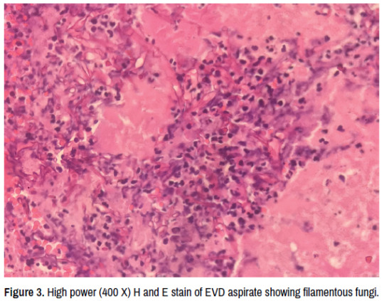
Intraparenchymal Hemorrhage Secondary to Rhino Orbitalcerebral Mucormycosis: A Case Report and Literature Review
Jasper Gerald R. Cubias* and Raymond L. Rosales
St. Luke’s Medical Center, Institute for Neurosciences, Section of Adult Neurology, Capital Towers, E. Rodriguez Ave., Quezon City, Philippines
Mucormycosis is a rare invasive opportunistic fungal infection with high morbidity and mortality, with Intraparenchymal Hemorrhage (ICH) being one of the less common complications cited in available literature. In this case report, we present a case of a 65 year old Filipino diabetic male with non-small cell lung cancer and recent history of steroid use who presented with headache. This patient developed proptosis, chemosis, and purulent discharge from the right eye, and eventually had sudden onset decrease in sensorium on the 6th day of admission. CT scan showed findings of ICH on the right frontal and patient underwent craniotomy and evacuation of hematoma. Histopathology and culture findings confirmed the diagnosis of Rhino-orbital-cerebral mucormycosis. The patient was treated with Amphotericin B initially then shifted to voriconazole based on culture and sensitivity testing. The patient was managed in-patient for 3 months and eventually survived 18 months from the diagnosis.
To date, no case reports of ICH secondary to Rhino-orbital-cerebral mucormycosis are available in our locale. Additionally, patients from all previous published case reports all succumbed to the infection. This case demonstrates that a high index of suspicion and early workup is warranted in patients presenting with headache and having risk factors for mucormycosis. Early diagnosis and prompt treatment are keys towards achieving a good outcome in these patients.
For citation:
Cubias, Jasper Gerald R. and Raymond L. Rosales. â??Intraparenchymal Hemorrhage Secondary to Rhino Orbital-cerebral Mucormycosis: A Case Report and Literature Review.â? Clin Neurol Neurosurg 5(2022): 131
New manuscript submission link: https://www.scholarscentral.org/submission/clinical-neurology-neurosurgery.html
or
E-mail: [email protected]
0 notes
Text
Music Tag
Music Tag
Rules: put your music on shuffle and write down the first 10 songs. Then tag people to do the same.
I was tagged by @cactus-spirit
1. The Beatles- Don't Bother Me
2. Family Force 5- Dance Or Die
3. The Monkees- Through The Looking Glass
4. The Monkees- The Kind Of Girl I Could Love
5. The Beatles- Strawberry Fields Forever
6. Buddy Holly- Everyday
7. Hank Green- I Know
8. Aerosmith- Remember
9. Yello- Oh Yeah
10. The Who- Substitute
A lot of old stuff popped up this time, I swear I listen to some modern music too lol.
If you see this on your dash, you’re tagged! I'm also gonna tag @theunidentifiedstoner @chemosis @full-metal-feelsies @godshideouscreation & @c-rotten-candy
#music tag#tag#tags#song tag#music shuffle#mine#shuffle#music#song#songs#classic rock#rock#folk#rock and roll#theunidentifiedstoner#chemosis#full-metal-feelsies#godshideouscreation#c-rotten-candy#the beatles#the monkees#family force 5#buddy holly#yello#aerosmith#hank green#the who
6 notes
·
View notes
Link
Conjunctival chemosis is the jelly-like swelling (edema) of the outer covering of the inner part of the eyelid, and the covering of sclera
0 notes
Text
basically i splashed insecticide in my eye and got chemosis so we walked to the er and it was scary for a bit but im fine and so is my eye !
10 notes
·
View notes
Text
Chemosis is the swelling (or edema) of the conjunctiva. It is due to the oozing of exudate from abnormally permeable capillaries. In general, chemosis is a nonspecific sign of eye irritation. The outer surface covering appears to have fluid in it. The conjunctiva becomes swollen and gelatinous in appearance.
2 notes
·
View notes
Text
I've genuinely been saying TIL way, WAY too often lately
100 days of productivity
Day 11
CVS/RS
HOCM: although beta-blockers/non-DHP Ca blockers are first line drugs to prevent or control symptoms, ACEis/ARBs MUST be started in all patients as there is evidence that they arrest the septal hypertrophy (or can even reverse it!) (angiotensin II and aldosterone are said to accelerate the hypertrophy; so are beta-1 agonists but with poorer evidence)
other than antiRAAS drugs and nitrate-hydralazine, ivabradine also has a mortality-reducing effect in CHF, specifically in patients with sustained HR > 70 bpm despite maximum beta blockade
atorvastatin fucks with digoxin, but ONLY atorvastatin fucksn't with warfarin
although verapamil is a contraindication to adenosine due to the risk of precipitating heart block, beta blockers are not
reversed split S2 in LBBB (P2 followed by A2)
Endo/Repro
haemochromatosis: pituitary dysfunction and hypogonadism are *not reversible*; hormone replacement is the mainstay of therapy
isolated FSH deficiency → ↓androgen binding proteins → oligospermia
toxic adenoma: 300-500 megabecquerels of radioiodine is 1st line; surgical excision is *second line* (radioiodine is preferentially taken up by adenomatous tissue bc it takes up iodine at a fast rate, and ↓TSH means the rest of the gland takes up much less iodine)
checkpoint inhibitors in oncology lead to persistent immune activation, but this may lead to loss of self tolerance especially against endocrine organs; this may lead to thyroiditis, adrenalitis, hypophysitis, endocrine pancreatitis etc; it is important to screen for the maintenance of the hormonal axes in patients on such drugs (eg, nivolumab, ipilimumab)
Rheum/Derm
increased BMI (without diabetes) is actually generally *protective* against osteoporosis d/t ↑oestrone (adipose oestrogen)
ANAs are mostly IgG; much less common are IgA and IgM ANAs
osteomyelitis: plain films are for screening, MRI is definitive
tennis-racquet Brisbeck granules on biopsy of a bone lesion → Langerhans cell histiocytosis
presence of pruritus is circumstantial evidence for bullous pemphigoid over pemhigus vulgaris
osteoporosis risk is high with steroid equivalent to prednisolone 7.5 mg/day over 3 months (or more); if it is anticipated that a patient might need such a long dose of steroids, start prophylaxis *immediately* with vitamin D + calcium + alendronate
Renal/Biochem/Toxo
natriuresis/hyponatraemia despite volume loss = suspect RAAS deficiency (eg, primary adrenal insufficiency)! SIADH does NOT explain profound hyponatraemia when volume depletion is evident, as aldosteronergic drive is usually sufficient to keep sodium levels as high as possible
FB GoT All Hyper about Liddle syndrome: Furosemide toxicity mimicks Bartter syndrome, Gitelman syndrome mimicks Thiazide toxicity and HyperAldosteronism mimicks Liddle syndrome
platinum compounds → hypomagnesaemia
GIT
pigment laden macrophages on colon biopsy = melanosis coli → stimulant laxative abuse
CNS/Ophthal/ENT
absent ankle jerk + positive Babinski: 3 major differentials: syrinx, motor neuron disease or subacute combined degeneration (SCD can have either exaggerated, decreased or absent reflexes)
sacral sparing = maintenance of sensation/power in central cord syndrome (eg, intramedullary metastatic tumour and syringes) and incomplete spinal cord injury due to peripheral arrangement of sacral spinothalamic fibres (test with lack of saddle anaesthesia; reduction deep touch sensation is sensitive for severity of cord injury)
orbital apex syndrome → optic neuropathy, 6th nerve palsy, proptosis, chemosis/injection, miosis, ptosis
Kearns-Sayer syndrome: MERT: Mitochondrial inheritance, External ophthalmoplegia, Retinitis pigmentosa, Teenage onset (<20 yrs)
Refsum disease: CANID: Cerebellar ataxia, Anosmia, Neuropathy, Icthyosis, Deafness
when deprescribing benzos, switch to equivalent dose of diazepam and reduce by 1/8ths every 2 weeks (typically 2 mg per 2 weeks); reinstate previous dose if patient withdraws, then after another 2 weeks try to reduce the dose again
4 notes
·
View notes
Photo
So today, a friend of mine, Erik, and I went to Chinatown to get ramen. Our favourite joint was closed because it was too early, so we went somewhere else.
It was sketchy af. And the food wasn’t that great.
So if I never blog again, it’s because I died. But at least we got ice cream after!
Also, I accidentally just gave myself something like chemosis due to rubbing my eyes so much thanks to my animal fur allergy. Yes, mom, “I am definitely not allergic to the cat.”
#Soffia rambling#Erik on my blog#My eyes hurt so much#I can't close them#I look like an alien#I can't medically say it's chemosis#But it's bulging of the eyes due to rubbing and/or allergies#God I'm not going to have to go to the hospital am I?#Holy hell it's getting worse and I'm not even touching my eyes and I washed them#GUYS I AM ACTUALLY IN SO MUCH PAIN
32 notes
·
View notes
Text

In Baltimore City, MD: Poor Carlton has been trying so hard and waited so patiently to find where he truly belongs- his forever family- almost a whole year to be exact. We are determined to help him find his perfect home.
Carlton- 5 years, altered male 80lbs
Meet Carlton, an all-around sweetheart and certified nature enthusiast. When he’s not by your side asking for pets and cuddles, you’ll find him on park ranger duty leading the way on the nearest path with his nose. Carlton really enjoyed going on a hiking trip with a volunteer; he was a good boy in the car and enjoyed looking out the window. It was his dream come true to have all the bushes and trees to sniff! He would sit very well with treats when pulling to the side to let other hikers pass.
Carlton is a very friendly dog that loves to be scratched behind his ears. He is treat motivated, and he takes them so gently. He knows “sit” and is very curious. He’d surely be a great student for training with his new family. He loves to play, and you can find him frolicking joyfully in the yard for a game of fetch. When playtime is over he is happy to snuggle up and soak up your affection.
Carlton spent some time with a short-term foster, where we learned he loves hugs (well we already knew that) and laying across your lap. He is housebroken and able to be crated. He would appreciate some space to run and play, as he loves to get out his zoomies and play fetch in a yard. Carlton has done well around children as young as 6. Unfortunately, he did recently have to be returned from foster due to being destructive in the home.
When playing off-leash with other dog's in the shelter's play yard, Carlton has been a rowdy and rough play mate! He is much better with females, and prefers not to have to engage with other males. Based on leash reactivity and prey drive that we've witnessed, Carlton would do best without cats or other small animals (including small dogs).
Upon examination, our vets noted that Carlton has mucoid discharge and mild chemosis, discharge in both eyes, and erythema and lichenification of the ventral abdomen, chest, thighs, and toes. He was given a Cytopoint injection, started on multiple meds to try to help his skin, and was also re-started on a hypoallergenic diet, which he very well might need the rest of his life. A full medical summary can be provided upon request.
Baltimore Animal Rescue & Care Shelter (BARCS)
New Address! 2490 Giles Rd, Baltimore, MD 21225
[email protected]| (410) 396-4695
Rescue pick-up hours:
Monday-Friday: 10:30 a.m.-6:30 p.m.
Saturday and Sunday: 8:30 a.m.-4:30 p.m
Adoption hours:
Monday-Friday: 2 p.m.-6 p.m.
Saturday and Sunday: 11 a.m.-4 p.m.
Baltimore Animal Rescue and Care Shelter, Inc. (BARCS) | 2490 Giles Rd, Baltimore, MD 21225
#dog rescue maryland#dog adoption maryland#dog rescue baltimore maryland#dog rescue#doglover#cute animals#pets#adopt a dog#senior dog rescue baltimore maryland#pitbull rescue#homeless animals adopt a dog foster a dog love a dog pitties pit bulls senior dogs maryland dogs#pitt bull rescue#pitties
1 note
·
View note
Text
Physical Examination of the Eye in Veterinary Medicine
A thorough ocular examination is an important diagnostic procedure in veterinary medicine. Primary and secondary ocular disease may occur. The changes seen in the eye may be related to another disease process, directing you to further diagnostics.
Before being able to perform a proper ocular examination, you must understand the anatomy of the species that you are examining. This post will largely focus on mammalian eyes.
Basic ocular anatomy includes:
The cornea - clear tissue covering the front of the eye.
The uvea (ciliary body, choroid, and iris) - vascular tissue of the eye. The iris is the coloured section surrounding the pupil.
The lens - clear disc within the eye that is used to focus light.
The retina - photoreceptors are present here. Light is converted into a signal for the brain to interpret as a visual.
The sclera - the white connective tissue surrounding the globe.
The optic nerve - carries nervous information to the brain.
Related structures include:
The third eyelid (nictitating membrane).
The eyelids and conjunctiva.
The lacrimal glands - produce the watery portion of tears.
The meibomian glands - produce the oily portion of tears.
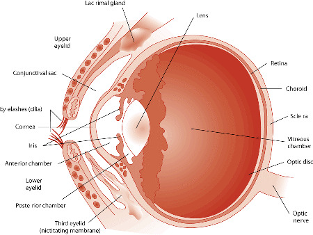
Image source: https://www.merckvetmanual.com/dog-owners/eye-disorders-of-dogs/eye-structure-and-function-in-dogs
Vision
The patient should be observed as they move around the examination room. Signs that the patient may be experiencing difficulty in seeing include reluctance to move, running into things, and high stepping.
Menace response:
This test is performed by performing a threatening hand gesture heading towards the eye. The contralateral eye should be covered when performing this test. It is important to avoid pushing air/creating a breeze toward the patient as air stimulus alone may cause the patient to blink. You must avoid contact with the whiskers. A positive menace response is achieved when the patient blinks.
The menace response is a learned response and is not present consistently in cats and dogs under the age of 3-4 months.
Tests: peripheral and central visual pathways, visual cortex, CN7.
Pupillary light reflex:
This test does NOT test vision as it is a subcortical reflex. This may be normal in a cortically blind animal.
This test is performed by shining a light into the eye and observing the response of pupil size. This should be done with a bright light in a dimly lit room. Both direct (the eye in which light is being shined) and indirect (the contralateral eye) PLRs are observed. Both direct and indirect should be present when light is shined into either eye.
Birds and reptiles do not have a PLR. They are able to control the constriction and dilation of their pupils independent of light.
Dazzle:
This test does NOT test vision as it is a subcortical reflex.
This test is performed by intensely illuminating the eye. A positive dazzle response is achieved when the patient reacts to the light. This reaction can be a blink, third eyelid protrusion, head movement, or globe retraction.
Tests: retina, CN2, CN7.
Periocular
Evaluate the orbits (bone surrounding the globe) for symmetry, deformity, or enlargement. Breed variations are important to understand during this portion of the examination. The orbital rim should be palpated to ensure no irregularities are missed.
The position and general movement of the eyes should be observed:
Strabismus - abnormal alignment of the eyes.
Nystagmus - rapid involuntary movement of the eyes.
Eyelids
Blink (palpebral) reflex:
This test is performed by touching the skin near both corners of the eye. This should produce a blink response. The finger should be gently dragged across the closed eye to ensure that the patient is capable of holding their eye closed.
Tests: CV7, orbicularis oculi muscle, CN2, CN5.
The eyelids should be observed for any abnormalities.
Abnormalities may include:
Masses or cysts.
Entropion - rolling in of the eyelid.
Ectropion - rolling out of the eyelid.
Ectopic cilia - eyelashes in abnormal locations.
Conjunctiva
The upper and lower eyelids should be manually everted. This allows for evaluation of the tissue.
Abnormalities may include:
Increased vascularity.
Lacerations or foreign bodies.
Growths.
Chemosis - edema.
Third eyelid
The third eyelid can often be extruded by placing gentle pressure on the tissues near the inner corner of the eye.
Abnormalities may include:
Prolapse of the gland - cherry eye.
Conjunctivitis.
Enlarged glands or lymphoid tissue.
Sclera
The sclera should be observed for changes in vasculature, colour, masses, or lacerations.
Cornea
The cornea should be avascular and unpigmented. It should be smooth.
The cornea should be examined for change in transparency/opacity, vascularization, pigmentation, laceration/ulceration, masses, and foreign bodies.
Fluorescein stain:
This stain is used to confirm the absence or presence of superficial ulcers. No corneal examination is complete without this test.
The dye does not stick to the outer layer of the cornea as it is lipid selective. The corneal stroma, when exposed, allows rapid diffusion of the dye. An area of fluorescein retention is indicative of an epithelial defect such as an ulcer or erosion.
Anterior Chamber
A focal light source should be shined through the anterior chamber of the eye to examine the aqueous humor. When increased protein is present, the beam of light should appear to be passing through smoke. This is called aqueous flare and is indicative of uveitis.
Lens
The lens should be transparent and avascular. The lens should be observed with an ophthalmoscope for opacities, position, and size.
Cataracts:
Focal - localized within various parts of the lens. Nuclear cataracts are usually stationary. Equatorial cataracts are usually progressive.
Immature cataract - involves more of the lens and causes blurred vision.
Mature cataract - entire lens affected, vision loss.
Lenticular sclerosis:
Begins to develop in dogs around 6 years of age. This is seen as a blue zone in the nucleus of the lens that does not impair visualization of the fundus.
Fundus
An ophthalmoscope is required for examination of the fundus. An examination can be performed without drug induced mydriasis (dilation of the iris) though it assists in complete examination.
Abnormalities may include:
Vascular patterns.
Retinal detachment.
Congestion or hemorrhage.
Changes in pigmentation.
Tear Production
Tear film is essential in maintaining normal corneal health. When the cornea is not appropriately moisturized, ulcers and other pathology can result.
The schirmer tear test is used to quantify the amount of aqueous tear portion present in each eye. This test does not evaluate the quality or make-up of the tears.
The test strip is placed in the lacrimal lake at the junction of the lateral and middle thirds of the lower eyelid. It is held in place for 60 seconds while the animal typically holds their eye closed. The test is not linear and therefore you cannot measure for 30 seconds and simply multiply by two.
Normal values are > 15mm for cats and dogs. Other species may have species-specific values published.
Intraocular Pressure
This is measured with tonometry. Applanation tonometers, such as the tonopen, are very accurate. Normal IOP is 16.8 ± 4.0 mm Hg in dogs; 20.2 ± 5.5 in cats.
Low IOP indicates uveitis while high IOP indicates glaucoma.
#veterinarian#veterinary medicine#veterinary#vet med#vet school#vet student#ophthalmology#vet ophthalmology#vetblr
56 notes
·
View notes
Last Seen Blogs