#cirripedia
Text
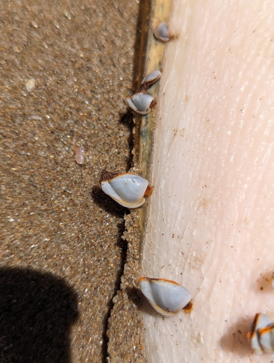
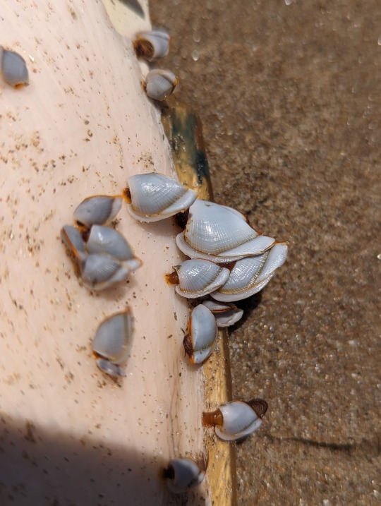
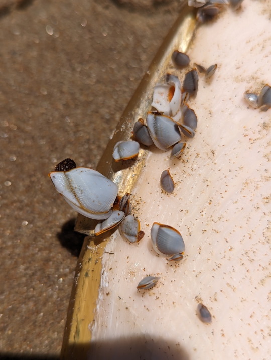
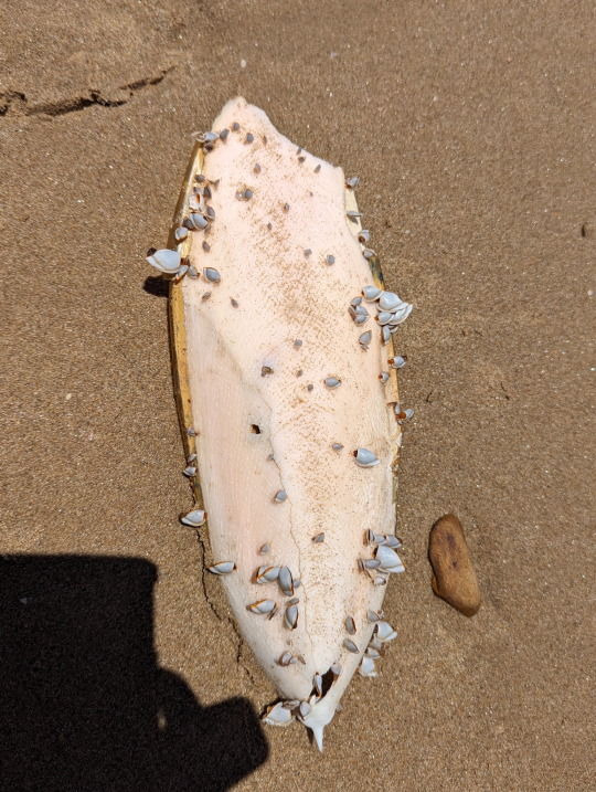
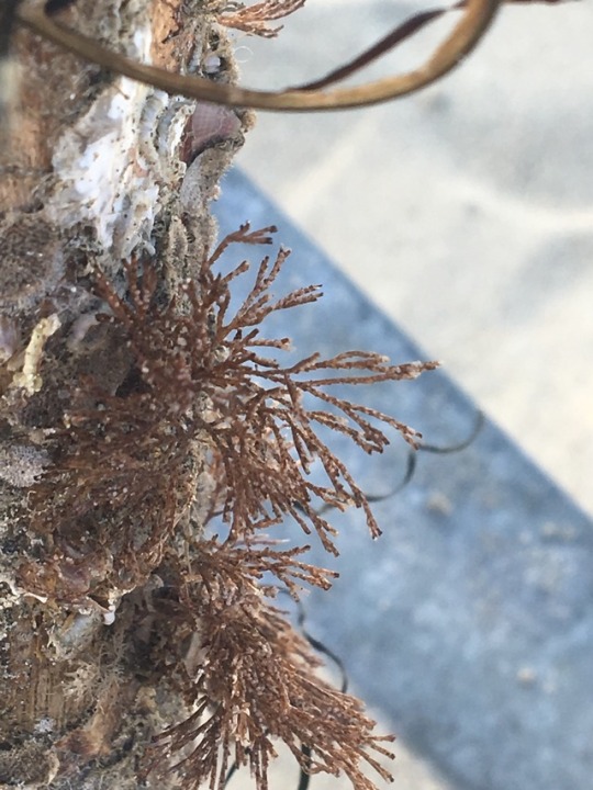
Goose Barnacles growing on a cuttlefish shell.
14/10/23 - Lepas sp.
QLD:WET - Garner's Beach
#Lepas#unidentified#invertblr#invertebrates#arthropoda#arthropods#Cirripedia#Barnacles#Crustacea#Crustaceans
13 notes
·
View notes
Text
Young girl behaved like a real whore and gets punished by her stepfather
Super Saliva Blowjob from MILF
damm white girl PAWG fucked by romemajor and don prince
Hairy Asian girl gets a pounding and a facial
Boys in love with other having gay sex and naked thai younger first
Straight men masturbating gay Public gay sex
Bdsm teens face spunky
Kesha from South Carolina riding that dick after the club!
WOW Sexy Teen Stepsister Fuck
Fun amateur fuck
#girlswholovegirls#Dartagnan#octopolar#traveltime#postfertilization#Anabaptistical#unfamed#matriliny#Cirripedia#swillbowl#jinkers#overhastiness#featness#mirled#steel-gray#reinjury#codetermine#hypophonous#belly-flopped#varec
0 notes
Text

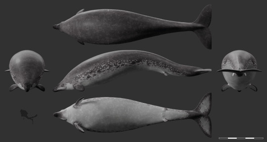
The archeocete Perucetus colossus dives through a coastal bloom of jellyfish in the Pisco Basin (southern Peru), some time during the Eocene (with bonus multiview).
I originally intended to add epibionts to this reconstruction (reflecting the specialized communities found on many living whales, especially baleen whales). Yet, interestingly, it appears that most animal epibionts and ectoparasites of modern cetaceans, such as whale barnacles (Hayashi et al. 2013) and remoras (Friedman et al. 2013), only appeared in the Neogene or late Paleogene, or have a poorly known (co-)evolutionary history, like whale lice (Pfeiffer 2009, Iwasa-Arai & Serejo 2018) and pennellids (large parasitic copepods) (Hermosilla et al. 2015). So, no epibionts* for big lad Perucetus!
References and notes about the reconstruction:
*animal epibionts. Unicellular eukaryotes like diatoms were most likely present on early cetaceans, given their prevalence on modern large marine animals (Ashworth et al. 2022). Of course, it is possible that other animals (i.e., early, less specialized representatives of modern groups, or different taxa altogether) were also already exploiting the surfaces offered by these early whales; however, this remains entirely speculative.
The reconstruction of Perucetus proposed in its original description (Bianucci et al. 2023) includes some rather odd (if interesting) choices about soft tissues, including limbs with webbed and distinguishable fingers, and a manatee-like tail. While these choices might be defendable in light of the rather basal status of Perucetus among cetaceans, I opted for a more derived look based on the assumption that fully marine cetaceans like basilosaurids would have probably rapidly acquired hydrodynamically favorable adaptations, pushing them towards a more familiar Neoceti-like appearance (even though Perucetus itself was likely a poor swimmer (Bianucci et al. 2023), it seems likely to me that this was a secondarily acquired trait, given the less extreme morphology of other basilosaurids).
Reconstruction in the multiview scaled to ~18 m in length after the estimations of Bianucci et al. (2023).
References:
Ashworth, M. P., Majewska, R., Frankovich, T. A., Sullivan, M., Bosak, S., Filek, K., Van de Vijver, B., Arendt, M., Schwenter, J., Nel, R., Robinson, N. J., Gary, M. P., Theriot, E. C., Stacy, N. I., Lam, D. W., Perrault, J. R., Manire, C. A., & Manning, S. R. (2022). Cultivating epizoic diatoms provides insights into the evolution and ecology of both epibionts and hosts. Scientific Reports, 12(1), Article 1. https://doi.org/10.1038/s41598-022-19064-0
Bianucci, G., Lambert, O., Urbina, M., Merella, M., Collareta, A., Bennion, R., Salas-Gismondi, R., Benites-Palomino, A., Post, K., de Muizon, C., Bosio, G., Di Celma, C., Malinverno, E., Pierantoni, P. P., Villa, I. M., & Amson, E. (2023). A heavyweight early whale pushes the boundaries of vertebrate morphology. Nature, 620(7975), Article 7975. https://doi.org/10.1038/s41586-023-06381-1
Friedman, M., Johanson, Z., Harrington, R. C., Near, T. J., & Graham, M. R. (2013). An early fossil remora (Echeneoidea) reveals the evolutionary assembly of the adhesion disc. Proceedings of the Royal Society B: Biological Sciences, 280(1766), 20131200. https://doi.org/10.1098/rspb.2013.1200
Hayashi, R., Chan, B. K. K., Simon-Blecher, N., Watanabe, H., Guy-Haim, T., Yonezawa, T., Levy, Y., Shuto, T., & Achituv, Y. (2013). Phylogenetic position and evolutionary history of the turtle and whale barnacles (Cirripedia: Balanomorpha: Coronuloidea). Molecular Phylogenetics and Evolution, 67(1), 9–14. https://doi.org/10.1016/j.ympev.2012.12.018
Hermosilla, C., Silva, L. M. R., Prieto, R., Kleinertz, S., Taubert, A., & Silva, M. A. (2015). Endo- and ectoparasites of large whales (Cetartiodactyla: Balaenopteridae, Physeteridae): Overcoming difficulties in obtaining appropriate samples by non- and minimally-invasive methods. International Journal for Parasitology: Parasites and Wildlife, 4(3), 414–420. https://doi.org/10.1016/j.ijppaw.2015.11.002
Pfeiffer, C. J. (2009). Whale Lice. In W. F. Perrin, B. Würsig, & J. G. M. Thewissen (Eds.), Encyclopedia of Marine Mammals (Second Edition) (pp. 1220–1223). Academic Press. https://doi.org/10.1016/B978-0-12-373553-9.00279-0
#'a heavyweight early whale pushes the boundaries of...' blablabla you've all read it by now#i have nothing to add#it's fat#look at it#that is all#perucetus#cetacean#mammal#vertebrate#eocene#cenozoic#paleontology#palaeoblr#paleoart#my art
412 notes
·
View notes
Text
Parasitic crustaceans are wild.
[cw: photos of parasites inside host bodies]
Here is Linguatula serrata, which lives inside the nose of dogs and other Carnivorans. It belongs to group Pentastomida, which has so shed its arthropod appendages that it was long classified as its own independent phylum (attested in fossils all the way from the Cambrian Explosion!), until molecular analyses showed they were in fact strange crustaceans -- closest relatives of the relatively normal-looking fish lice (Branchiura).
And then there’s Pennellidae...

Do those horn-things look like crustaceans to you? And yet the family Pennellidae is fully part of Copepoda, the chief component of crustacean plankton. Its body is simply one elongated trunk and a tiny head biting onto the fish, with two long egg cords trailing behind.
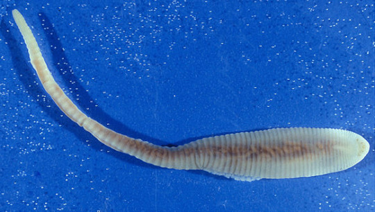
Look at the cod worm (Lernaeocera branchialis), another Pennellidae, hanging from the gills of a fish:
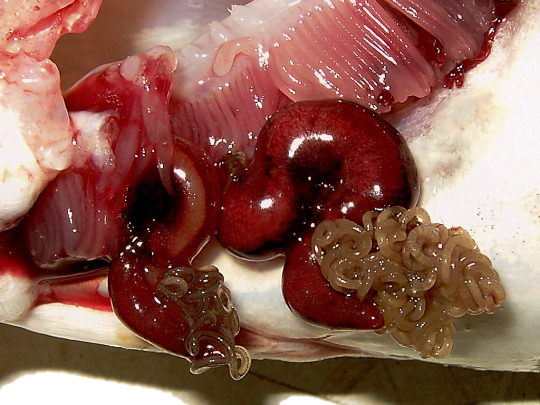
It’s those two red things that look like slugs wearing a wig made of soy noodles. Here’s what it looks like on its own, extracted and preserved:

(source) The coiled strings are egg masses. The slug-like part is the copepod’s trunk. The thin branching thing at the bottom is its head, converted into a sort of root system that no longer does head-like things, but rather burrows into the fish host’s blood vessels to feed its eggs. Incidentally, this is just the female; the male still looks like a regular planktonic crustacean.
Now, regular barnacles (Cirripedia) are strange enough...

(source; picture them as shrimps lying on their back, with a digestive system that fell out of the body wall but is still contained by the outer shell, and feathery legs poking out to filter water)
... but parasitic barnacles of clade Rhizocephala go much further:

Here, on the left, is Sacculina carcini. No, not the crab; the yellow sac poking out of the crab’s belly. On the right, its relative Clistosaccus paguri shows what it might look like once extracted.
Sacculina carcini is fun. A larva looks much like any other crustacean planktonic larva, until it finds a suitable host. It stings the unfortunate crab in a vulnerable spot between armor plates, and effectively injects itself into the host, leaving its own shell outside, and transferring only soft tissues.
Once inside, it grows more like a fungus than an animal, turning into a root-like web that infests the crab’s entire body, down to its leg tips. Then it takes over not only the crab’s digestive system, leeching nutrients for its own eggs, but also its nervous system, effectively controlling it like a puppet.
When the parasite is mature, its egg sac starts bulging out of the crab’s body: that’s the yellow part you see in the photo. Male Sacculina stay larvae their whole life: they just mate with the female’s egg sac and then die. The parasite makes the crab take care of itself as if it was the crab’s own eggs. There’s no competition, since the host is sterilized; to leave more food for the parasite, it also stops molting and regenerating lost limbs. If the host is male, and therefore poorly suited to carrying egg sacs under its tail, Sacculina messes with its hormones and effectively turns it female.
Finally the eggs are released and the whole cycle starts again, with the only purpose of making more eggs whose purpose is making more eggs.
(all pictures from Wikipedia unless specified otherwise)
173 notes
·
View notes
Photo

🌊 A monograph on the sub-class Cirripedia,. London, Ray society, 1851-54.
8 notes
·
View notes
Text
this is cirripedia I got termed for saying kys too many times on the kys website
5 notes
·
View notes
Text
Part II of my spec project Batrachiterra, this is a lesser post simply containing the seeded species list.
These are the frogs seeded, note that D. auratus represents the genus Dendrobates who also includes two other species.

Seeded species
Many species were added in order to make the planet habitable for human and frog colonists such as:
Photosynthetic organisms
Pinus spp.
Passiflora spp.
Bromelia spp.
Bryophytes
Algae
Lichens
Grasses
Fungi
Polyporales
Tuber spp.
Agaricus spp.
Suillaceae
Amanitaceae
Animalia
Isopoda
Oniscideans
Cirolanidae
Centipedes
Scolopendromorpha
Geophilomorpha
Millipedes
Oniscomorpha
Platydesmida
Polyzoniida
Siphonophorida
Diptera
Culicidae
Asilidae
Sialis spp.
Solenopsis spp.
Xylocopa spp.
Gastropoda
Eupulmonata
Systellomatophora
Styllomatophora
Oligochaeta
Cnidaria
Cubozoa
Scyphozoa
Cirripedia
Tunicata
Nematoda
Bacteria
Zooplankton
#worldbuilding#art#illustration#digital art#spec bio#frog#frogblr#spec evo#speculative biology#speculative zoology#speculative evolution#batrachiterra
42 notes
·
View notes
Text


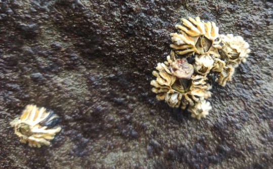

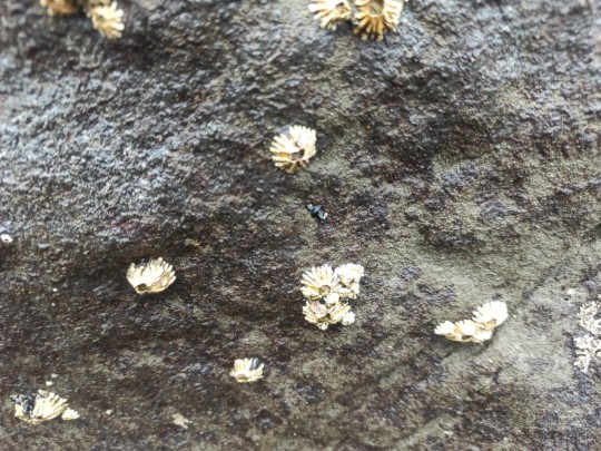
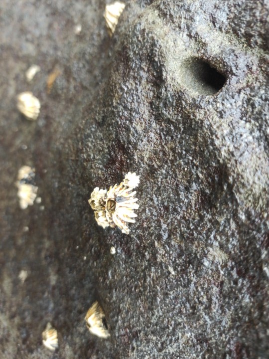
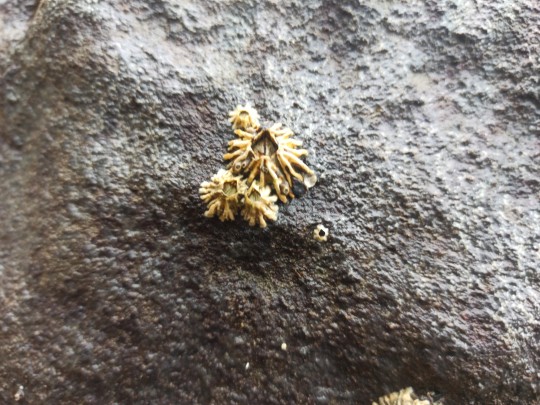
Epopella plicata is a species of symmetrical sessile barnacle in the family Tetraclitidae. It is found in New Zealand. On mid-tidal rocks.
Phylum: Arthropoda
Subphylum: Crustacea
Class: Thecostraca
Subclass: Cirripedia
Order: Balanomorpha
Family: Tetraclitidae
Genus: Epopella
Browns Bay, Auckland 0630
7QQ2+MPF Auckland
-36.7108220, 174.7518110
0 notes
Text
carapace
noun
A hard bony or chitinous outer covering, such as the fused dorsal plates of a turtle or the portion of the exoskeleton covering the head and thorax of a crustacean.
A protective, shell-like covering likened to that of a turtle or crustacean.
The shell of a turtle or tortoise; specifically, the upper shell, the under shell being called the plastron.
In Mammalia, the shell of an armadillo.In Cirripedia, the multivalvular shell, test, or case.
In higher Crustacea, the shield covering the cephalothorax, sometimes separable into a cephalostegite and an omostegite. See cut under Apus.
One of the many hard cases, tests, or shells which are likened to a carapace, as those of certain infusorians; a lorica.
The thick shell or shield which covers the back of the tortoise, or turtle, the crab, and other crustaceous animals.
A hard protective covering of bone or chitin.
0 notes
Text
Honestly, while he held certain... unfortunate beliefs, I do think someone should do art of videogame enemies in Ernst Haeckel's drawing-style.
Like, Outer Worlds got close, and Mobestiary has a similar (albeit more anatomical) feel, but I mean things like all the Headcrab variants drawn like the Cirripedia plate.
1 note
·
View note
Photo
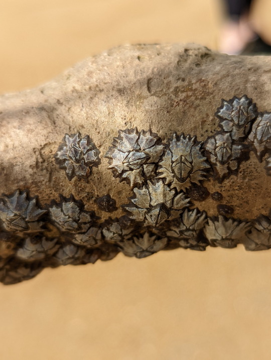


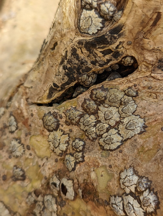
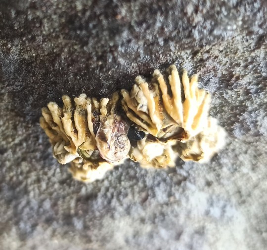
Barnacles at home on a Mangrove tree
many people don’t seem to realize that barnacles are a kind of crustacean. Well, you do now.
Why they’ve called a Mangrove tree home, is because they simply look for any sturdy surface to latch onto. Permanently. That’s why they’re often found on man-made structures.
Unidentified, Family Chthamalidae
15/06/22
#Chthamalidae#Barnacles#Chthamaloidea#Balanomorpha#Thoracicalcarea#Thoracica#Normal Barnacles#Cirripedia#Thecostraca#Hexanauplia#Multicrustacea#Crustacea#crustaceans#tidepooling#outside entomology
31 notes
·
View notes
Text
Biomed Grid | Heavy Metals in Barnacles Balanus Sp.: From Biomonitoring to Coastal Management
Introduction
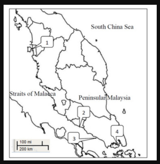
The first preliminary study on the heavy metal concentrations in the soft tissue and shells of Balanus sp. (Subphylum: Crustacea; Class: Cirripedia) from Malaysia was reported by Yap et al. [1]. As part of regular monitoring of heavy metal contamination and bioavailability’s in the coastal waters of Peninsular Malaysia, the data of this study should serve as biomonitoring data for longterm reference in the future. Barnacles have been used to assess the bioavailability of heavy metals in the coastal waters in many countries including India [2, 3], Mexico, Hong Kong [4, 5], China [6] and Poland [7]. According to [8], barnacles are among crustaceans which appear most able to fulfil the characteristics of ideal bio monitors. The objective of this study is to determine the levels of heavy metals in the soft tissues and shells of Balanus sp. collected from four sites in Peninsular Malaysia.
Materials and Methods
Samples of barnacles Balanus sp. and their habitat surface sediments were collected at the same time in 2008. Sampling locations and site descriptions are shown in (Figure 1) & (Table 1), respectively. The collected samples were placed in polyethene bags and stored in the low temperature cold iceboxes and taking back to the laboratory. In the laboratory, the samples were stored at -10°C. The measurements of length-width-height of the biological specimens were recorded by using a Vernier caliper to an accuracy of 0.01cm. The identification of the barnacles was based on the book authored by [9] and George (1979). For metal analysis, the Balanus samples were thawed at room temperature on a clean tissue paper to defrost. After cleaning, the soft tissues of the barnacles were dissected from the hard tissues. They were dried in 60°C for 72 hours in an oven until constant dry weights. Dried samples were weighed for 0.5g and triplicates were analyzed for each pooled sample. They were digested with 10ml concentrated HNO3 (Analar grade. BDH 69%) in a hot-block digester first at low temperature (40°C) for 1 hour and were completely digested at a high temperature (140°C) for 3 hours [10]. The digested samples were diluted up to 40 ml with DDW and filtered with Whatman filtered paper No. 1 into acid-washed polyethene bottles.
Figure 1: Map showing the sampling sites for barnacles in the west coast of Peninsular Malaysia (1 = Kuala Juru; 2= Sebatu; 3= Sg. Ayam; 4= Kg. Pasir Puteh).
Table 1: Global Positioning System (GPS), date of sampling and description of all sampling sites for Balanus sp.
The collected surface sediments were oven-dried and were sieved using 63μm mesh size. For the surface sediment samples, the geochemical fractions of easily, freely, Exchangeable or Leachable (EFLE), acid-reducible (AR), Oxidizable-Organic (OO) and Resistant (Res) were fractionated by sequential extraction technique as suggested by [11]. The concentrations of Cd, Cu, Fe, Ni and Zn were determined by an air-acetylene flame Atomic Absorption Spectrophotometer (AAS) Perkin-Elmer Model Analyst 800. The data were presented in μg/g dry weight. Multiple-level calibration standards were analyzed to generate calibration curves against which sample concentrations were calculated. Standard solutions for each metal were prepared from 1000mg/L per stock solution of each metal (MERCK Titrisol).
For quality control, all the glassware and equipment used were acid-washed with 10% diluted nitric acid solution for one night long. Procedural blanks were prepared for every digestion made for standardization. Quality control samples made from standard solution for Cd, Cu, Fe, Ni and Zn were analyzed after five to ten samples to check for accuracy of the samples. The recoveries percentages were acceptable at 80-110% for each of the heavy metal analyses. The blank samples were subtracted from the results to avoid the contamination possibility. The analytical procedures for the samples were checked with the Certified Reference Material (CRM) for dogfish liver (DOLT-3, National Research Council Canada) (Table 2).
Table 2: Analytical results for the certified reference material (DOLT-3 Dogfish liver) and the measured values for each metal (μg/g dry weight).
Results and Discussion
(Table 3) shows the allometric data for all populations of Balanus sp. from the four sampling sites in Peninsular Malaysia. The levels of Cd, Cu, Fe, Ni and Zn in the soft tissues and shells of Balanus sp. collected from the four sites are presented in (Table 4). (Table 5) shows the levels of the five heavy metals in the four geochemical fractions of surface sediments collected from the four sampling sites. For Cd levels in the shells: Sg. Ayam> Sebatu> Kuala Juru > Kg. Pasir Puteh. For Cd levels in the soft tissues: Sg. Ayam> Kg. Pasir Puteh> Sebatu> Kuala Juru. This highest bioavailable Cd levels to the Balanus collected from Sg. Ayam is well supported by the highest contamination level of Cd in the surface sediments represented by the geochemical fractions of EFLE and AR, and the highest percentage of non-resistant faction (59.3%) (Table 5).
Table 3: Allometric parameters for all Balanus sp. populations.
Table 4: Heavy metal concentration (mean μg/g dry weight ± SE) in the total Soft Tissues (ST) and shells of barnacles Balanus sp. collected from the intertidal areas of Peninsular Malaysia.
Table 5: Concentrations (μg/g dry weight) of heavy metals in the four geochemical fractions of surface sediments collected from the 4 sampling sites.
For Fe in the shells and soft tissues: Sg. Ayam> Sg. Sebatu> Kg. Pasir Puteh> Kuala Juru. However, these Fe bioavailability results do not agree with the Fe levels in the geochemical fractions in the surface sediments from Sg. Ayam (Table 5) due to Fe is not an anthropogenic metal and complications of other factors affecting the Fe accumulation in the Balanus and physicochemical factors being involved in the sedimentation processes. For Cu, the highest Cu level was found in the Balanus soft tissues collected from Kuala Juru, followed by Kg. Pasir Puteh, Sebatu and Sg. Ayam. The highest Cu level in the shells was also found in the Balanus from Kuala Juru. These Balanus results are well supported by the Cu levels in the surface sediments from Kuala Juru in the geochemical fractions of EFLE, AR and OO, with the highest non-resistant fraction (48.2%) (Table 5). This indicated the high bioavailable of Cu to the Balanus collected from Kuala Juru, which is a Cu contaminated site as evidenced in the surface sediment results.
Inconsistent results are found for Ni levels between soft tissues and shells of Balanus. For Ni the shells: Sg. Ayam> Kg. Pasir Puteh> Sg. Sebatu> Kuala Juru. The reverse pattern was found for the Ni in the soft tissues: Kuala Juru> Sebatu> Kg. Pasir Puteh> Sg. Ayam. Only Balanus soft tissues in the Kuala Juru is supported by the highest Ni levels in the EFLE and AR geochemical fractions ((Table 5)). For Zn levels in the shells: Kg. Pasir Puteh> Sg. Ayam> Kuala Juru> Sebatu. For Zn levels in the soft tissues: Kg. Pasir Puteh> Sebatu > Sg. Ayam> Kuala Juru. The highest Zn levels in both shells and soft tissues are supported by EFLE fraction in the surface sediments (Table 5). Although Balanus sp. is not a direct food source to the human, they are feeding source for birds and ducks which become a potential food chain to the end consumers- human. They are potential bio monitor of heavy metal pollution the surrounding environment [7, 8, 12, 13] because they provide integrated measures of the metals supply available to them in the local environment, accumulating the metal taken up from food [12]. According to [13, 14] Cu is accumulated by barnacles in Cu- and sulphur-rich deposits, probably representing end products of the lysosomal breakdown of Cu-containing metallothionein’s.
The metal concentrations (μg/g dry weight) in the soft tissues of Balanus sp. collected from four sites in Peninsular Malaysia ranged from 2.93-4.17 for Cd, 20.2-92.5 for Cu, 480-1193 for Fe, 6.40-18.0 for Ni, and 224-414 for Zn (Table 4). The present results are comparable to and lower than those in the Balanus Amphitrite for Hong Kong coastal waters reported by [5] (Cd: 0.69-9.45; Cu: 52.4-1810; Fe: 313-1470; Ni: 1.25-98.9; Zn: 2860-23300). Our results are also comparable to and within those in Balanus sp. collected from Penang’s Bridge and Semilang (Peninsular Malaysia) reported by [1, 15, 16, 17, 18, 19] (Cd: 4.72-6.66; Cu: 10.3-20.3; Fe: 633-670; Ni: 21.6-23.2; Zn: 361-434).
Conclusion
This preliminary study on heavy metal levels provides a new baseline against which future local changes can be assessed. This is highly recommended that further studies should be focused on the genetic structures and taxonomy on this potential Balanus sp. that can be established as a good bio monitor in Malaysian coastal waters in the future. Overall, this preliminary baseline data can be used for regular ecological monitoring for the effective management of the coastal area in Malaysia.
Acknowledgement
The authors wish to acknowledge the financial support provided through the Research University Grant Scheme (RUGS), [Vote no: 91229], by University Putra Malaysia and through e-Science Fund [Vote no: 5450338], by the Ministry of Science, Technology and Innovation, Malaysia.
Read More About this Article: https://biomedgrid.com/fulltext/volume7/heavy-metals-in-barnacles-balanus-sp.001128.php
For more about: Journals on Biomedical Science :Biomed Grid | Current Issue
#biomedgrid#american journal of biomedical science & research#medical and medicinal journal#list of open access medical journal
0 notes
Text


Num mundo subaquático, Rio de Janeiro.
#nature#nature photopragpy#brasil#rio de janeiro#cirripedia#crustaceans#barnacles#oceanlife#sea#atlantic ocean#traveller#Mangaratiba
3 notes
·
View notes
Photo

Red thatched barnacle (Tetraclita rubescens)
Photo by Marlin Harms
#red thatched barnacle#barnacle#tetraclita rubescens#tetraclita#tetraclitinae#tetraclitidae#tetraclitoidea#balanomorpha#sessilia#thoracica#cirripedia#thecostraca#hexanauplia#crustacea#pancrustacea#arthropoda#panarthropoda#ecdysozoa
25 notes
·
View notes
Photo
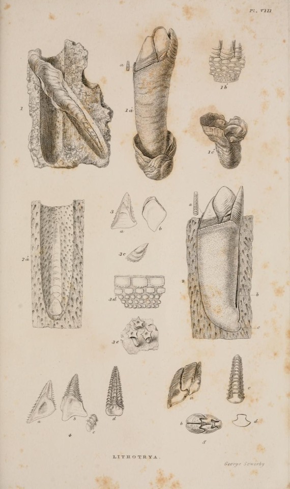
🌊 A monograph on the sub-class Cirripedia,. London, Ray society, 1851-54.
5 notes
·
View notes
Photo


2 notes
·
View notes
Last Seen Blogs

monstrenke
Esperpento En Una

mikeissavage
Mike Savage

obsessed-with-london
Joannie

amica-rattis
And I’ll love the littler things
origami-princess
Origami Tutorials!