
rev-medicine
REV MED | Anatomy Lessons
Online Medical Education Platform Simple & Effective e-Learning using diagrams, videos, notes Q&A’s & more! Sign up now to early access list! IG @rev.med
125 posts
Don't wanna be here? Send us removal request.
Last Seen Blogs


Photo

🩺 Clinical points & high yield info about the appendix - read below 👇🏼 @rev.med 🧠 Keep learning with us! Tap the link in bio & follow us @rev.med ✅ Overview The large intestine consist of several parts including the cecum. Attached to the posteromedial end of the cecum is a narrow tube referred to as the Appendix! The appendix has no vital function and it is supported by the mesoappendix, which is a fold that suspends it from the terminal ileum. @rev.med ✅ Clock Face Positions *Pre-ileal: Anterior to the terminal ileum at 1 or 2 o’clock *Post-ileal: Posterior to the terminal ileum at 1 or 2 o’clock *Sub-ileal: Parallel with the terminal ileum at 3 o’clock *Pelvic: Descending over the pelvic brim at 5 o’clock *Subcecal: Below the cecum at 6 o’clock *Paracecal: alongside the lateral border of the cecum at 10 o’clock *Retrocecal: behind the cecum at 11 o’clock (also known as the most common position of the appendix) @rev.med ✅ High Yield Facts Embryonic origin: derived from midgut (so we will see superior mesenteric vessels) *Arterial supply: appendicular artery - from ileocolic artery - from superior mesenteric artery. *Venous drainage: appendicular vein *Innervation: Ilecolic branch of the superior mesenteric plexus (travels with the ilecolic artery) *Lymphatic drainage: lymph drains from the appendix into the lymph nodes within the mesoappendix & into ilecolic lymph nodes (which surround the ilecolic artery) @rev.med @rev.med #appendix #REVMED #REVupyourbrain #revmedicine https://www.instagram.com/p/Cfb4PybOtxn/?igshid=NGJjMDIxMWI=
1 note
·
View note
Photo

Are you invited to the Knee anastomosis party? Everyones going, see the invite list below... ⤵️ @rev.med 💭 This is one of the best diagrams to use when studying the knee anastomosis. Spend time understanding and seeing the true beauty of its simplicity. Follow along for more 👉🏽 @rev.med ✅ Overview The anastomosis around the knee is a network of arterial supply that provides collateral circulation. It’s situated around the patella & around the lower end of the femur & upper end of the tibia. It is an important anastomosis because it compensates for the narrowing of the popliteal artery during prolonged flexion of the knee. In these diagrams, we look at the right knee with both an anterior and a posterior view. The first artery we have is the base of this anastomosis, known as the Popliteal artery. This artery gives off the 5 genicular branches. @rev.med ✅ Genicular Branches The lower limb is supplied by the femoral artery. This artery lies in the femoral triangle and it goes through the adductor canal. It then enters the posterior aspect of the knee after passing through the adductor hiatus of the adductor magnus muscle. The femoral artery becomes the popliteal artery and goes through a space known as the popliteal fossa, a diamond shaped space on the posterior aspect of the knee. Popliteal artery gives off the lateral superior genicular & medial superior genicular arteries, the lateral inferior genicular & medial inferior genicular arteries. It also gives off the middle genicular branch (supplying the cruciate ligaments of the knee joint). @rev.med The genicular branches enter into the anterior aspect of the knee joint and form an anastomosis in front of the patella bone. ✅ More Party People Femoral artery in the adductor canal gives off the descending genicular branch which divides into superficial and deep branches. The superficial branch goes off with the saphenous nerve and the deep branch descends down and contributes to the anastomosis. ❗️*Notes continued in the comments*❗️ @rev.med @rev.med https://www.instagram.com/p/Cetdd5lKjhT/?igshid=NGJjMDIxMWI=
3 notes
·
View notes
Photo

Mnemonics to help you learn the Femoral Triangle👇🏼 @rev.med 💭 I promise you nothing is as chaotic as it seems. Nothing is worth diminishing your health. Nothing is worth poisoning yourself into stress, anxiety, and fear. ✅ Overview This lesson walks you through the right anterior thigh showcasing the Femoral Triangle and its content. We’d like for you to always think the ‘Navy Sails’ when labeling the femoral triangle. For the borders recall the mnemonic ‘SAIL’ and for the content inside you’ll use the mnemonic ‘NAVEL’. Let’s see them here: ✅ Mnemonic The borders of the Femoral Triangle are made up the superior border which is the ‘Inguinal Ligament (IL)’, the medial border which is the ‘Adductor longus muscle (A)’, and the lateral border which is the ‘Sartorius muscle (S)’. @rev.med S A I L ⛵️ S: Sartorius muscle - medial border of the sartorius muscle. A: Adductor longus muscle - medial border of this muscle forms the border and the rest of this muscle forms part of the floor of the triangle. IL: Inguinal ligament - which is a ligament that runs from the anterior superior iliac spine to the pubic tubercle. @rev.med The content of the Femoral Triangle contains some of the major neurovascular structures of the lower limb. This mnemonic is used from Lateral to Medial in order. N A V E L ⚓️ N: Femoral nerve - it innervates the anterior compartment of the thigh, and provides sensory branches to the leg and foot. A: Femoral artery - its responsible for the majority of the arterial supply to the lower limb. V: Femoral vein and the great saphenous vein which drains into the femoral vein within the triangle. E: Empty Space - nothing special here L: Lymph vessels and deep inguinal lymph nodes are found here 🧠 Clinical stuff? We’ll discuss in another post. That femoral canal has a lot to teach us. @rev.med @rev.med https://www.instagram.com/p/CewCQtROtQJ/?igshid=NGJjMDIxMWI=
2 notes
·
View notes
Photo

These cutaneous nerves aren’t so bad! Have a swipe & read🦶🏼 @rev.med 🗣 Are you still not following @rev.med ? Well you’re missing out on some learning! ✅ Overview Cutaneous innervation refers to the area of the skin supplied by a specific nerve. These cutaneous nerves are coming from the sacral & lumbar plexuses, supplying the lower limb. @rev.med *The anteromedial side is supplied by the Saphenous nerve. *The posterolateral side by the Sural nerve. *The anterolateral part by the superficial fibular nerve (aka peroneal nerve). Superficial fibular nerves supply the cutaneous nerve distribution of dorsum aspect of the foot. The cutaneous branches of medial & lateral plantar nerves distribute the cutaneous nerves of the sole (plantar) of the foot. @rev.med ✅ Index Plantar Surface *Medial plantar nerve (L4 & L5) & Lateral plantar nerve (S1 & S2) are branches of tibial nerve. *Saphenous nerve (L3 & L4) is the terminal branch of the femoral nerve. It provides innervation for medial leg & foot. Accompanies great saphenous vein. @rev.med *Sural nerve (S1 & S2) made up of branches of the tibial nerve & common fibular nerve. Supplies sensation to the skin of lateral margins and lateral margins of little toe. *Calcaneal branches (S1 & S2) branch of tibial nerve. Dorsal Surface *Superficial fibular nerve (L4-S1) is the terminal branch of the common fibular nerve. Becomes dorsal digital nerves. @rev.med *Dorsal lateral cutaneous nerve (L2-L3) is a continuation of the sural nerve along the lateral side of the foot & little toe. *Common fibular nerve (L4-S2) a terminal branch of the sciatic nerve. @rev.med *Deep fibular nerve (L4 & L5) originates from sciatic nerve. Innervates the cleft between 1st & 2nd toes. *Saphenous nerve supplies medial margins & the head of the first metatarsal. @rev.med @rev.med #feet #foot #humanbody #anatomia #anatomie #anatomy #medico #foamed https://www.instagram.com/p/Ce1E_JFPk6u/?igshid=NGJjMDIxMWI=
1 note
·
View note
Photo
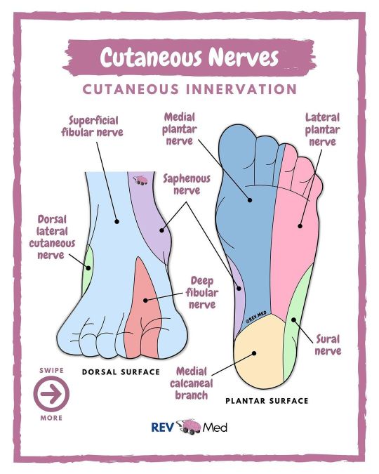
Must know these notes on the Foot. Read below ⤵️ & SWIPE to see more feet! 🦶🏼 @rev.med 🗣 You should be following @rev.med to learn quickly and efficiently. ✅ Overview Cutaneous innervation refers to the area of the skin supplied by a specific nerve. These cutaneous nerves are coming from the sacral & lumbar plexuses, supplying the lower limb. @rev.med *The anteromedial side is supplied by the Saphenous nerve. *The posterolateral side by the Sural nerve. *The anterolateral part by the superficial fibular nerve (aka peroneal nerve). Superficial fibular nerves supply the cutaneous nerve distribution of dorsum aspect of the foot. The cutaneous branches of medial & lateral plantar nerves distribute the cutaneous nerves of the sole (plantar) of the foot. @rev.med ✅ Index Plantar Surface *Medial plantar nerve (L4 & L5) & Lateral plantar nerve (S1 & S2) are branches of tibial nerve. *Saphenous nerve (L3 & L4) is the terminal branch of the femoral nerve. It provides innervation for medial leg & foot. Accompanies great saphenous vein. @rev.med *Sural nerve (S1 & S2) made up of branches of the tibial nerve & common fibular nerve. Supplies sensation to the skin of lateral margins and lateral margins of little toe. *Calcaneal branches (S1 & S2) branch of tibial nerve. Dorsal Surface *Superficial fibular nerve (L4-S1) is the terminal branch of the common fibular nerve. Becomes dorsal digital nerves. @rev.med *Dorsal lateral cutaneous nerve (L2-L3) is a continuation of the sural nerve along the lateral side of the foot & little toe. *Common fibular nerve (L4-S2) a terminal branch of the sciatic nerve. @rev.med *Deep fibular nerve (L4 & L5) originates from sciatic nerve. Innervates the cleft between 1st & 2nd toes. *Saphenous nerve supplies medial margins & the head of the first metatarsal. @rev.med @rev.med #foot #REVmed #REVupyourbrain #revmedicine #anatomy #anatomyandphysiology #physiology #physicaltherapy #feet #meded #medico #neetpg #pance #physicianassociate #nursingstudent #medstudent #studystudy #anatomydrawing #medicine #studygram #studynotes #anatomia https://www.instagram.com/p/CTfHcYFsZ2J/?utm_medium=tumblr
#foot#revmed#revupyourbrain#revmedicine#anatomy#anatomyandphysiology#physiology#physicaltherapy#feet#meded#medico#neetpg#pance#physicianassociate#nursingstudent#medstudent#studystudy#anatomydrawing#medicine#studygram#studynotes#anatomia
1 note
·
View note
Photo
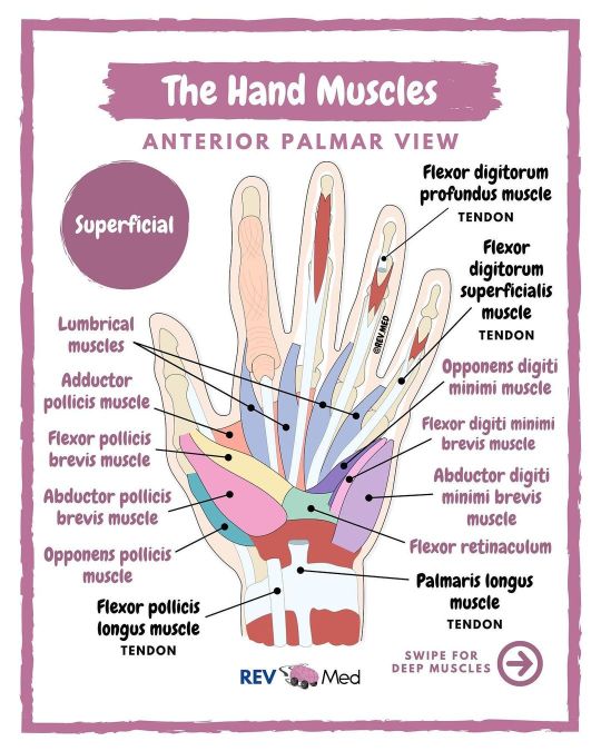
Hand Muscles brought to you by @rev.med ✋🏽 Also notes here ⤵️ 🧠 Tell us below in the comments - which class are you currently struggling with? ✅ Overview Today: Intrinsic muscles, which means they are located within the hand, and responsible for fine motor functions of the hand. See if you want to really memorize and know them.. then follow this method: 1️⃣ Thenar (thumb) muscles 2️⃣ Hypothenar (pinky) muscles 3️⃣ Other muscles: Interossei, palmaris brevis & Lumbricals @rev.med ✅ Intrinsic Muscles ✋🏾 1️⃣ Thenar (Thumb) Muscles All Innervated by Median nerve - Except Adductor Pollicis Muscle *Adductor pollicis muscle (Ulnar nerve) Insertion - Both attach into base of proximal phalanx of the thumb Action - Adductor of the thumb *Flexor pollicis brevis muscle Insertion - Base of the proximal phalanx of thumb Action - Flexes MCP joint of the thumb *Abductor pollicis brevis muscle Insertion - Lateral side of proximal phalanx of thumb Action - Abducts the thumb *Opponens pollicis muscle Insertion - Lateral margin of the first metacarpal of thumb Action - Opposes thumb, medially rotates and flexes metacarpal on the trapezium @rev.med 2️⃣ Hypothenar (Pinky) muscles All innervated by Ulnar nerve *Abductor digiti minimi brevis muscle Insertion - Base of proximal phalanx of little finger Action - Abducts little finger *Flexor digiti minimi brevis muscle Insertion - base of proximal phalanx of little finger Action - Flexes MCP joint of little finger *Opponens digiti minimi muscle Insertion - Medial margin of 5th metacarpal Action - Rotates metacarpal of little finger towards palm, producing opposition. 3️⃣ Other muscles *Lumbricals (4) - Index & middle fingers innervated by median nerve. Little and ring finger innervated by ulnar nerve. *Interossei (2) - Dorsal and palmar interossei innervated by ulnar nerve *Palmaris Brevis muscle - innervated by ulnar nerve @rev.med @rev.med #handmuscle #REVMED #REVupyourbrain #REVmedicine https://www.instagram.com/p/CTclvrwM5Tu/?utm_medium=tumblr
1 note
·
View note
Photo

🧠 Your Organoscope ‘What Organ are you?’ Choose & Comment below ⤵️ @rev.med 👉🏽Tag a friend and tell them they are a ____(organ)____ and let them see why! 😆 🫀C H O O S E 🫁 Y O U R 🧠 O R G A N This may not be your most conventional horoscope or personality test, but let’s have fun with it. Remember to save the post! ➡️ Swipe and choose your organ. Once you got it, share it on your story or share it with friends and colleagues. Get a good laugh and see who’s the same as you! @rev.med Each organ has specific traits that make it unique and purposeful. Here @rev.med, all organs matter, but the brain 🧠 matters just a little bit more 😉 Humans have five vital organs that are essential for survival. These are the brain, heart, kidneys, liver and lungs. There are also a number of other organs that work together with these vital organs to ensure that the body is functioning well. @rev.med The vital organs are those that a person needs to survive. A problem with any of these organs can quickly become life threatening. It is not possible to live without these organs. But in the case of kidneys and lungs, a person can live without one of the pair. 💭 We are keeping it light and fun today. Normal programming is back on for tommorow. Hope you are doing well and not letting anything bring you down. Just always remember this... strength grows in the moments when you think you can’t go on but you keep going anyway. All the best to you 🧠 @rev.med @rev.med #organs #REVMED #REVupyourbrain #REVmedicine #organoscope #horoscope #zoadiacsigns #personality #vitalsigns #organ #anatomy #anatomia #anatomyandphysiology #foamed #motivation #personalitytest #premed #medicine #medschool #nursingschool #medstudent #nursingstudent #studygram #medschoolmemes #nurselife (at New York City) https://www.instagram.com/p/CTXYhZ1MGi4/?utm_medium=tumblr
#organs#revmed#revupyourbrain#revmedicine#organoscope#horoscope#zoadiacsigns#personality#vitalsigns#organ#anatomy#anatomia#anatomyandphysiology#foamed#motivation#personalitytest#premed#medicine#medschool#nursingschool#medstudent#nursingstudent#studygram#medschoolmemes#nurselife
1 note
·
View note
Photo

Swipe and master the forearm muscles. Continue reading below for more info! ⤵️@rev.med 👉🏼 Be sure to follow @rev.med for the latest and Tag someone who will find these diagrams helpful! Muscles of the Forearm (Anterior Compartment) ✅ Overview 8 muscles that allow for wrist and/or finger flexion, are anterior to the interosseous membrane, and arranged in 3 layers, listed below! @rev.med ✅ Superficial Layer *Flexor carpi radialis muscle Origin: Medial epicondyle of humerus Insertion: Bases of second and third metacarpals *Pronator teres muscle Origin: Medial epicondyle and coronoid process of ulna Insertion: Middle of lateral side of radius *Flexor carpi ulnaris muscle Origin: Medial epicondyle; medial olecranon, and posterior border of ulna Insertion: Pisiform, hook of hamate, and base of fifth metacarpal @rev.med *Palmaris longus muscle Origin: Medial epicondyle of humerus Insertion: Flexor retinaculum, palmar aponeurosis ✅ Middle Layer *Flexor digitorum superficialis muscle Origin: Medial epicondyle, coronoid process, oblique line of radius Insertion: Middle phalanges of fingers @rev.med ✅ Deep Layer *Flexor pollicis longus muscle Origin: Anterior surface of radius, interosseous membrane, and coronoid process Insertion: Base of distal phalanx of thumb *Flexor digitorum profundus muscle Origin: Anteromedial surface of ulna, interosseous membrane Insertion: Bases of distal phalanges of fingers *Pronator quadratus muscle Origin: Anterior surface of distal ulna Insertion: Anterior surface of distal radius @rev.med @rev.med #forearm #REVMED #REVupyourbrain #REVmedicine #anatomy #anatomia #anatomyandphysiology #physiology #medschool #humananatomy #medicalschool #medstudent #medico #neetpg #pance #usmlestep1 #usmle #nclex #premed #nursingschool #paschool #healthcare #muscles #arm #studynotes #studygram https://www.instagram.com/p/CTNFQ5gM1DF/?utm_medium=tumblr
#forearm#revmed#revupyourbrain#revmedicine#anatomy#anatomia#anatomyandphysiology#physiology#medschool#humananatomy#medicalschool#medstudent#medico#neetpg#pance#usmlestep1#usmle#nclex#premed#nursingschool#paschool#healthcare#muscles#arm#studynotes#studygram
12 notes
·
View notes
Photo

This is your go-to diagram for the 💀foramen. We got some mnemonics to help you out below! ⤵️ @rev.med 📚 If you aren't following @rev.med then you're clearly missing out! WE got some exciting new content coming your way very soon. Comment below 'Video' or 'Diagram' so we can know what you'd like to see more of! ✅ Mnemonics and Ways to remember 3 SMALL Sized Opening (foramina of middle) *Stylomastoid Foramen *Foramen Spinosum *Foramen Ovale 3 LARGE Laceration of Artery and Vein (foramina of middle) *Foramen Lacerum *Carotid Canal *Jugular Foramen Foramen Ovale contents: “OVALE���” *Otic ganglion *V3 cranial nerve *Accessory meningeal artery *Lesser Petrosal nerve *Emissary veins @rev.med Trigeminal Nerve relations (foramina) “Standing Room Only” *Superior Orbital Fissure V1 *Foramen Rotundum V2 *Foramen Ovale V3 Superior Orbital Fissure (structures passing through) “Live Free To See No Insult At All” *Lacrimal Nerve CN V1 *Frontal Nerve CN V1 *Trochlear CN 4 *Superior division of Oculomotor nerve CN 3 *Nasociliary nerve CN V1 *Inferior division of Oculomotor nerve CN 3 *Abducens Nerve CN 6 🧠 Save this diagram and come back to it. Just needs some recall. @rev.med @rev.med #cranialfossa #REVmed #REVupyourbrain #revmedicine https://www.instagram.com/p/CTFXNReM1k3/?utm_medium=tumblr
1 note
·
View note
Photo

Here’s the segments of the Lungs! 🫁 Mnemonics & notes below ⤵️ @rev.med Tag and share this post with a classmate that would benefit from this! Don’t miss a post or video! Be sure to follow 👉🏼 @rev.med ✅ Overview The lungs 🫁 are divided into bronchopulmonary segments, each supplied with air by a tertiary bronchi. Bronchopulmonary segments have their own distinct region making each a discrete functional and anatomical unit. This property allows an individual segment to be surgically removed without affecting other segments. @rev.med ✅ Mnemonic Right Lung: A PALM Seed Makes Another Little Palm Right upper lobe *A: apical segment *P: posterior segment *A: anterior segment Middle Lobe *L: lateral segment *M: medial segment Right lower lobe *S: superior segment *M: medial segment *A: anterior segment *L: lateral segment *P: posterior segment @rev.med Left Lung: ASIA ALPS Left upper lobe *A: apicoposterior segment *S: superior lingular segment *I: inferior lingular segment *A: anterior segment Left lower lobe *A: anteromedial segment *L: lateral segment *P: posterior segment *S: superior segment 💭... and sometimes, life is just hard, and some days are just rough... and some times you just gotta cry before you can move forward... and guess what? ...All of that is okay. Be like your left lung 🫁 and make room for your heart. You got this. Trust me you do ☺️ @rev.med @rev.med #lungs #REVMED #REVupyourbrain #revmedicine https://www.instagram.com/p/CS9p22DsYmk/?utm_medium=tumblr
1 note
·
View note
Photo

Let’s go over the arteries of the Hand 🖐🏿 Read on👇🏽 @rev.med 🫀 Tag a friend or classmate so you both can study this diagram and begin conquering anatomy. @rev.med ✅ Palmar arterial supply Arteries of the hand come from the radial & ulnar arteries. Divided into palmar & dorsal components. 👉🏼 Palmar Superifical part *Superficial palmar arch: this is the superficial branch of the ulnar artery which anastomoses with a SUPERFICIAL branch of the radial artery. In front of the flexor tendons and middle of the metacarpal bones. It is superficial to long flexor tendons of the digits and deep to palmar aponeurosis. @rev.med *Palmar digital artery: branches off the superficial palmar arch and runs on the ulnar side of the little finger. *Common palmar digital arteries: there are 3 palmar digital arteries which branch off the superficial palmar arch and run in the webs between fingers. They split into two proper palmar digital arteries that supply adjacent fingers. *Proper palmar digitals: From the common palmar digitals and run along the sides of the fingers, but not the thumb. *Superficial palmar branch: Off the radial artery and superficial to the thenar muscles. 👉🏼 Palmar Deep part *Radial artery: passes into the wrist and hand along the roof of the anatomical snuffbox and then dorsally around the scaphoid and trapezium bone. Passes between 2 heads of 1st dorsal interosseous muscle and then between 2 heads of adductor pollicis to enter into deep palm & form the deep palmar arch. @rev.med *Princeps pollicis artery: Starts at the radial artery at the palm & descends past 1st metacarpal to proximal phalanx of the thumb. *Radialis indicis artery: from the radial artery & runs along the lateral side of index finger. *Deep palmar arch: Continuation of radial artery which anastomoses with the deep branch of ulnar artery. It originates between oblique & transverse heads of the adductor pollicis muscle. @rev.med @rev.med #hand #REVMED #REVupyourbrain #revmedicine https://www.instagram.com/p/CSrsEXZM3Aa/?utm_medium=tumblr
1 note
·
View note
Photo
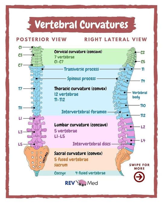
🤓 We have 4 main curvatures of our Spine! Swipe & read on ⤵️ @rev.med 🧠 Save this post! You might need it later when studying and be sure to follow along @rev.med ✅ Overview The adult vertebral column does not form a straight line, but instead has 4 curvatures along its length. These curves increase the vertebral column’s strength, flexibility, and ability to absorb shock. When the load on the spine is increased, by carrying a heavy backpack for example, the curvatures increase in depth (become more curved) to accommodate the extra weight! They then spring back when the weight is removed. @rev.med The four adult curvatures are classified as either primary or secondary curvatures. Primary curves are retained from the original fetal curvature, while secondary curvatures develop after birth. ✅ Primary curvatures Thoracic and sacral curvatures are concave anteriorly and are referred to as kyphoses, believe it or not they actually appear during the fetal development time. These two curvatures are considered the primary curvatures. @rev.med ✅ Secondary curvatures Secondary are the cervical and lumbar curvatures which concave posteriorly and convex anteriorly, referred to as lordoses. They are highlighted to support the head and the upright position of the human posture. Important about the Lumbar curvature is that it comes into play when an infant begins to present the upright posture when standing or walking. Lumbar curvature is more pronounced in women and ends at the lumbosacral angle (L5 & Sacrum level). @rev.med Sacral curvature differs in men and women in that women sacral curvature is reduced so that the coccyx protrudes less into the pelvic outlet. 🤓 We’ll speak more about the Clinical Conditions soon, such as: Osteoporosis, compression/wedge fracture, chance fracture, axial burst fracture, curvature abnormalities such as Kyphosis (hunchback) - Lordosis - Scoliosis and MORE! Let us know if you’d like to see this in the comments below! @rev.med @rev.med #spine #REVmed #REVupyourbrain #revmedicine https://www.instagram.com/p/CSo-t-wsLtI/?utm_medium=tumblr
1 note
·
View note
Photo
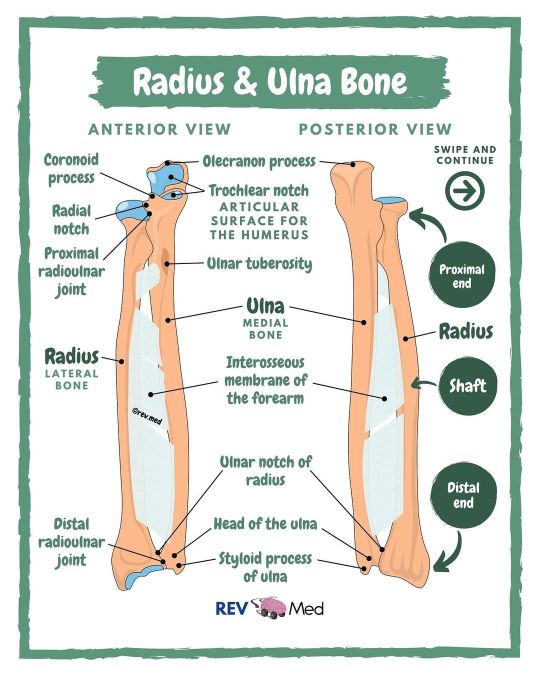
Here we have a simple diagram, swipe and spend some time to answer! 🧠 @rev.med 🤓 See the latest from us here on Instagram and try our daily quiz in our story. Don't miss out - be sure to follow @rev.med ✅ Overview *The radius and ulna are the bones of the forearm. The forearm is the region of the upper limb that extends from the elbow to the wrist. *The radius bone supports the lateral (thumb) side of the forearm and the ulna bone supports the medial (little finger, pinky) side. *At the elbow, the radius and ulna articulate with the trochlea and capitulum of the humerus bone. @rev.med ✅ Movement *The junction between the humerus and radius is a hinge joint, which permits flexion and extension movements (front to back) of the elbow. *At the wrist, the radius articulates with the proximal row of carpal bones to form an ellipsoidal joint. *The junction between the humerus and radius is a hinge joint, which permits flexion and extension movements (front to back) of the elbow. *At the wrist, the radius articulates with the proximal row of carpal bones to form an ellipsoidal joint. *This junction permits the wrist to move in two planes, flexion and extension (front to back) and abduction and adduction (side to side). *The forearm bones also articulate with each other. Proximally, the head of the radius forms a joint with the radial notch of the ulna, and distally, the head of the ulna forms a joint with the ulnar notch of the radius. *These pivot joints allow the radius to rotate around the ulna, which turns the palm of the hand (pronation and supination). @rev.med ✅ Also *An interosseous membrane spans the distance between the medial edge of the radius and the lateral edge of the ulna. *This thin sheet of connective tissue separates the forearm into anterior and posterior compartments. It also transfers tension from the radius to the ulna and serves as an attachment point for some of the muscles that move the wrist and hand. @rev.med @rev.med #Bones #REVmed #REVupyourbrain #revmedicine https://www.instagram.com/p/CSmfSCxrjK2/?utm_medium=tumblr
1 note
·
View note
Photo

🦵🏽Lower limb Cutaneous innervation @rev.med Read notes below ⤵️ 📚 Download our latest book! See the link in our bio ... and it’s free! Click! These are the specific cutaneous nerves that cover the skin of Lower Limb. Don’t confuse these with Dermatomes, even though they are similar, a dermatome specifies the area covered by the spinal nerve. @rev.med ✅ Cutaneous Nerves of Lower Limb Saphenous Nerve *From Femoral nerve in Femoral triangle (watch video). Skin on medial side of leg and foot. Deep Fibular Nerve *Anterior muscles of leg and foot, skin of first and second toes Superficial Fibular Nerve *Skin on lateral side of lower leg and dorsum of foot. Divides into Medial & Intermediate dorsal cutaneous nerve. Sural Nerve *Medial sural + Lateral sural = Sural Nerve, skin of the back of the leg, lateral side of ankle, heel, and foot. Lateral Sural Cutaneous Nerve *From common fibular nerve at popliteal fossa (watch our video). Skin on the posterolateral side of the leg. @rev.med Medial Sural Cutaneous Nerve *From Tibial nerve in popliteal fossa. Skin on back of leg and lateral side of ankle, heel and foot. Lateral Femoral Cutaneous Nerve *Skin on anterior and lateral aspects of thigh. Cluneal (Your booty) Nerves *Skin of gluteal region, contains superior, middle and inferior branches. Posterior Femoral Cutaneous Nerves *From Sacral plexus, skin of booty, thigh and calf. 👀 You know more than you think you do! Head over to our YouTube Channel & subscribe! Type in REV MED and begin learning 🧠 @rev.med @rev.med #cutaneousnerves #REVmed #REVupyourbrain #REVmedicine https://www.instagram.com/p/CSj4CHFMwVg/?utm_medium=tumblr
1 note
·
View note
Photo
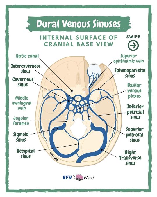
Swipe & label the diagram. Place your answers below in the comments👇🏿@rev.med 🧠 Share this post and test your friends - studying together is a great method to become better friends 😉 ✅ Overview The Cerebrum, Cerebellum and brainstem are all drained by veins which empty into the dural venous sinuses. Within the dura mater - the dural venous sinuses lie between the periosteal & meningeal layers. The scalp, face and CNS are all drained into this system of sinuses. These sinuses are unique however as they do not contain valves! @rev.med ✅ Venous Sinuses These valveless veins are 11 in total. The straight, superior, and inferior sagittal sinuses are found in the falx cerebri of the dura mater. They meet at the confluence of sinuses. The straight sinus is a continuation of the great cerebral vein of Galen and the inferior sagittal sinus. From the confluence, the transverse sinus continues bi-laterally and curves into the sigmoid sinus to meet the opening of the internal jugular vein. @rev.med The cavernous sinus drains the ophthalmic veins and can be found on either side of the sella turcica. From here, the blood returns to the internal jugular vein via the superior or inferior petrosal sinuses. 👀 Critical stuff to know! Sketch out just the sinuses on a paper and label it, repeat till you get it! Much more coming soon, stay focused, keep at it, and don’t give up no matter what! @rev.med @rev.med #venoussinuses #REVMED #REVupyourbrain #revmedicine https://www.instagram.com/p/CSe4BTLLWos/?utm_medium=tumblr
2 notes
·
View notes
Photo
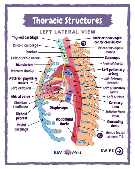
🤓 A look into to Thoracic Cavity in the lateral view - swipe & read below! ⤵️ @rev.med 👀 Take a look and study this! We also have a mental check in and acronym below ⤵️ ✅ High Yield Structures related to the vertebral levels. Remember these important structures to score some points on exam day. *T1 Apex of the lungs *T2 Sternoclavicular joint *T3 Top of the arch of the aorta *T4/T5 Bifurcation of the trachea; sternal angle *T5 Top of the heart *T6 Center of the roots of the lungs *T8 Inferior vena cava opening in the diaphragm; ventricles of the heart *T9 Inferior angle of the scapula, xiphoid process, inferior extent of the lungs anteriorly at the mid-clavicular line *T10 Esophageal opening in the diaphragm, inferior extent of lungs posteriorly *T12 Aortic opening in the diaphragm, posterior extent of parietal pleura @rev.med *L3 Attachment of the crura of the diaphragm 🧠 Mental Check-in S = Stop Stop what you are doing: Press the pause button on your thoughts and actions. T = Take Take a few deep breaths to center yourself and bring yourself fully into the present moment. O = Observe Observe what is going on with your: Body What physical sensations are you aware of (touch, sight, hearing, taste, smell)? Emotions What are you feeling right now? Mind What assumptions are your making about your feelings? What is the story you’re telling yourself about why you are having them? P = Proceed Proceed with whatever you were doing, making a conscious, intentional choice to incorporate what you just learned. Hope this helps you center your self and begin the studying process! @rev.med @rev.med #heart #REVMED #REVupyourbrain #revmedicine https://www.instagram.com/p/CSZm7rULB6N/?utm_medium=tumblr
1 note
·
View note
Photo

Extremely HIGH YIELD Neuro notes👇🏼Read on and share 🧠 @rev.med Make sure to save this post so you have it when you study! Be sure to follow us for more Neuro Anatomy @rev.med 👀 Please be sure to read these notes - we have literally made it so effectively simple! ✅ 3 Major white matter column systems Think of the tracts found in these columns as highways & dirt roads. The Dorsal Column system is a highway, and the Anterior & Lateral Column systems are like dirt roads. Its because the dorsal column is heavily myelinated and fast, and antero-lateral column is poorly myelinated. So the advanced system (the dorsal column) is faster because cars go faster on a highway. Dorsal Column (Dorsal Funiculus) *Only ascending tracts. *Advanced & Modern sensations *Highway - high velocity *Fine touch *2-point discrimination *Fast & More accuracy *Proprioception - sense of position *Better localized sensations from specific areas - highway lanes are consistent @rev.med Anterior & Lateral Column systems *Both ascending and descending tracts *Primitive & Old School sensations *Dirt Roads - low velocity *Crude touch *Pain & Temperature *Less accuracy & Slower *Sexual sensation * Poorly localized sensations - dirt road lanes vary ✅ Table to know Ascending Tracts *Dorsal Column - Vibration, proprioception, two-point discrimination *Spinocerebellar (Anterior & Posterior) - Proprioception in joints and muscles *Spinothalamic: —Lateral spinothalamic tract - Pain and temperature —Anterior (ventral) spinothalamic tract - Pressure and crude touch @rev.med Descending Tracts *Corticospinal (Anterior & Lateral) - Voluntary, discrete, skilled motor activities *Reticulospinal (Medial & Lateral) - Regulation to voluntary movements and reflexes *Tectospinal - Postural movements from visual stimuli *Vestibulospinal - Inhibition of flexor and promotion of extensor muscle activity *Rubrospinal - Promotion of flexor and inhibition of extensor muscle activity @rev.med @rev.med #spinalcord #REVMED #REVupyourbrain #revmedicine https://www.instagram.com/p/CSR4aR_LKR0/?utm_medium=tumblr
1 note
·
View note