#brown sequard syndrome
Text
He posted this on his Instagram and captioned it with "Omw to kill and then eat the mf who severed my spine ☆(ノ◕ヮ◕)ノ*o(〃^▽^〃)o" and nobody questioned it
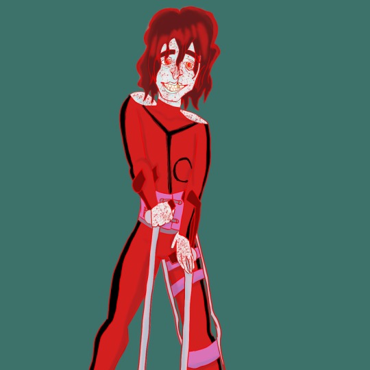
#demigod oc#angel oc#half demon#half angel#genderfluid#genderfluid oc#my oc art#my ocs#artblr#original character#teacher oc#russian oc#ustrashkin happenings#disabled oc#disabled#leg braces#crutches#brown sequard syndrome
1 note
·
View note
Note
Does Leo have Posterior Cord syndrome or Brown-Sequard syndrome? The way you've depicted him seems to indicate the former but I wanted to get your opinion 0w0
I was afraid someone would ask about that...
I don't have any medical background and spines have always squicked me out. So when I tried researching back injuries, I got overwhelmed by medical jargon and grossed out from the pictures.
I kind of just settled on Leo having a vague lumbar injury and hoped no one would ask about it (heh).
Sorry, this is a disappointing answer.
Posterior Cord syndrome actually fits really well, though. I might use that.

6 notes
·
View notes
Text
What is the most effective treatment for spinal cord injury?
People with SCI may benefit from rehabilitation
Physical therapy is geared toward muscle strengthening, communication, and mobility.
Use of assistive devices such as wheelchairs, walkers, and leg braces.
Use of adaptive devices for communication.
Occupational therapy focused on fine motor skills.
What is incomplete spinal cord injury?
Incomplete spinal cord injury is a condition in which there is some kind of neural pathways which remain functional below the affected site.
There may be the possibility of gaining neuromuscular function in the body after neurorehabilitation for several months.
Moreover, the body tries to rejuvenate the neural pathways by itself- a process known as neuroplasticity.
In a complete spinal cord injury, the sensation and motor function are completely lost below the site of injury. The neural pathways are completely damaged and there is less chance of recovery.
What are the causes of incomplete spinal cord injuries?
The causes of incomplete spinal cord injuries are below
Vehicle accident
Fall
Violence
Surgical Incidents
What are the different types of incomplete spinal cord injuries?1. Anterior Cord Syndrome
In anterior cord syndrome, the anterior spinal artery ( which runs through the anterior part of the spinal cord) is damaged and there is no muscle function below the point of injury.
However, there may be some sensation of touch below the point of injury, since the anterior part of the spinal cord is only affected. Anterior Cord Syndrome cases are difficult to treat.
2. Central Cord Syndrome
In Central Cord Syndrome, the central part of the spinal cord is affected. This may happen due to hyperextension of the neck ( abrupt forward and then backward movement of the neck).
There is paralysis of the upper limb region, but lower limbs may not be affected. Loss of bladder control can happen due to this syndrome. There is a need for long time hospital stay as a result of this syndrome.
3. Posterior Cord Syndrome
Posterior Cord Syndrome is less common than anterior and central cord syndrome. In this syndrome, the back part of the spinal cord gets injured.
There are fewer problems related to motor functions and muscle movement, but sensory function is often lost due to the injury.
4. Brown-Sequard Syndrome
In this type of syndrome, either the left side or the right side of the spinal cord is affected.
One side of the body remains functional with motor activities while the affected side may not have sensory and motor activities.
What are the treatment options for incomplete spinal cord injury?General Treatment
Medication is an important treatment option to control pain and inflammation. Surgery may be required to relieve the nerves from the pressure of the damaged vertebral column or vertebral bones.
An immobilization therapy using traction or neck braces and collars may be provided to stabilize the spinal cord.
Recent development like hypothermia or lowering the body temperature to control inflammation is also recommended
Rehabilitation Treatment
The physiotherapist will help you to improve your motor skills through various therapies including exercises.
Nowadays, advanced wheelchairs are electronically operated to climb heights or ride on rough roads. Some devices are voice-controlled and can switch on computers or electric devices easily. Electrical stimulators can help to control hand and leg movement to some extent.
Unfortunately, there is no cure for incomplete spinal cord injury in the conventional treatment regimes.
Sometimes, people respond to exercises and physical therapy but that can take a long period of time.
Stem cell therapy for incomplete
spinal cord injury treatment
Stem cells is isolated from the bone marrow cells. These cells are sorted in the laboratories and along with growth factors, they are injected into the cerebrospinal fluid.
Stem cells can form neuronal cells that can form new connections in the neurological circuit below the point of injury and can help completely recover from paralysis.
The motor skills and the sensation will start coming within 2 to 3 months.
What is the Recovery Outlook for incomplete spinal cord injury?
Recovery depends on the severity of the injury and varies from individual to individual. However, there are a few things that need to be taken care of.
Sometimes due to inflammation of the spinal cord, there is a condition called spinal shock that damages completely all the sensations and motor skills of the body. This condition should be prevented at an early stage in the emergency room.
With regular exercises, and the use of orthotics, there may be chances of recovery within 18 months, but there is no guarantee for that.
You can try stem cell therapy to cure your symptoms of spinal cord injury permanently.
We provide a List of the top 10 spine surgeons in India who are experts in spine surgeries with a history of great success. Take a look at our top surgeons with detailed information about them
0 notes
Text
Questions
Describe the pathophysiology and etiology of low back pain.
Explain the mechanisms of nerve compression and radiculopathy related to low back pain.
Compare and contrast the motor, sensory, reflex changes and the dermatomes involved.
What mode of treatment may you consider for these conditions.
Compare and contrast complete spinal cord transection, Brown-Sequard and Cauda Equina Syndromes.
View On WordPress
0 notes
Photo

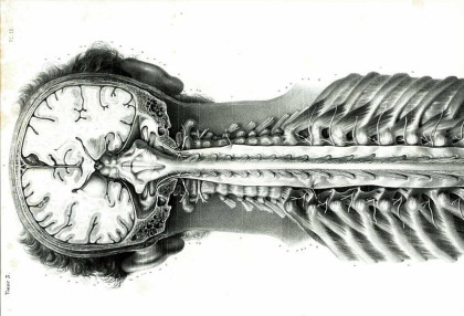
Extra medullary compression of the spinal cord. For discussion of these images, follow episode five and six of podcast ‘Clinical neurology with KD’ in Apple podcast, Spotify or Google podcast. An extramedullary compressive lesion goes through three stages, according to Oppenheim. An initial stage of radicular pain and segmental motor and sensory symptoms. The initial phase is followed by a Brown-Sequard syndrome and finally a complete transection of the cord. The rapidity of development of these stages depends on the aetiology and can be acute or chronic. Some of the cardinal features of extramedullary compression include Radicular or root pain, which is a unilateral lancinating pain down the dermatome on coughing, straining, or Valsalva. The patient will have early corticospinal tract involvement with lower limb spasticity more than the upper limb. The leg is more involved as leg fibres are laterally placed in the corticospinal tract than arm fibres. The LMN findings are rare and, if present, occur at the segmental level at the site of compression. The patient will have only late bladder involvement. They have ascending paresthesia as sacral fibres are laterally placed in the spinothalamic tract. Funicular or tract pain is less common. The patient can have vertebral pain and tenderness, which suggest extramedullary extradural lesion. #clinicalneurologywithkd #neurologyteachingclub #paediatricneurology #NTC #neurology #neurosciences #neuro #clinicalneurology #medicine #clinicalmedicine #criticalcaremedicine #mbbs #medicos #doctors #neuroanatomy #casediscussion #medicineresidents #residency #medschool #futureneurologist #neuroimages #NEET #finalmbbs #neetpg #neetsuperspeciality #neetmedicine #eanneurology #nejm #emergencymedicine #spinalcord https://www.instagram.com/p/Cds4asLpst1/?igshid=NGJjMDIxMWI=
#clinicalneurologywithkd#neurologyteachingclub#paediatricneurology#ntc#neurology#neurosciences#neuro#clinicalneurology#medicine#clinicalmedicine#criticalcaremedicine#mbbs#medicos#doctors#neuroanatomy#casediscussion#medicineresidents#residency#medschool#futureneurologist#neuroimages#neet#finalmbbs#neetpg#neetsuperspeciality#neetmedicine#eanneurology#nejm#emergencymedicine#spinalcord
0 notes
Text
I hate spinal cord lesions. Neuro in general wasn’t taught well in my school. I have to learn it on my own. 😫 Eventually I’ll review all the neuro videos in OnlineMedEd.
Anterior cord syndrome occurs when the blood supply to the anterior portion of the spinal cord is interrupted. Anterior spinal cord syndrome has been known to occur in patients undergoing thoracoabdominal aneurysm repair. The anterior spinal cord blood supply is mainly for the anterior spinal, intercostal, and lumbar arteries. This blood supply becomes compromised when ligation of intercostal and lumbar arteries occur. Anterior cord syndrome is characterized by loss of motor function below the level of injury, loss of sensations carried by the anterior columns of the spinal cord (pain and temperature), and preservation of sensations carried by the posterior columns (fine touch and proprioception). A type of incomplete spinal cord injury, anterior spinal syndrome is produced by injury of the motor and sensory paths of the anterior part of the spinal cord. Patients cannot feel coarse sensations such as pain and temperature, which are carried through the anterolateral pathways. However, proprioceptive sense and sensation of fine touch are preserved.
Brown-Séquard syndrome occurs with a hemisection injury to the spinal cord, which involves a relatively greater ipsilateral loss of proprioception and motor function, with contralateral loss of pain and temperature sensation.
Cauda equina syndrome involves injury to the lumbosacral nerve roots in the spinal canal and is characterized by an areflexic bowel and/or bladder, with variable motor and sensory loss in the lower limbs.
Central cord syndrome usually involves a cervical lesion, with greater motor weakness in the upper extremities than in the lower extremities, with sacral sensory sparing.
Posterior cord syndrome is a condition caused by lesion of the posterior portion of the spinal cord. It can be caused by an interruption to the posterior spinal artery. This condition involves loss of proprioception and vibration sensation only. Unlike anterior cord syndrome, it is a very rare condition. It is possible for it to present similar to Brown-Séquard syndrome.
#spinal cord syndromes#Brown Sequard syndrome#Brown Sequard#cauda equina#central cord syndrome#anterior cord syndrome#posterior cord syndrome
1 note
·
View note
Link
Brown-Séquard syndrome (also known as Brown-Séquard's hemiplegia, Brown-Séquard's paralysis, hemiparaplegic syndrome, hemiplegia et hemiparaplegia spinalis, or spinal hemiparaplegia) is caused by damage to one half of the spinal cord, resulting in paralysis and loss of proprioception on the same (or ipsilateral) side as the injury or lesion, and loss of pain and temperature sensation on the opposite (or contralateral) side as the lesion. It is named after physiologist Charles-Édouard Brown-Séquard, who first described the condition in 1850.[1]
#brown sequard syndrome#hemiparaplegic syndrome#spinal hemiparaplegia#spinal cord injury#sci#medical
1 note
·
View note
Text
Today we hit the three week mark in knowing your spinal cord! I’m hoping we can do a full four weeks, that would be quite the collection of knowledge. For those of you just joining in, you can find all of our posts in the neuroanatomy category ordered in reverse chronological order. As per the last few posts, we’ve covered the majority of the anatomy and now we are looking at different disorders of the spinal cord. Today we’re going to cover another type of injury, this one called anterior spinal artery syndrome, so let’s get started!
In our post yesterday we saw an image which sort of gave some spoilers to the more unique kinds of spinal cord injury. For those who haven’t read it, I suggest you do since it is very interesting, but if you’re only here for the anterior spinal artery syndrome, below is the image I’m referencing from yesterday. The middle image is what we will be covering today, tomorrow we will (probably) cover central cord syndrome.
Anterior spinal artery syndrome, labeled as anterior cord syndrome above, is somewhat similar to Brown-Sequard syndrome in that it almost a coronal version of a hemisection (Brown-Sequard). Like the name suggests, it is caused by a blood flow issue.
This type of lesion is caused by ischemia of the anterior spinal artery. Ischemia is a medical term meaning the flow of blood is restricted, this can happen anywhere on the body (one of the more common areas is the feet in fact). The anterior spinal artery is the artery that supplies blood to the anterior portion of the spinal cord. The anterior spinal artery follows the anterior median fissure and below we can see an actual cross section of cord showing the artery and because we tend to show the cord alone, we have included an image showing how blood is supplied to the cord.
Spinal cord cross section with the anterior artery showing (top)
This image shows how blood is supplied to the spinal cord
Like Brown-Sequard, anterior cord syndrome can happen at any time and is not congenital (born with it). Because it disrupts the corticospinal tract, typically when it occurs you will have complete motor loss below the lesion. This will also disrupt the spinothalamic tract, you will also lose pain and temperature sensing below the lesion as well. What makes this type of injury interesting is that the medial lemniscus tract remains intact. Therefore, you still have proprioception (as well as vibration sensing). This means that you know where your extremities are in space, even below the injury; however, you have no real control over the affected extremities.
Causes of anterior cord syndrome are just as multifaceted as the causes of Brown-Sequard. These range from things like an aortic aneurysm, direct trauma to the aorta, surgery, disc herniation, damage to the spinal cord, sickle cell, decompression sickness, or even from infections like vasculitis. Like I said, it’s a long list and those are just a few of the things that comprise it.
Because this is caused by a blood flow issue, when it happens symptoms come on fast (10-15 minutes). Unfortunately, when diagnosed there isn’t much that can be done and the prognosis is not good. You can expect that while symptoms will (most likely) not get worse, they will not get better either. In fact, over 50% of people with anterior cord syndrome see little or no change to their condition and the mortality rate is approximately 20%.
Like with each of these I would like to remind you that treatment is in its infancy and unfortunately we haven’t seen much of an improvement for spinal cord injury outcomes since the 80’s really. However, that is slowly changing and with time (and a lot of luck) we will find ways to treat spinal cord injury. So while things are somewhat bleak now, they won’t always be. Who knows, maybe now that you’ve read this you will be inspired to find the fix.
Until next time, don’t stop learning!
Day #178: Know your spinal cord - Anterior spinal artery syndrome #neuroscience #neuroanatomy -- Today we hit the three week mark in knowing your spinal cord! I'm hoping we can do a full four weeks, that would be quite the collection of knowledge.
1 note
·
View note
Text
Spinal Cord Injuries, Types, Symptoms, Causes, Stem Cell Therapy Application In Treating Spinal Cord Injuries
The Spinal Cord is an absolutely vital part of the nervous system. It carries electrical information to and from the brain which allows an individual to feel what’s going on in the hands and feet, moving arms and legs, keep breathing, and ultimately keeping alive.
However, damage to it can result in loss of sensation in both voluntary and involuntary functions of huge swathes of the body, it can be a life-changing injury.
Causes: Damage to the spinal cord can occur in lots of different ways but most often it involves trauma, road traffic accidents and falls are the most common. Sometimes it can involve a slipped disc pressing on the spinal cord or a fractured vertebra either damaging or pushing on the spinal cord itself.
Other less common causes include infection or ischemia which is when spinal cord tissue dies due to lack of blood flow. Tumors can also cause spinal cord injury. Over half of all spinal cord injuries occur in the cervical spine or in the neck.
Symptoms: Symptoms can vary depending on the location of the damage but the most common usually experienced by most of the patients are:
Loss of muscle movement (voluntary/involuntary)
Loss of sensation either proximal or distal or both
Loss of bladder or bowel control or both
Problems in inhale and exhale
Upper or lower back pain or both at the same time
Types: Some of the most common types are called incomplete injuries because not all of the spinal cord is damaged and there are still some functions left these include :
Central Cord Syndrome
Anterior Cord Syndrome
Brown Sequard Syndrome
Conus Medullaris Syndrome
Common incomplete spinal cord injuries and even complete ones occur from over flexing the neck in young people and overextending the neck in the elderly.

Treatment: Managing and treating a spinal cord injury depends on the mechanism of its location, if it’s caused by trauma then immobilization of the spine as soon as possible is vital in most cases along with either conservative or surgical correction of any additional spinal injuries.
Likewise, if it’s in a certain location the actual surgical procedures used to fix the damage might be different. Surgical correction typically involves removing any blood, fragments of the disc that are putting pressure on the spinal cord and the nerves.
The commonest of such procedure is known as “Decompression” or a “Laminectomy”, but after that sometimes screws and rods might need to scaffold and stabilize the spine.
Any compressive damage caused by the tumors may involve an operation to remove them.
Once the problem is fixed surgically or non-surgically, the next step is spinal rehabilitation which is complex and different for each individual patient and it should be clear by the team looking after the patient that the goals are set to achieve the best outcome.
Rehabilitation is multi-disciplinary and doctors often discharge the patient to a team of specialist who supplies pain relief and focused on relieving nerve pain or providing laxatives to help open the bowels if the patient has lost control of that.
The patient’s recovery would be treated with both physical and psychological components taken into account. A physical therapist will work on the mobility of the patient that involves using walking aids, low-level electrical stimulation to help muscle readjust after the injury.
The occupational therapist will work on the function of the upper limbs and how well a patient can use them to do normal activities such as cooking, eating, cleaning.
Lastly, specialist rehab nurses will help to move the patient to stop developing pressure sores which affect up to a quarter of chronic spinal cord injury patients, if the loss of bladder or bowel control is present they will also help monitor these functions so the patients don’t have accidents and infections.
The psychologist can help the patient with any emotional or behavioral concerns whilst the speech and language therapist can help the patient in swallowing and communication.
Spinal cord injury is a beast with many faces and positive outcomes cannot be guaranteed even after surgery and recovery can take as long as a minimum of 18-24 months.
Role Of Stem Cell Therapy In Spinal Cord Injury: It has been seen that even with the most advanced treatment and physical, speech, and psychological therapy, recovery of spinal cord injury is bleak and unpromising for a lot of patients with first 24 hours of survival is important but still their life expectancy is not more than 10 years.
Scientists have discovered that stem cells are not differentiated cells and can regenerate the nervous system important cells like nerve axons and myelin sheath which are vital for the functioning of the nervous system.
How Stem cells can treat spinal cord injury? Stem cells are introduced directly into the spinal cord at the site of the injury, where they help repair damaged nerves and produce more myelin in the spinal cord.
Stem cell therapy is the cells found naturally in the body and harness the unique qualities of reinsulating and repair nerves that are damaged.
With the help from stem cell therapy, dysfunctional nerves get functional abilities to conduct motor and sensory signals between the brain, hand, arm, and fingers and hopefully reverse the devastating paralysis that affects many patients with spinal cord injuries.
By utilizing the latest stem cell-based therapy scientists have significantly improved the quality of life and restore nerve functionality of spinal cord injury patients.
#stemcell#stemcells#shifarejuvenation#spinal#spinalcord#spinalcordinjury#spinalcordinjurytreatment#cervicalspine#thoracicspine#lumbarspine#stemcellpakistan#stemcelltherapy
0 notes
Text
Could never remember this, but I guessed and got it right!
True Brown-Sequard syndrome may occur after injury to one half of the spinal cord. This lateral hemisection syndrome results in injury to corticospinal tract, spinothalamic tract and the dorsal column unilaterally. Symptoms include ipsilateral weakness (corticospinal tract), ipsilateral loss of vibration and proprioception (dorsal column tract) along with contralateral loss of pain and temperature sensation (spinothalamic tract). Posterior column and spinothalamic loss occur on opposite sides of the body in this syndrome secondary to spinothalamic decussation near the level of the lesion (one or two levels below the injury). This injury may occur due to penetrating trauma (knife or bullet), disc herniation, vasculitis, infarction and demyelination. The prognosis and potential for recovery are variable depending on the injury.
2 notes
·
View notes
Note
My character suffers from (among other things) hemiparesis as a result of spinal cord damage. At the moment the cause of the damage is a gunshot wound, but how plausible is that? And if it's entirely improbable, what sort of injury might cause that type of spinal cord damage or just the hemiparesis? The injury occurred five years before the story begins and she's received plenty of medical care in that time.
The answer you’re looking for is yes, this is wholly probable, and I’m happy that I can give it!
The spinal cord lesion you’re looking for is a particular type of incomplete spinal cord injury, or iSCI, called Brown-Séquard Syndrome. It can definitely be caused by a gunshot wound.
In Brown-Sequard, you lose two things: motion and sensation on the ipsilateral side (same side) as the injury, and temperature and pain sensation on the contralateral (opposite) side. Of course, the level of the body to which this paralysis occurs depends on the level of the spine at which the injury occurred; to understand this, you need a map of dermatomes, which looks like this:

So an injury to the character’s left at T4, for example, would cause loss of motor function on the left side below the nipple, and loss of pain and temperature sensation on the right side below the same point.
Specifically, on the left side, they would have a type of paralysis called spastic paralysis, in which the muscles get unusually tight, stiff, and “pull.” Because of this they’re likely to take a muscle relaxer on a regular basis to help loosen the muscles; baclofen is a common medication.
Note that sensation on the ipsilateral side to the injury will actually be mostly preserved, though they’ll lose the ability to tell light touch from strong touch, the ability to feel vibration, and proprioception. Because of Reasons(TM) , the sensory side and motor side of the spinal cord switch sides in the ascending neurons. This is why the damage will include right-sided loss of pain sensation and left-sided motor paralysis.
There’s a shockingly good Wikipedia article on Brown-Sequard Syndrome [here].
Go forth and shoot your character right in the spine!
xoxo, Aunt Scripty
[disclaimer]
[Maim Your Characters is selling out! Get your signed copy while you can!]
[Patreon: where the inbox never closes!]
146 notes
·
View notes
Text
Day #177: Know your spinal cord - Brown-Sequard Syndrome
Day #177: Know your spinal cord - Brown-Sequard Syndrome #neuroanatomy #neuroscience --
We’ve made it to day twenty in our little series on knowing your spinal cord. As always, you can find each and every post in this series through our neuroanatomy category. Since we’ve covered all the major neuroanatomy, the latest posts have been on spinal cord diseases and disorders. That said, today we’re covering Brown-Sequard syndrome, so let’s take a look at what this is.
(more…)
View On WordPress
1 note
·
View note
Text
Caernarfon teenager defies the odds to walk again after horror motorbike crash
Cian suffered a crushed spine and broken vertebrae and has been diagnosed with Brown Sequard Syndrome (BSS), a rare spinal disorder which ...
from Google Alert - Spine News https://ift.tt/2v1tFSv
0 notes
Text
The Migration of Kirschner Wire from Left Distal Clavicle to the Intradural Anterior Thoracic Spine
Authored by Douglas Gonsales

Abstract
We report a 42 year old man who had undergone osteo synthesis to treat left clavicle fracture with a Kirschner wires (K-wire). After 1 month of surgery, began to feel pain in left clavicle region. He was informed that the symptom was expected by the surgical procedure and just analgesics were prescribed. Three months after surgery the pain in left clavicle and upper thoracic spine region continue and an x-ray shows the K-wire had migrated medially. It was attempted to remove with locally anesthetic, but unsuccessfully.
Thoracic spine CT scan showed the wire in the T2-T3 level of the spinal canal. No sensory or motor disability. There was submitted a surgical procedure with a right lateral trans-thoracic approach with elevation of the scapula to remove the wire around 105mm of length successfully. The 6 months of follow-up were neurologically asymptomatic.
Keywords: Kirschner wire migration; Clavicular fracture; Thoracic spine
Go to
Introduction
Surgical Procedure to the clavicle fracture is incomum. It just accepted to unstable fracture that can be treated surgically with plates, pins or wires [1]. Kirschner wires (K-wire) are simple tools to manage some fractures. A notable concern is the potential for these devices to migrate to distant anatomic sites. Migration of K-wires used for fixation of the clavicle fracture into the thoracic cavity or thoracic and cervical spine is a rare but serious complication, including lethal cardiovascular events [2-16]. Migration of K-wire to the another cavity (thoracic, mediastinum and spine) is refer with a neurologic symptom. There are a few cases and it has been reported since 1943 [16,17]. It was found just one case report with asymptomatic spinal migration [18]. The symptom are sensitive disability, horner’s syndrome, diplopia, headache, subarachnoid hemorrhage, sporadically arachnoiditis [19,20] and Brown-Sequard syndrome [21]. The follow-up of K-wire fixation is recommendated and whether observed signal of migration it will be necessary to be remove immediately [22,23].
Go to
Case Report
A 42 year old man has been involved in a motorcycle accident presenting with fractured of his left clavicle, and underwent of k-wire fixation. He came to the emergency department complaining of back pain some days after the procedure. After 1 month of the surgery, began to feel pain in the left clavicle region treated with just an analgesics. Three months after surgery a pain in the left clavicle and upper thoracic spine region continue and an x-ray were obtained showing an osteosynthesis device had migrated medially to the spinal. Simple removing were attempted unsuccessfully. Physical examination shows a surgical scar on the left clavicle region and pain at the palpation of the thoracic spine and with a high mobilization of the trunk. No neurological symptoms were observed.
Thoracic spine CT scan which showed a foreign body in the spinal canal at the T2-T3 level (Figure 1&2). Left lateral trans-thoracic with elevation of the scapula approach with visualization of the heart, spine and lung (Figure 3) were performed to remove the K-wire. Immediate post procedural were in the intensive care unit with a chest drain (Figure 4) with no neurological symptoms. The chest drain were removed in 48 hours. The 6 months follow-up has observed no neurological symptoms.
Go to
Discussion
Migration of the osteosynthetic device with penetration into the spinal canal is rare but it is a well-known complication since the first report in 1943 [17], and the infection is the most common complication. The spinal canal migrating site are more frequently to thoracic, mediastinum, abdome, heart, pelvis, liver [4,24-26] and occur frequently when use the no bending technique. Distant migrations of K-wires have been reported, for example, from the finger to the heart, pelvis to the abdomen, pelvis to the heart, and hip to the liver [25,27-29]. The diagnostic were necessary a good physical examination, complete neurologic examination, and the work-up with x-ray, CT. K-wire migration from the shoulder into the abdomen can compromise the spleen [12] abdominal aortic lumen [30] the neck and spine [14,16].
The most commonly migrating site from the shoulder region are the chest wall with invasion of the thorax, ending up in the pleural space [31], pulmonary parenchyma [8] mediastinum [5], esophagus [3], cavities, pericardial space, subclavian artery, ascending aortic wall or pulmonary artery and this migration can result in serious complications, including lethal cardiovascular events [9,10,32]. Once the correct diagnosis has been made the surgical remove were obligatory. There are no consensus regarding the best surgical technique.
It was decided by left lateral trans-thoracic surgical approach with elevation of the scapula, pulling the rib greatly facilitating the visualization of the entire thoracic cavity and complete access to the K-wire near the T2 vertebra. Several methods to prevent or promptly detect such migration have been devised. Patients undergoing wire osteo synthesis should receive regular plain radiographic follow-up after a complete instruction of the complications possibilities. However, threaded wires cannot completely eliminate the possibility of migration [33] even after years of being firmly in place. It is recommended that patients with K-wires are monitored and that if any sign of moving these wires should be removed mainly before symptoms [22,23].
For more open access journals in juniper publishers please click https://juniperpublishers.com/
For more articles on Open Access Journal of Neurology & Neurosurgery Please click on https://juniperpublishers.com/oajnn/
Open Access Journal of Neurology & Neurosurgery in Full text in Juniper Publishers
https://juniperpublishers.com/oajnn/OAJNN.MS.ID.555595.php
#Juniper Video Articles#juniper publishers peer review#juniper publishers Reviews#Juniper Publishers group
0 notes
Text
LS football player, CV cheerleader 'back home' after sustaining serious injuries at Friday's game
Brown-Sequard Syndrome is characterized by a lesion on the spinal cord which resuls in weakness or paralysis on one side of the body and a loss of ...
from Google Alert - Spinal https://ift.tt/2UVh24D
0 notes
Photo

Brown-Sequard syndrome (BSS) is a rare neurological condition characterized by a lesion in the spinal cord which results in weakness or paralysis (hemiparaplegia) on one side of the body and a loss of sensation (hemianesthesia) on the opposite side. #mbbsinrussia #medical #mbbslife #medicalassistant #medicalstudent #medicalmedium #medicalmarijuana #bams #bamschool #bhms #neet #mbbsdiaries #mbbsabroad #mbbsdiaries #medicine #mbbs #mbbshoes #mbbslife📚💊💉 #bamsavage #doctor #doctors https://www.instagram.com/p/Bw0vDSzFNzo/?utm_source=ig_tumblr_share&igshid=1gd0n6dlqigto
#mbbsinrussia#medical#mbbslife#medicalassistant#medicalstudent#medicalmedium#medicalmarijuana#bams#bamschool#bhms#neet#mbbsdiaries#mbbsabroad#medicine#mbbs#mbbshoes#mbbslife📚💊💉#bamsavage#doctor#doctors
0 notes
Last Seen Blogs

sakura-stories
Fanfic meadow

moo-savr
★M00

robobirdie
Robobirdie

changeangora35
کمپرسور باد ویکی کمپرسور

toky502
TOKY!