#Neurosurgical
Text
Neuro Doctor in Greater Noida
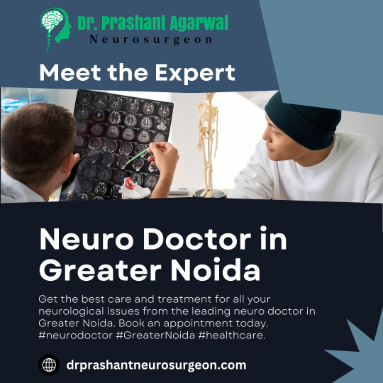
In the sprawling expanse of Greater Noida, a burgeoning city with a blend of modernity and tradition, the pursuit of specialized medical care can sometimes feel like navigating a labyrinth. When it comes to neurology, a discipline at the intersection of science and compassion, finding the right neuro doctor is paramount. With the intricacies of the nervous system at stake, it's essential to embark on this quest armed with knowledge and a clear plan of action.
Understanding Neurology and Its Importance: Neurology, a branch of medicine concerned with disorders of the nervous system, holds significant importance in the realm of healthcare. From debilitating conditions like Parkinson's disease and epilepsy to the complexities of stroke and neuropathies, neurologists play a pivotal role in diagnosis, treatment, and management. Their expertise spans a spectrum of disorders affecting the brain, spinal cord, nerves, and muscles.
Initial Research and Referral: The journey to find a competent neuro doctor in Greater Noida begins with thorough research. Seek recommendations from trusted sources such as primary care physicians, family, friends, or online platforms with credible reviews. Utilize professional networks or local medical associations for referrals to renowned specialists in the field.
Verification of Credentials and Expertise: Once a list of potential neurologists is compiled, delve into verifying their credentials and expertise. Look for board certification, affiliations with reputable medical institutions, academic achievements, and specialized training in neurology subspecialties. Assessing their experience in treating specific neurological conditions relevant to your needs is crucial.
Accessibility and Location Considerations: Consider the accessibility and location of the neuro doctor's practice. Greater Noida's vast geographical spread warrants attention to proximity, ease of transportation, and availability of parking facilities. Opt for a clinic or hospital with convenient access, minimizing travel-related stress, especially in emergencies or for follow-up appointments.
Evaluation of Communication and Patient-Centered Care: Effective communication between patient and physician forms the cornerstone of quality healthcare delivery. Evaluate the neuro doctor's approachability, attentiveness to patient concerns, and willingness to engage in shared decision-making. A compassionate and patient-centered approach fosters trust and promotes collaboration in navigating complex neurological conditions.
Assessment of Facility and Technology: Assess the facility where the neuro doctor practices, ensuring it meets standards for cleanliness, safety, and modern infrastructure. Consider the availability of advanced diagnostic equipment, such as MRI or CT scanners, and specialized neurology services like electromyography (EMG) or neurophysiological studies. A well-equipped facility enhances diagnostic accuracy and facilitates comprehensive care.
Consultation and Second Opinions: Schedule initial consultations with shortlisted neurologists to discuss your medical history, symptoms, and treatment goals. Use this opportunity to gauge the doctor's communication style, thoroughness of evaluation, and alignment with your preferences. Don't hesitate to seek second opinions if uncertain or if the diagnosis and treatment plan are complex.
Continuity of Care and Follow-Up: Establishing a long-term relationship with a neuro doctor fosters continuity of care, essential for managing chronic neurological conditions. Discuss follow-up protocols, medication management, and strategies for ongoing symptom monitoring. Clear communication channels and a collaborative care approach ensure seamless transitions between consultations and necessary interventions.
Community Feedback and Reviews: After consultations or treatments, contribute to the collective knowledge base by providing feedback and reviews about your experience with the neuro doctor. Honest appraisals assist others in making informed decisions and contribute to continuous improvement in healthcare delivery.
Conclusion: In the pursuit of neurological care in Greater Noida, navigating the landscape of specialists demands diligence, research, and discernment. By prioritizing credentials, communication, accessibility, and patient-centered care, individuals can forge partnerships with neuro doctors who embody excellence and compassion in the field of neurology. Remember, your journey toward neurological wellness begins with informed choices and a commitment to advocacy for your health and well-being.
0 notes
Text
Dr. Prashant Agarwal – The Best Neurosurgeon in Greater Noida
Introduction
In the realm of healthcare, the importance of finding a skilled and compassionate neurosurgeon cannot be overstated. When it comes to neurological concerns, the residents of Greater Noida are fortunate to have access to one of the finest practitioners in the field – Dr. Prashant Agarwal. In this blog, we will delve into the expertise and commendable achievements that make Dr. Agarwal stand out as the best neurosurgeon in Greater Noida.
Impressive Credentials and Qualifications: Dr. Prashant Agarwal is a distinguished neurosurgeon with an impressive educational background and qualifications. Holding degrees from renowned institutions, his journey in neurosurgery has been marked by a commitment to excellence and continuous learning. Patients can rest assured knowing that their neurological health is in the hands of a highly qualified professional.
Extensive Experience: Experience is often the best indicator of a healthcare professional’s proficiency, and Dr. Agarwal boasts a wealth of experience in neurosurgery. Having successfully treated a myriad of complex cases, his seasoned approach instills confidence in patients facing neurological challenges. His ability to navigate intricate procedures with precision reflects the depth of his expertise.
Cutting-edge Technology and State-of-the-Art Facilities: Dr. Prashant Agarwal’s commitment to providing top-notch care is evident in the advanced technology and state-of-the-art facilities available at his clinic. Keeping pace with the latest advancements in neurosurgical techniques, patients benefit from the most modern diagnostic and treatment modalities. This ensures accurate diagnoses and optimal outcomes for a wide range of neurological conditions.
Comprehensive Range of Neurosurgical Services: What sets Dr. Agarwal apart is his ability to address a comprehensive array of neurological issues. From brain tumors and spinal disorders to nerve-related conditions, he offers a holistic approach to neurosurgical care. Patients appreciate the convenience of finding a one-stop solution for their diverse neurological needs.
Patient-Centric Approach: Beyond technical proficiency, Dr. Prashant Agarwal is celebrated for his patient-centric approach. His empathetic and communicative style fosters a sense of trust and understanding. Neurological issues can be daunting, and Dr. Agarwal ensures that patients and their families are well-informed and supported throughout the treatment journey.
Positive Patient Testimonials: The true measure of a healthcare professional’s impact lies in the testimonials of satisfied patients. Dr. Agarwal’s track record is adorned with glowing reviews commending his skill, compassion, and the positive outcomes he has achieved. These testimonials serve as a testament to his dedication to improving the lives of those grappling with neurological challenges.
Community Involvement and Outreach: Dr. Agarwal goes beyond the confines of his clinic to contribute to the well-being of the community. His involvement in educational initiatives, awareness campaigns, and outreach programs underscores his commitment to not only treating neurological disorders but also preventing them and promoting overall neurological health in the community.
Conclusion
In the realm of neurosurgery in Greater Noida, Dr. Prashant Agarwal stands tall as the epitome of excellence. His stellar credentials, extensive experience, patient-centric approach, and commitment to staying at the forefront of technological advancements make him the best neurosurgeon in the region. For those seeking unparalleled neurosurgical care, Dr. Agarwal’s clinic is undoubtedly the destination of choice, offering a beacon of hope for individuals navigating the complex landscape of neurological health.
0 notes
Text
Best Neurosurgeon in Patna | Nirog Hospital
Discover unparalleled neurosurgical care at Nirog Hospital, Patna – your trusted destination for the best neurosurgeon in Patna. With a decade of medical excellence, our dedicated team is committed to your well-being. Experience compassionate treatment and cutting-edge interventions for neurological conditions. At Nirog Hospital, we prioritize your health above all, ensuring comprehensive care that aligns with the latest advancements in neurosurgery.
0 notes
Text

𝑨𝒏𝒆𝒔𝒕𝒉𝒆𝒔𝒊𝒂 𝑨𝒅𝒎𝒊𝒏𝒊𝒔𝒕𝒓𝒂𝒕𝒊𝒐𝒏: Anesthesiologists are responsible for administering anesthesia to patients undergoing surgery or certain medical procedures.
𝑻𝒚𝒑𝒆𝒔 𝒐𝒇 𝑨𝒏𝒆𝒔𝒕𝒉𝒆𝒔𝒊𝒂: Anesthesiologists use different types of anesthesia based on the nature and duration of the procedure.
Visit @ https://symbiosisonlinepublishing.com/anesthesiology-painmanagement/
#anesthesiology#PainManagement#painmanagementsolutions#Anesthetist#anesthesiologist#anesthesialife#epidural#thoracicsurgery#Pharmacokinetics#ultrasoundanesthesia#rheumatology#plasticsurgery#vascularsurgery#Neurosurgical#colorectalsurgery#emergencycare#chronicpain#journals#journal#pubmed#peerreview#symbiosisonlinepublishing
0 notes
Text
What is minimally invasive spine surgery
The main aim of minimally invasive spine surgery is to treat spine conditions with the least amount of incision and a faster recovery time. The minimally invasive surgery uses the latest and most specialized instruments, like endoscopes and lasers, to access the spine and perform the necessary procedures.
0 notes
Link
#neurosurgery #neurosurgeon #neurosurgical #neurology #neurologist #testimonial #brainsurgery #ApolloHospitals #surgery
0 notes
Text
Delayed onset of intracerebral tension pneumocephalus 2 years after an anterior skull base fracture: Case report by Sokchan Sim in Journal of Clinical Case Reports Medical Images and Health Sciences
ABSTRACT
Pneumocephalus, the presence of air within the cranial cavity, is most commonly caused by trauma, tumor, infection and fistulation into the intracranial cavity or secondary to neurosurgery. We describe an unusually delayed neurological deficit from intracerebral tension pneumocephalus, 2 years following a head trauma with anterior skull base fracture. A 22-year-old man presented to our neurosurgical consultation with recurrent seizures and progressive right hemiparesis. The brain CT scan without iv contrast revealed an intracerebral tension pneumocephalus in the left frontal lobe, and a persistent hole in the left anterior frontal skull base connecting to pneumocephalus. We performed a left frontal craniotomy, and dura-plasty using galea flap to cover the skull-base bone defect. The patient has recovered gradually from his motor deficit after this surgery, finally to the level that he could play his favorite guitar. This is a rare case of a delayed development neurological deficit due to pneumocephalus from a “ball-valve” effect secondary to an old anterior skull base fracture.
Key words: Pneumocephalus, hemiparesis, craniotomy, dura-plasty
INTRODUCTION
Pneumocephalus is an air entrapment in the cranial cavity. It is commonly seen after head and facial trauma, ear infections, and tumors of the skull base or neurosurgical interventions. In some extremely rare cases, it happens spontaneously. Pneumocephalus is a complication of head injury in 3.9–9.7% of the cases. The accumulation of intracranial air can be acute (<72 h) or delayed (≥72 h). In tension pneumocephalus, the continuous accumulation of intracranial air is thought to be caused by a “ball-valve” mechanism. In turn, this may lead to a mass effect on the brain, with subsequent neurological deterioration and signs of herniation. Delayed tension pneumocephalus is extremely rare and requires proper neurosurgical attention. Surgical treatment involves aspiration of air into a syringe and closure of the dura defect through a cranial surgery.
CASE REPORT
A 22-year-old male presented to our neurosurgical consultation with chronic headaches, progressive right-sided weakness and occasional seizures. Two years prior to this visit, he suffered a severe traumatic brain injury by motorcycle accident. He had lost his consciousness for three days, and hospitalized in a provincial hospital for two weeks without any surgical intervention. He was then discharged home with persistent rhinorrhea for 10 months before it ceased spontaneously. 18 months after his injury, this patient began having progressive weakness on his right side of the body, and some episodes of seizures. He also reported occasional headaches. He was otherwise healthy before this accident. On examination, the young man had full consciousness, was alert and oriented. He had grade 3 out of 5 hemiparesis on his right side. A brain CT scan without iv contrast was obtained revealing a large pneumocephalus in the left frontal lobe. We noted a continuity of the air and the anterior skull base defect. (Figure.1)
CSF examination and culture were negative for infection, as well as the nasal swab.
Figure 1: A. Axial view of the CT scan showing hypodensity area in the left frontal lobe, pneumocephalus. B. Sagittal view presenting the large air space with its connection to the frontal skull base. C. Coronal view showing the bony defect of the anterior skull base.
We decided to perform the surgery by doing bi-coronal approach for a left frontal craniotomy and repair of the dura defect on the frontal skull base using the pedunculated galea flap. (Figure.2)
Figure 2 :A. Bi-coronal incision with preservation of large frontal galea. B. Galea still attached to the frontal base is lifted up.
The surgery went well without any complication. The post-operative course was without any significant event. No sign of infection was noticed. The patient recovered gradually from his motor deficit on his right side. The post-operative CT scan showed complete resorption of the intracerebral pneumocephalus. (Figure.3). Intravenous prophylactic antibiotics were used to prevent meningitis.
Figure 3: Post-operative CT scan showing no hypodensity area in the left frontal lobe, complete disappearance of the pneumocephalus A. Axial view B. Sagittal view C. Coronal view. Noted the small bone defect from craniotomy site.
At one-month follow-up, his motor function on the right body became normal that he could play his favorite guitar again. At three-month follow up, he had an episode of new seizures, we controlled his seizures with anti-epileptic drugs for two years afterward.
DISCUSSION
The term “pneumocephalus” was first coined more than one century ago by Luckett and Wolff independently. The term “tension pneumocephalus” was proposed by Ectors, Kessler, and Stern in 1962. Pneumocephalus or also known as pneumatocele or intracranial aerocele is defined as the presence of air in the epidural, subdural, or subarachnoid space, within the brain parenchyma or ventricular cavities. It is a complication of head injury in 3.9 – 9.7% cases. It also appears after supratentorial craniotomy surgery. The accumulation of intracranial air can be acute, less than 72 hours, or delayed, more than 72 hours.
Two mechanisms have been proposed to explain pneumocephalus. In the first mechanism, the pathophysiologic process starts with Cerebro-Spinal Fluid (CSF) leak in the presence of associated discontinuity of the cranium and leptomeningeal disruption. Subsequent development of relative negative Intra-cranial Pressure (ICP) results in a sufficient “vacuum effect” to cause additional accumulation of air within the cranial cavity. This air is generally distributed in the subarachnoid space. The second mechanism is based on the presence of a “one-way valve” at the site of the leptomeningeal tear. In this case, we found on the CT scan images a bone and dura defect in the left anterior skull base, in connection with intracerebral air collection. The air went in, and was trapped inside the frontal cerebral parenchyma. Slowly it became larger and more significant, putting mass effect into the brain tissue of the patient’s frontal lobe. The patient had experienced rhinorrhea (CSF leak through the nose) after the head trauma but disappeared spontaneously after 10 months. He then developed right hemiparesis and experienced episodes of seizures. Recurrent headaches were also a main complaint. These signs and symptoms were described in previous reports about tension pneumocephalus.
The diagnostic imaging for pneumocephalus is CT scan. “Mount Fuji sign” is described when there are bilateral hypoattenuation collections, causing compression and separation of the frontal lobes on CT scan. In our case, an intraparenchymal air-filled long cavity was seen in the left frontal lobe, with its tip connecting to the frontal skull base.
Most cases of pneumocephalus tend to resolve spontaneously with conservative management. Nonoperative management involves oxygen therapy, maintaining the patient supine or in Trendelenburg position, prophylactic antimicrobial therapy (especially in posttraumatic cases), adequate analgesia, frequent neurologic checks, and repeated CT scans. The use of continuous high concentration inspired oxygen as a treatment modality for traumatic pneumocephalus may have certain theoretical benefits. Prompt decompression of intracranial air is the initial treatment of symptomatic pneumocephalus. The principles of subsequent treatment parallel those for a CSF leak. It is important to identify the site where the communication between the air cavity and the external environment occurs. If the site can be identified, the passage should be sealed off, thereby decreasing the possibility of worsening or recurrent pneumocephalus. Effective therapy of tension pneumocephalus through a controlled decompression using a closed water-seal drainage system has also been described. In our case, we performed a full scale left frontal craniotomy to evacuate air from the intraparenchymal cavity, closure of the skull base defect by using pedunculated galea flap, re-enforced by bio-glue as a sealing material.
CONCLUSION
Tension pneumocephalus is a life-threatening neurosurgical case. Although the development of this massive intracerebral air trap was delayed in this case, it caused significant neurological deficit. The patients who suffer from head trauma, with CSF leak should be subject for long term follow up.
Disclosure: Nothing to disclose, and there was no conflict of interest among the authors.
Research ethics: Informed consent has been obtained from the patient.
For more information: https://jmedcasereportsimages.org/about-us/
For more submission : https://jmedcasereportsimages.org/
#Pneumocephalus#hemiparesis#craniotomy#dura-plasty#CT#neurosurgical#headaches#Cerebro-Spinal Fluid#ICP#Intra-cranial Pressure#CSF#Sokchan Sim#jcrmhs
0 notes
Text
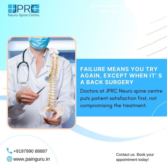
FAILURE MEANS YOU TRY AGAIN, EXCEPT WHEN IT'S A BACK SURGERY
FAILED BACK SURGERY SYNDROME IS WHEN YOU STILL HAVING PAIN AFTER A SURGERY MEANT TO TREAT IT
AND REDO SURGERY ONLY DOES WORSE
In many cases, most doctors prefer another surgery to correct the previous failed attempt but often it causes more pain and discomfort. Why go to all that length when you've easier options, that too at your hometown?
Doctors at JPRC Neuro spine centre puts patient satisfaction first, not compromising the treatment.
We can help, contact us now. Book your appointment ASAP
#pain#painguru#neuro#neuroscience#neurosurgeon#neurosurgical#painfree#painfreelife#painfreeliving#jprc#painrelief#painkiller#treatment#recover#recovery#recoverispossible#test#reports#sufferings#suffer#sufferingpain#spine#spinepain#spinepainrelief
0 notes
Text
youtube
Testimonial of Successful Neurosurgery by Vydehi Hospital | The Story of Basanti Majumdar
A traffic road accident on 26th December 2022 in West Bengal seriously injured Basanti Majumdar with an odontoid fracture (At the junction of head and neck). She developed weakness in all four limbs; as a result, she was bedridden. She was brought from West Bengal to VIMSRC 2 weeks later with a lot of hope of recovery. She was evaluated by the specialized team of Dr. Anantha Kishan from Vydehi Hospital and underwent a major neurosurgical procedure to fix the fracture, and she gradually mobilized. Just 10 days after surgery, the lady was able to walk.
Vydehi for better healing you…
#neurology#vydehihospital#vydehi hospital#healthcare#medical care#treatment#neurosurgery#neurosurgical#spinehealth#spinesurgeon#neurosurgeon#Youtube
0 notes
Text
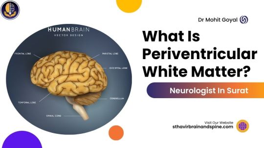
What Is Periventricular White Matter?
White-counted sickness encompasses brain white-be-counted harm due to diminished blood go with the flow, impacting memory, stability, and mobility. Those at risk for cardiovascular issues face an extended likelihood of growing this condition, emphasizing the want for expert neurologist care.
White remember contains a complex network of axons facilitating brain communication. The myelin sheath, giving it a white hue, protects nerve fibers. Gray matter in surface and deep mind areas derives color from neuron cellular bodies.
Optimal blood glide and vitamins are vital for white rely fitness. Ischemia and nutrient deficiency can damage axons, main to swelling or loss. Analogous to a garden desiring water and vitamins, insufficient blood drift and an unhealthy diet can harm the mind, highlighting the importance of neurologist steerage.
What is white matter disease?
White count number disorder, additionally referred to as cerebral microvascular modifications, refers to alterations inside the nerve fibers connecting areas of your brain. Age-associated and vascular shifts contribute to its improvement, ranging from mild to extreme.
Detectable as brilliant spots on mind MRI, those lesions may be inconspicuous with aging or disrupt vital pathways, affecting reminiscence and balance. The severity of symptoms correlates with the extent of white be-counted lesions. Strongly associated with cardiovascular hazard elements, researchers take into account it as a critical biomarker for lifelong dangers of stroke, dementia, and disability.
Whom does white matter disease impact?
White be counted sickness can impact individuals universally, but it is accepted amongst those elderly 60 and above, especially in individuals with cardiovascular issues. While more youthful people may showcase minimal white rely lesions in conditions like migraines, growing older and unmanaged cardiovascular danger factors elevate the likelihood of more lesions. Additionally, genetic factors can contribute to the predisposition for white count number sickness.
What are the indications of white matter disease?
Cognitive challenges, including memory issues.
Reduced walking speed.
Problems with balance lead to frequent falls.
Difficulty multitasking, such as walking and talking simultaneously.
Mood alterations, such as depressive symptoms.
Urinary incontinence.
These signs may additionally accentuate in individuals with superior white matter disorder. Although some symptoms align with normal growing old or other clinical conditions (e.g., arthritis, diabetes-associated neuropathy, Alzheimer’s dementia, and sleep troubles), sudden onset and fast development warrant attention.
White remember disease may be by the way discovered at some stage in a mind MRI for unrelated motives, and in some cases, it can be asymptomatic. Consult your healthcare company to explore whether your signs and symptoms can be attributed to white count number disease or different underlying reasons.
What leads to the development of white matter disease?
Researchers continue to resolve the reasons for the white count number ailment, linking it to chronic discounts in the blood that goes with the flow to nerve fibers, resulting in damage. Aging-triggered arterial stiffening and decreased elasticity contribute to diminished blood flow, harming white matter.
Atherosclerosis, characterized by artery wall thickening and plaque buildup, is any other component, affecting arteries throughout the body, inclusive of the mind. Cardiovascular hazard elements like high blood strain, diabetes-related accelerated blood sugar, increased nutritional fat consumption (high cholesterol), and smoking extend the prevalence of white depend spots or lesions within the brain.
Additional contributors to the development of white matter lesions encompass:
Genetic predispositions.
Autoimmune disorders.
Chronic inflammation.
Infectious diseases affecting the central nervous system.
Traumatic brain injury.
Exposure to toxins or environmental pollutants.
Certain medications and treatments.
Metabolic disorders.
Radiation therapy.
Neurodegenerative conditions.
Any alteration in the chemical composition, damage, or decreased blood glide impacting myelinated fibers can appear as white remembered lesions on MRI. While vascular issues make contributions to white matter sickness, non-vascular reasons, like demyelinating disorders (e.g., multiple sclerosis) and genetic conditions consisting of leukodystrophy, can result in comparable lesions.
Consultation with neurologists and neuro-radiologists, mainly the high-quality in Surat, aids in distinguishing the beginning of white be-counted lesions through MRI. In some instances, extra exams may be essential for a comprehensive assessment.
0 notes
Text
Brain Neurosurgeons in Greater Noida
Greater Noida, a city synonymous with rapid development and technological advancements, has become a hub for various specialized healthcare services. Among these, the field of neurosurgery stands out as an essential component, catering to individuals dealing with complex neurological conditions. In this article, we delve into the world of brain neurosurgeons in Greater Noida, exploring their expertise, advancements in the field, and the impact they have on the lives of their patients.
The Expertise of Brain Neurosurgeons in Greater Noida:
Brain neurosurgeons in Greater Noida are renowned for their extensive expertise and advanced training in the intricate field of neurosurgery. These specialists undergo rigorous education and training to acquire the skills necessary for dealing with a wide array of neurological disorders, ranging from brain tumors and spinal abnormalities to traumatic injuries and degenerative diseases.
The neurosurgical teams in Greater Noida are often equipped with state-of-the-art technology and cutting-edge surgical techniques, ensuring that patients receive the highest standard of care. Many neurosurgeons in the region have gained national and international recognition for their contributions to research, innovation, and patient outcomes.
0 notes
Video
Neurosurgery is the surgical treatment of the nervous system. This medical specialty focuses on the diagnosis and care of patients who have diseases or injuries to the brain, spinal cord, spinal column, or peripheral nerves in any part of the body.
0 notes
Text
All major neurosurgery/ microscopic surgeries
Treatment of all diseases related to the Brain.
Call Now: 7070991111, 7070991116, 7070991129....
Alam Hospital & Research Centre Ranchi
.
.
.
.
.
.
.
.
.
.
.
Alam hospital& Research Centre Pvt. Ltd.
Booty Road, Bariatu Ranchi JharKhand - 834009
Call no: 7070991111, 7070991116, 7070991129
#neurosurgery#neurosurgeon#neurosurgical#neurosurgicaltv#neurosurgeontobe#neurosurgerynurse#neurosurgeondoctor#neurosurgeonsuniteplease#NeurosurgeryAwarenessMonth#neurosurg#neuroexpert#neuroexperts#neuroexpertos#neuroexpertise#neuroexpertindia#neuroexpertsareas#neuroexpertsinchennai#neuroexpertinrajasthan#Ranchi#ranchi#ranchin#ranching#RanchiNews#ranchigirl#ranchiupdates#ranchiblogger#ranchidiaries#ranchi_the_heart_of_jharkhand#alamhospital
1 note
·
View note
Photo
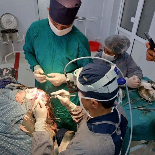
University staff performed a high-tech neurosurgical operation in the Dzhambay region Representatives of the departments of neurosurgery and resuscitation and anesthesiology, as well as leading employees of the clinic No. 1 of the Samarkand State Medical #University in the Jambay district medical association of the region, performed a complex high-tech neurosurgical operation. #Surgical treatment was recommended for the patient's diagnosis of intracerebral tumor in the area of the left frontal lobe of the brain, confirmed in the district medical association. Taking into account the place of residence and social status of the patient, at the suggestion of the administration of the Dzhambay RMO, on July 22, a high-tech complex operation was successfully performed by representatives of the Samara State Medical University. The operation was attended by residents of the departments of traumatology and #orthopedics, resuscitation and surgery of the Dzhambay RMO, as well as medical students passing through the district medical association, who received practical knowledge about a complex #neurosurgical operation. As a result of the surgical intervention, a complete absence of negative consequences of a neurological nature in a patient with a complex diagnosis was achieved. #samarkhand #mbbs (at Samarkand State Medical Institute) https://www.instagram.com/p/Ch_yYKlpzJR/?igshid=NGJjMDIxMWI=
0 notes
Text
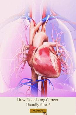
"Cancer can only control a body but our will and our spirit are forever our own".
Let's fight… "Cancer may have started the fight but we will finish it".
Check here to learn more about lung cancer:
How Does Lung Cancer Usually Start?
#cancer#cancersurvivor#lungcancer#pain#chemotherapy#health#acutepain#chronicpain#breakthroughpain#PainAndCancer#Cancerreduction#radiationtherapy#CancerMedications#Neurosurgical#CancerTreatment#PainTreatment#science
0 notes
Last Seen Blogs

birdeeandthebeat
Birdee And The Beat
spoiledberry
hiatus

xo-so-3-mien-xs3mien
KQ XS 3 miền - Trực tiếp kết quả xổ số 3 miền

nassfau
EH I EH

sweetstwawbewwymilk
My avatar points at gay people