
Last Seen Blogs

nenebewet
Untitled

abi-kat
Life.as.I.see.it.

darlingriver
Darling River

radpeacharbiter
Appealing Readers

ilmiotempo
Party Hard
Photo

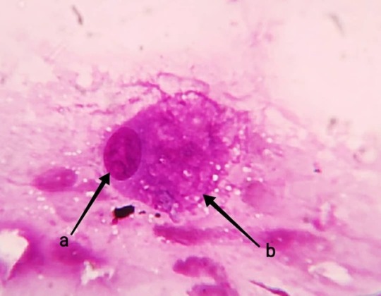
Canine #hepatocytes Location: #liver Characteristics and functions: #epithelialcells forming the #parenchyma of liver Shape and size: Large #polygonal cells with N:C ratio Nucleus: Round, centrally placed, compact and fine reticular chromatin(a), single #nucleolus (b), #binucleation commonly observed Cytoplasm: Eosinophilic and granular(c), some #lipofuscin pigments(d) or sometimes dark-brown black #bile Cytoarchitecture: Small clusters with pavement(e) or large clusters with trabecular morphology https://www.instagram.com/p/Bxwd3g3hyWP/?igshid=y6c5fmfnn52q
0 notes
Photo

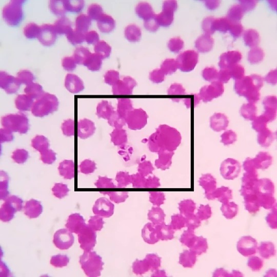
#merozoite 's of #babesiacanis in a #caninebloodsmear: Babesia canis are #intercellular #haemoprotozoa belonging to the family of #piroplasmida e. The babesial organisms are transmitted by the dog #browntick #rhipicephalussanguineus , which are present as #sporozoites in the salivary glands. During their blood meal the sporozoites are passed on to the vertebrate host and attaches itself on the erythrocytic membranes. The parasites enter the RBCs and multiply by asexual reproduction to form merozoites which are seen as leaf like structures. While feeding again the organisms are transferred to the ticks and the organisms enter the gut mucosa and undergo gametogony to form male and female forms which reproduce by sexual and by asexual reproduction to form more sporozoites. And the cycle continues!! Most common babesial organisms seen in canine are Babesia canis and Babesia Gibsoni. Babesia Gibsoni are more smaller, seen as small ring/ circular structures and lack a pyriform shape. Babesia canis on the other hand have large pyriform merozoites and pointed at one end. #vetpathology #vetpath #vetparasitology #veterinaryparasitology #veterinaryparapathologist #bloodparasite #vetcasestudy #vetcases #vetstudies #vetmed #vetmedicine #vetlife #vetknowledge #vetclinicalpathology #vetcliniclife #vetdiagnosis #vetlabdiagnostic https://www.instagram.com/drdashvetpath/p/Bw82BXIh07-/?utm_source=ig_tumblr_share&igshid=15ay9kvz7f12y
#merozoite#babesiacanis#caninebloodsmear#intercellular#haemoprotozoa#piroplasmida#browntick#rhipicephalussanguineus#sporozoites#vetpathology#vetpath#vetparasitology#veterinaryparasitology#veterinaryparapathologist#bloodparasite#vetcasestudy#vetcases#vetstudies#vetmed#vetmedicine#vetlife#vetknowledge#vetclinicalpathology#vetcliniclife#vetdiagnosis#vetlabdiagnostic
0 notes
Photo
#merozoite 's of #babesiacanis in a #caninebloodsmear: Babesia canis are #intercellular #haemoprotozoa belonging to the family of #piroplasmida e. The babesial organisms are transmitted by the dog #browntick #rhipicephalussanguineus , which are present as #sporozoites in the salivary glands. During their blood meal the sporozoites are passed on to the vertebrate host and attaches itself on the erythrocytic membranes. The parasites enter the RBCs and multiply by asexual reproduction to form merozoites which are seen as leaf like structures. While feeding again the organisms are transferred to the ticks and the organisms enter the gut mucosa and undergo gametogony to form male and female forms which reproduce by sexual and by asexual reproduction to form more sporozoites. And the cycle continues!! Most common babesial organisms seen in canine are Babesia canis and Babesia Gibsoni. Babesia Gibsoni are more smaller, seen as small ring/ circular structures and lack a pyriform shape. Babesia canis on the other hand have large pyriform merozoites and pointed at one end. #vetpathology #vetpath #vetparasitology #veterinaryparasitology #veterinaryparapathologist #bloodparasite #vetcasestudy #vetcases #vetstudies #vetmed #vetmedicine #vetlife #vetknowledge #vetclinicalpathology #vetcliniclife #vetdiagnosis #vetlabdiagnostic https://www.instagram.com/drdashvetpath/p/Bw82BXIh07-/?utm_source=ig_tumblr_share&igshid=1l7cvdwrcv8o9
#merozoite#babesiacanis#caninebloodsmear#intercellular#haemoprotozoa#piroplasmida#browntick#rhipicephalussanguineus#sporozoites#vetpathology#vetpath#vetparasitology#veterinaryparasitology#veterinaryparapathologist#bloodparasite#vetcasestudy#vetcases#vetstudies#vetmed#vetmedicine#vetlife#vetknowledge#vetclinicalpathology#vetcliniclife#vetdiagnosis#vetlabdiagnostic
0 notes
Photo
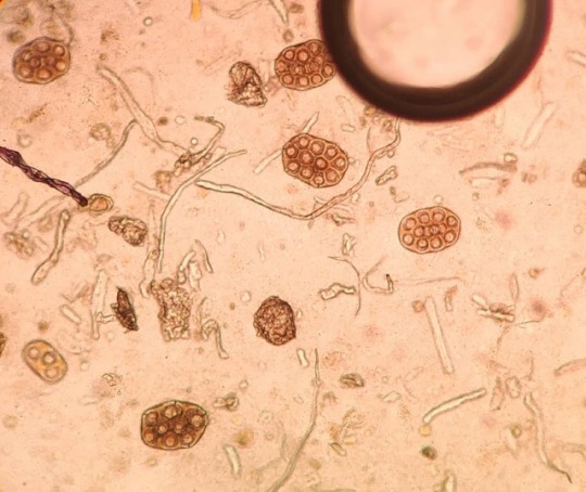
#diphylidiumcaninum (dog and cat #fleatapeworm ) Eggs: Also known as double pored #tapeworm is a principle parasite of canines and felines but is also seen in a wide variety of carnivores including humans. The main life cycle of the tapeworm is centred on larval stages of dog /cat fleas (#ctenocephalides spp.) which act as an intermediate host for transmission of the parasite. The larva migrates to surrounding tissues in the flea larvae and moult to a cyst. The adult flea harbours the infective #cysticercoid stages of the worm. Consumption of such infective fleas propagates the life cycle of the parasite. The cysticercoid matures into tapeworm in the gut of the vertebrate host where they attach themselves, feed, mature and reproduce. The eggs are passed in the feces in form of egg pockets containing many fertilized eggs which are all seen in the gravid proglottid segments of the adult tapeworm. #vetlearn #vetstudies #vetcasestudy #vetpath #vetparasitology #veterinaryparapathologist #stoolsample #parasitesofdogs #tapewormeggs #microscopicworld #microscopiclife #underthemicroscope #cestodes #gutparasites #intestinalparasites https://www.instagram.com/drdashvetpath/p/BwyXZgfBdD1/?utm_source=ig_tumblr_share&igshid=1odv6mw8jw34p
#diphylidiumcaninum#fleatapeworm#tapeworm#ctenocephalides#cysticercoid#vetlearn#vetstudies#vetcasestudy#vetpath#vetparasitology#veterinaryparapathologist#stoolsample#parasitesofdogs#tapewormeggs#microscopicworld#microscopiclife#underthemicroscope#cestodes#gutparasites#intestinalparasites
0 notes
Photo
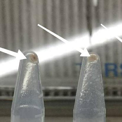
Happy #dnaday to all. A few images and work of my past lab life. Really missing those hard molecular work days... 1 and 2 : DNA extraction and precipitation 3. Electrophoresis 4 and 5. Gel documentation #lablife #vetpath #dnaisolation #dnawork #molecularpathology #vetclinicalpathology #dna #molecularlaboratory #moleculartestinglabs #pcr #vetlabdiagnostic #ethidiumbromide #uvflourescence #dnaladder #deoxyribonucleicacid #researchwork #vetstudies #vetstudent https://www.instagram.com/drdashvetpath/p/Bwt6gdkhLRV/?utm_source=ig_tumblr_share&igshid=1ldf0jbpduy37
#dnaday#lablife#vetpath#dnaisolation#dnawork#molecularpathology#vetclinicalpathology#dna#molecularlaboratory#moleculartestinglabs#pcr#vetlabdiagnostic#ethidiumbromide#uvflourescence#dnaladder#deoxyribonucleicacid#researchwork#vetstudies#vetstudent
0 notes
Photo

#salivaryglandcells Location: #salivaryglands Characteristics and functions: #epithelialcells secreting #saliva . Shape and size: Pear shaped #mediumsized cells with low nuclear: cytoplasmic ratio. Nucleus: Small round and peripherally placed with compact chromatin. Cytoplasm: Filled with numerous small vacuoles and #foamycytoplasm Cytoarchitecture: Pavement, clusters and #3-Dcytoarchitecture . Background: Light eosinophilic salivary contents. Ddx: #macrophages ,#adipophage #sebocytes #vetpathology #veterinarypathology #vetpath #vetclinicalpathology #vetcytology #cytology #salivaryglandcells #vetmed #vetmedicine #vetmedworld #medvetlife #lablife #normalcaninecell #veterinarylabdiagnosis #pathology #clinicalcytology #cytopathology #cytopath #vetcytopath https://www.instagram.com/drdashvetpath/p/Bwq_5t_Bg62/?utm_source=ig_tumblr_share&igshid=7rnval8n66dd
#salivaryglandcells#salivaryglands#epithelialcells#saliva#mediumsized#foamycytoplasm#3#macrophages#adipophage#sebocytes#vetpathology#veterinarypathology#vetpath#vetclinicalpathology#vetcytology#cytology#vetmed#vetmedicine#vetmedworld#medvetlife#lablife#normalcaninecell#veterinarylabdiagnosis#pathology#clinicalcytology#cytopathology#cytopath#vetcytopath
0 notes
Photo
#salivarygland cells: Location: #salivaryglands Characteristics and functions: #epithelialcells secreting #saliva . Shape and size: Pear shaped #mediumsized cells with low nuclear: cytoplasmic ratio. Nucleus: Small round and peripherally placed with compact chromatin. Cytoplasm: Filled with numerous small vacuoles and #foamycytoplasm Cytoarchitecture: Pavement, clusters and #3-Dcytoarchitecture. Background: Light eosinophilic salivary contents. Ddx: #macrophages ,#adipophage #sebocytes #vetpathology #veterinarypathology #vetpath #vetclinicalpathology #vetcytology #cytology #salivaryglandcells #vetmed #vetmedicine #vetmedworld #medvetlife #lablife #normalcaninecell #veterinarylabdiagnosis #pathology #clinicalcytology #cytopathology #cytopath #vetcytopath https://www.instagram.com/drdashvetpath/p/Bwq_PuNBiWI/?utm_source=ig_tumblr_share&igshid=1pvzj4hw5x1ey
#salivarygland#salivaryglands#epithelialcells#saliva#mediumsized#foamycytoplasm#3#macrophages#adipophage#sebocytes#vetpathology#veterinarypathology#vetpath#vetclinicalpathology#vetcytology#cytology#salivaryglandcells#vetmed#vetmedicine#vetmedworld#medvetlife#lablife#normalcaninecell#veterinarylabdiagnosis#pathology#clinicalcytology#cytopathology#cytopath#vetcytopath
0 notes
Photo
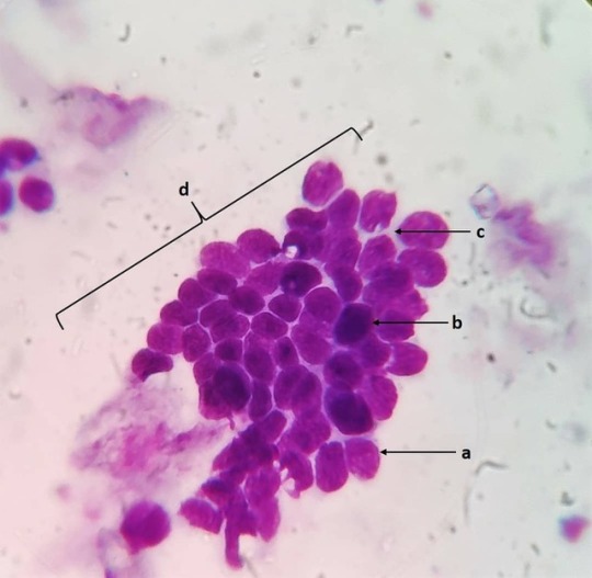
Basal cells: Location: Basal layer of all epithelia. Characteristics and function: Stem cell regenerative compartment for epithelial structures. Shape and size: Small round cells with a high N:C ratio. Nucleus: Round (a) to ovoid (b) centrally placed with compact chromatin. Cytoplasm: Scant with mild basophilia (c). Cytoarchitecture: Pavement (d) or columnar. Ddx: Matured lymphocytes. #vetpath #vetpathology #vetpathologist #vetcytology #vetcyto #veterinaryclinicalpathology #vetclinpath #veterinarycytology #veterinarycytopathology #veterinarymedicine #vetlabdiagnostic #vetcases #vetlife #normalcaninecell #basalcell #vetlablife #veterinary_medicine #vetmed https://www.instagram.com/drdashvetpath/p/BweUHo2Bi7J/?utm_source=ig_tumblr_share&igshid=1hu6g0s917um0
#vetpath#vetpathology#vetpathologist#vetcytology#vetcyto#veterinaryclinicalpathology#vetclinpath#veterinarycytology#veterinarycytopathology#veterinarymedicine#vetlabdiagnostic#vetcases#vetlife#normalcaninecell#basalcell#vetlablife#veterinary_medicine#vetmed
0 notes
Photo

#ancylostomacaninum eggs: #nematode also known as #hookworms of dogs, a principle parasite of the dogs, can also be seen in many other animals including wolves, foxes, cats and humans. #larvae of this spp. of nematode cause #cutaneouslarvamigrans (CLM) in humans by transdermal migration. One special feature of this parasite is it can cause #transplacentaltransmission in neonates by crossing the placenta from the bitch. It can also infect the young ones by #transmammarymigration through colostrum and milk. DOC - #benzimidazoles group of anthelmintics (#fenbendazole ) or #ivermectin #vetpath #vetpathology #vetpathologist #vetparasitology #veterinaryparapathologist #veterinaryparasitology #vetparasite #hookwormsofinstagram #doghookworms #cutaneouslarvamigran #dogparasites #nematodeeggs https://www.instagram.com/drdashvetpath/p/BwY5Q2SBm80/?utm_source=ig_tumblr_share&igshid=35gs398xwaec
#ancylostomacaninum#nematode#hookworms#larvae#cutaneouslarvamigrans#transplacentaltransmission#transmammarymigration#benzimidazoles#fenbendazole#ivermectin#vetpath#vetpathology#vetpathologist#vetparasitology#veterinaryparapathologist#veterinaryparasitology#vetparasite#hookwormsofinstagram#doghookworms#cutaneouslarvamigran#dogparasites#nematodeeggs
0 notes
Photo

Melanocytes Location: Pigmented areas of body. Characteristics and function: Melanin producing cells of the body. Associated with epidermis, mucosae and serosal surfaces. Shape and size: Medium sized with pleomorphic morphology and no defined cellular limits (a) Cytoplasm: Abundant, faint Basophilic (c), large numbers of dark fine melanin granules (d). Nucleus: Large, round, central with fine stippled chromatin (b). Background: Free melanin pigment. Differential: Melanophage. #vetpath #vetpath #veterinaryclinpath #veterinarypathology #veterinarycytopathology #veterinarycytology #vetcytology #cytology #cytologyvet #cytologystuff #normalcaninecell #normalcell #melanocytes #melaningranules https://www.instagram.com/drdashvetpath/p/BwRCJnNhCkM/?utm_source=ig_tumblr_share&igshid=v1hiuv0kzaws
#vetpath#veterinaryclinpath#veterinarypathology#veterinarycytopathology#veterinarycytology#vetcytology#cytology#cytologyvet#cytologystuff#normalcaninecell#normalcell#melanocytes#melaningranules
0 notes
Photo
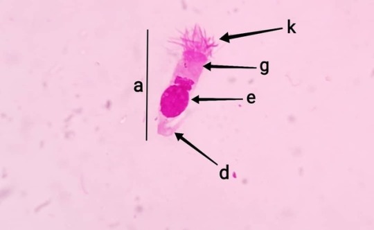
Ciliated epithelial cells: Location: Respiratory mucosa, conjunctiva Function: Lining of mucous membrane. Sweeping action of cilia helps in clearing out dust particles and debris. Shape: Elongated, columnar and wavy (a). Size: Medium sized cells seen sometimes with nucleus protuding out (b). Cytoplasm: Moderately Basophilic (g), apical ciliary apparatus (k), focal hyper basophilic area (h), superficial eosinophilic zone (i) and cilia (j). Nucleus: Round (e), oval (f), subterminal with fine stippled chromatin. Cytoarchitecture: Exfoliate as individual cells. Background: Mucinous matrix #vetpath #veterinarypathology #veterinarycytopathology #vetcytology #veterinarycytology #normalcell #cytopathology #normalcaninecell #respiratoryepithelialcells #ciliatedepithelium #ciliated #ciliatedcolumnarepithilium #hairycells https://www.instagram.com/drdashvetpath/p/BwHM6l0hvmM/?utm_source=ig_tumblr_share&igshid=1xzqjs1fbr0mv
#vetpath#veterinarypathology#veterinarycytopathology#vetcytology#veterinarycytology#normalcell#cytopathology#normalcaninecell#respiratoryepithelialcells#ciliatedepithelium#ciliated#ciliatedcolumnarepithilium#hairycells
0 notes
Photo

Conjunctival squamous: Location: Bulbar conjunctiva. Shape and size: Intermediate to superficial cells. Polygonal with a low N:C ratio. Characteristics: Epithelial cells of the bulbar conjunctiva. Nucleus: Centrally placed with compact fine chromatin (a) Cytoplasm: Clear, amphophilic with melanin pigments in perinuclear area (b). Cytoarchitecture: Single or in clusters. #vetclinicalpathology #vetpath #vetpathology #vetcytology #veterinarypathology #vetopthalmology #veterinaryclinicalpathology #veterinarycytology #veterinarycytopathology #normalcaninecell #normalcell #normalcytology #conjunctivalsquamouscell #eyecell https://www.instagram.com/drdashvetpath/p/BwB1X7OBjRi/?utm_source=ig_tumblr_share&igshid=lohfxwr2i8cg
#vetclinicalpathology#vetpath#vetpathology#vetcytology#veterinarypathology#vetopthalmology#veterinaryclinicalpathology#veterinarycytology#veterinarycytopathology#normalcaninecell#normalcell#normalcytology#conjunctivalsquamouscell#eyecell
0 notes
Photo
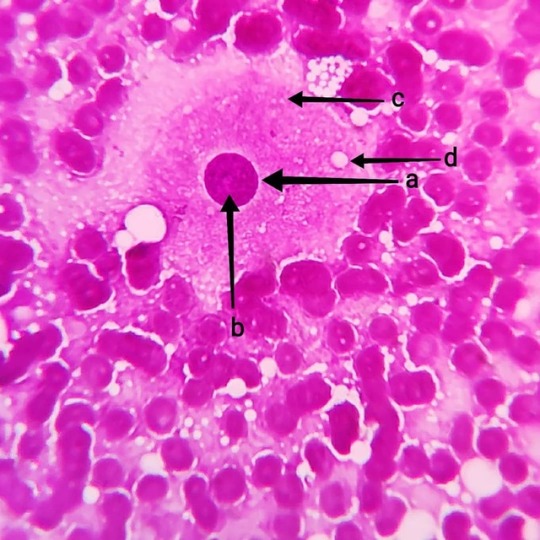
Luteal cell: Location- Female genital tract, #ovary #corpusluteum Characteristic and function - Production of #progesterone and formation of #corpusluteum Shape - Pear shaped Size - Vert large to large Cytoplasm - Basophilic (c) and abundant with empty vacuoles (d). Nucleus - Large (a), oval round, central to paracentral with finely stippled chromatin and prominent nucleolus (b). Cytoarchitecture - Pavement clusters or single cells. #vetpath #veterinarypathology #cytology #vetcytology #veterinarycytology #veterinarycytopathology #cytopathology #cytopath #normalcell #vetdiagnosis #impressionsmear #diffquick #diffquikstain #ovariancell https://www.instagram.com/drdashvetpath/p/Bv3Da3fhMdM/?utm_source=ig_tumblr_share&igshid=1pugr3qr10uxs
#ovary#corpusluteum#progesterone#vetpath#veterinarypathology#cytology#vetcytology#veterinarycytology#veterinarycytopathology#cytopathology#cytopath#normalcell#vetdiagnosis#impressionsmear#diffquick#diffquikstain#ovariancell
0 notes
Photo

Fleas Ctenocephalides Felis: Most commonly seen fleas in household cats and dogs. Principle host of the cats but also seen infesting dogs and other mammals. Fleas go through 4 life cycle phases egg, larva, pupa and adult. It's is estimated that a female lays somewhere between 5000 to 8000 eggs during her life. The fleas need to feed on a blood meal before they start producing eggs. The eggs develop to larvae in around three to six weeks. The larvae are negatively phototrophic and hide from the light unto the ground substrates. They again moult to pupae by spinning a Coccon and waiting for suitable environmental conditions and for a suitable host. The adult fleas are capable of detecting the CO2 and temperature from the hosts and this helps in lodging of the fleas to the hosts body. Clinical signs and symptoms develop due to allergic reactions towards the substances present in the fleas saliva (Flea allergy dermatitis - FAD). Morphological differentiation between C. felis and C. canis: a. The first spine of genal comb of C. felis is almost the same length as the second, in C. canis is almost half the length. b. Head of C. felis is more rounder and longer than that of C. canis. c. C. felis - tibiae of all 6 legs have 7 to 8 bristles C. canis - tibiae of all 6 legs have 4 to 5 bristles. To effectively control fleas, both the fleas on the animalss as well as the fleas and eggs in the surroundings have to treated. Adulticides along with insect development inhibitors (Lufeneuron) and insect growth regulators (Methoprene) have been used effectively to control flea population. Spot on (Imidacloprid, fipronil, Methoprene) solutions are more effective alternative in pets rather than the commercial available sprays and dips. #vetpath #vetpathology #vetclinpath #vetclinicalpathology #vetlabdiagnostic #veterinarypath #veterinaryclinicalpathology #veterinarypathology #veterinaryparapathologist #vetdermatology #veterinarydermatology #veterinaryentomology #vetlaboratory #vetdiagnostics #vetparasitology #veterinaryparasitology #vetparasite #flea #catflea #dogflea #ctenocephalidesfelis #externalparasites #fleaallergy #fleaallergydermatitis #wetmount #parasites https://www.instagram.com/drdashvetpath/p/BvyuuHyhN9f/?utm_source=ig_tumblr_share&igshid=mnofp94i184l
#vetpath#vetpathology#vetclinpath#vetclinicalpathology#vetlabdiagnostic#veterinarypath#veterinaryclinicalpathology#veterinarypathology#veterinaryparapathologist#vetdermatology#veterinarydermatology#veterinaryentomology#vetlaboratory#vetdiagnostics#vetparasitology#veterinaryparasitology#vetparasite#flea#catflea#dogflea#ctenocephalidesfelis#externalparasites#fleaallergy#fleaallergydermatitis#wetmount#parasites
0 notes
Photo
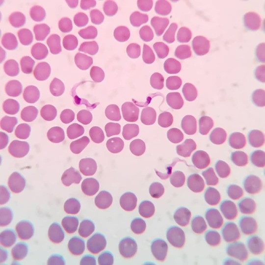
Trypanosoma spp. in canine blood smear: Seen are trypomastigote stages trypanosoma parasites in the blood smear of a dog. They are extracellular parasites seen feeding on blood and also on lymph. Trypanosoma spp. are flagellate parasites bearing a undulating membrane observed in the blood of mammals. The parasites also posses a kinetoplast, a small disc like structure containing mitochondrial DNA seen close to the nucleus. T. evansi and T. cruzi are seen most commonly infecting the domestic dog. Other species like T. congolensis, T. vivax, T. equiperdum, T. brucei and T. rangeli are also seen infecting dogs, humans and other mammals. Dogs serve as reservoir hosts and helps in spreading the infection to humans. The common vectors involved in the transmission of Trypasnoma spp. parasites are the Tse-tse flies and kissing bugs (Triatoma spp.). #vetpath #veterinarypathology #veterinaryclinpath #vetclinpath #veterinaryhaematology #haematology #clinicalhaematology #veterinaryparasitology #veterinaryparapathologist #veterinaryparasites #vetparasite #bloodsmears #parasitology #wetmount #bloodparasite #flagellates #parasitism #parasiticinfection #trypanosoma #trypanosomiasis #caninetrypanosoma #trypanosomes #trypanosome #kissingbugs #tsetsefly #triatoma https://www.instagram.com/drdashvetpath/p/BvlncHxBPfM/?utm_source=ig_tumblr_share&igshid=16yzwb5d6esr3
#vetpath#veterinarypathology#veterinaryclinpath#vetclinpath#veterinaryhaematology#haematology#clinicalhaematology#veterinaryparasitology#veterinaryparapathologist#veterinaryparasites#vetparasite#bloodsmears#parasitology#wetmount#bloodparasite#flagellates#parasitism#parasiticinfection#trypanosoma#trypanosomiasis#caninetrypanosoma#trypanosomes#trypanosome#kissingbugs#tsetsefly#triatoma
0 notes
Photo

And again!!!! Lipoma in a dog: Seen are plump adipocytes with optically empty cytoplasm due to the stored fats/lipids in the cells. The nucleus is pushed to the periphery to accomodate the maximum available space for storage of fats/lipids. Adipose tissue are usually found intertwined with collagenous fibres and blood vessels forming a 3D cytoarchitecture. #vetpath #vetpathology #cytopathology #vetdiagnosis #vetdiagnostics #fna #fnac #cytology #cytologyvet #veterinarycytopathology #vetlabdiagnostic #fineneedleaspirationcytology #fineneedleaspirate #lipoma #subcutaneous #veterinaryclinicalpathology #cytologicalexamination #aspirationcytology https://www.instagram.com/drdashvetpath/p/Bvenfy5hfrW/?utm_source=ig_tumblr_share&igshid=306j3bci9mbr
#vetpath#vetpathology#cytopathology#vetdiagnosis#vetdiagnostics#fna#fnac#cytology#cytologyvet#veterinarycytopathology#vetlabdiagnostic#fineneedleaspirationcytology#fineneedleaspirate#lipoma#subcutaneous#veterinaryclinicalpathology#cytologicalexamination#aspirationcytology
0 notes
Photo

Granulosa cells: Location: Female genital tract, ovaries. Function: Oocyte maturation and support. Nucleus: Eccentric, polygonal with reticular chromatin (a) Cytoplasm: Scant and basophilic (b), sometimes seen with empty vacuoles (c) Cytoarchitecture: Small pavement clusters (d) Ddx: Lymphoblasts #vetpath #veterinarypathology #vetcytology #veterinarycytology #cytology #cytologyvet #cellcytology #cellsundermicroscope #ovariancells #granulosa #granulosacell #cytodiagnostic #normalcell #cytologicalexamination #impressionsmear https://www.instagram.com/drdashvetpath/p/BvbzxP_hfQb/?utm_source=ig_tumblr_share&igshid=j6zcavgyj9ew
#vetpath#veterinarypathology#vetcytology#veterinarycytology#cytology#cytologyvet#cellcytology#cellsundermicroscope#ovariancells#granulosa#granulosacell#cytodiagnostic#normalcell#cytologicalexamination#impressionsmear
0 notes