#Lupine Publishers Group
Text
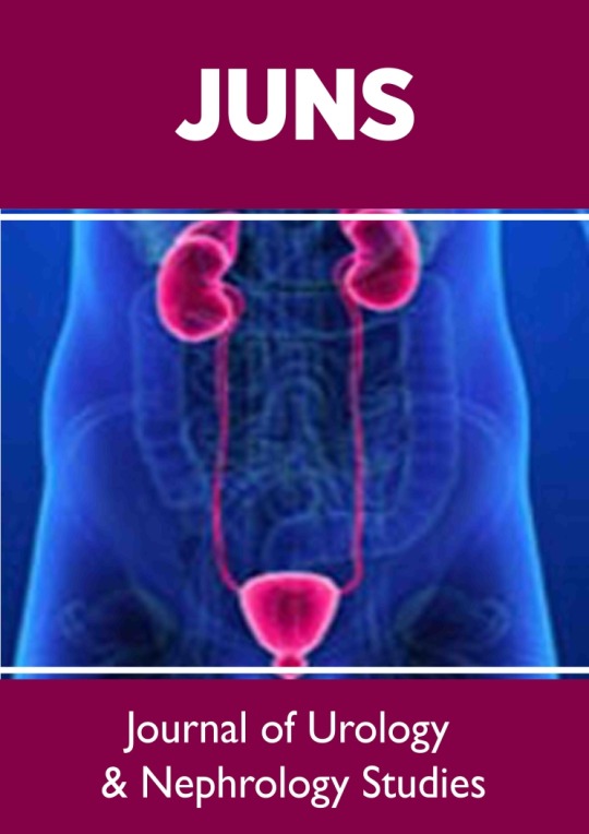
Lupine Publishers|Radiology; USG and Colour Doppler of Post Renal Transplant Complications
Abstract
Kidney transplant is the treatment of choice for patients with end-stage renal disease. Kidney transplant offers better quality of life. It is more cost effective than hemodialysis. Advances in surgical technique, along with improvement in organ preservation and immunosuppression have improved patient outcomes. Post-operative complications, however, can limit this success. Ultrasound and Doppler study is the primary imaging modality for evaluation of renal transplant, providing real –time information about complication in graft. A multimodality approach including CT scan, MRI or conventional angiography may be necessary in cases when sonography and Doppler are inconclusive to diagnose the etiologies of these complications. Radiologists play an integral role within the multidisciplinary team in care of transplant patient at every stage of the transplant process. Therefore, the radiologist should always be aware when evaluating the failing renal graft, whether the cause is renal or extrinsic. In this pictorial essay we tried to gather the most common complication of transplant kidney in different cases that occurred in our hospital, with an emphasis on Ultrasound and Doppler study.
Keywords: USG; Colour Doppler; Post renal transplantation; Complications
Introduction
The preferred modality for renal replacement is renal transplantation, and its superiority in prolonging the longevity of patients with end-stage renal disease is well established [1]. Kidney transplantation is typically classified as deceased-donor (formerly known as cadaveric) or living-donor transplantation depending on the source of the donor organ. Due to improvement in transplantation technology, advancement in immunosuppressant and graft preservation techniques the 1-year survival rates for grafts, are reported to be 80% for mismatched cadaveric renal grafts; 90% for nonidentical living related grafts; 95% for human lymphocyte antigen-identical grafts [2]. Radiologists play a major role at every stage of the transplant process with transplantation team. Ultrasonography with colour Doppler is the principal modality used for evaluation of renal allograft. USG is a relatively cheap, noninvasive, and non-nephrotoxic modality. It is applied for diagnostic and monitoring purposes in the post-transplant period. This pictorial essay describes USG and Doppler imaging appearances of the major complications that may occur in renal transplantation. All our images have been obtained from a single our center which is major transplantation center in India. All post renal transplant patients undergo a USG and comprehensive Doppler evaluation on post-operative day one. The sonographic examination algorithm includes gray-scale evaluation of the graft and spectral Doppler. Further imaging is based on clinical follow-up including daily monitoring of laboratory values. If clinical parameters are abnormal, repeat sonography is performed and depending on the results, CT, MRI, or angiography may be requested.
Surgical Technique
Surgical technique and location of placement of renal allograft depends on the variation in arterial and venous anatomy of donor. The transplanted kidney is usually placed extraperitoneally in the patient’s right iliac fossa (less commonly in left iliac fossa), with end-to-side or end to end anastomosis to the external iliac vasculature. The currently preferred method for restoring urinary drainage is ureteroneocystostomy, a procedure by which the ureter is implanted directly into the dome of the bladder (Figure 1).
Figure 1: Diagrammatic representation of anatomical and anastomotic arrangement (end to side fashion) in renal transplantation in right renal fossa
Post renal transplantation complications
Urologic Complications
The prevalence of urologic complications varied from 10% to 25% with a mortality rate ranging from 20% to 30%. Incidence rate is decreased range between 3% and 9% in the current era because of advancement in surgical techniques and frequent use of ureteral stents [3,4].
Urinary Obstruction
a) Incidence: 2%-5% of kidney transplant recipients.
b) Site of obstruction: Approx. 90% of stenoses occur in the distal third of the ureter due to its poor vascular supply.
c) Imaging appearance: US can easily confirm the diagnosis of hydronephrosis and dilated renal pelvis and thus determine the level of ureteral obstruction (Figure 2). When contents of pelvic calyceal system are echogenic and weakly shadowing, fungus balls should be considered, whereas low-level echoes suggest pyonephrosis (Figure 3).
Figure 2: Grey scale USG image shows (A) dilated pelvic calyceal system, (B) dilated ureter may favors urinary obstruction.
Figure 3: Grey scale USG image shows echogenic material within (A) dilated pelvic calyceal system, (B) dilated renal pelvis may favors debris.
Urine Leaks and Urinomas
a) Incidence: up to 6 % of renal transplant recipients [5]
b) Location: extraperitoneal or intraperitoneal, if intraperitoneal may present with ascites.
c) Imaging appearance: USG findings are nonspecific, well defined anechoic fluid collection with septa or without septation, adjacent to the lower pole of the transplant in most of the cases (Figure 4).
Figure 4: Grey scale USG image shows anechoic collection (A) near urinary bladder, (B) near lower pole of transplanted kidney may favors urinoma.
Drainage of fluid under ultrasound guidance and testing the fluid for creatinine helps to differentiate it from seromas or lymphoceles. High concentration of creatinine will be found in case of urinoma if we compare with serous fluid.
Calculous Disease
a) Incidence: 1% to 2 % of post-transplant cases develops clinically relevant stones as compared to general population [6]. As the kidney is denervated patient will not suffer typical renal colic.
b) Imaging appearance: Calculus appears as strongly reactive focus of variable size producing acoustic shadowing on USG and twinkling artifact on colour Doppler (Figure 5). Other rare urologic complications are ureteric necrosis and vesico-ureteric reflux.
Figure 5: Grey scale USG image of transplanted kidney show echogenic focus with acoustic shadowing (A) in lower calyceal system, (B) in upper calyceal system, (D) in mid part of transplanted ureter and (C) with twinkling artifact on colour Doppler favoring calculus.
Peritransplant Fluid Collections
Fluid collection in peritransplant region has been reported in up to 50 % of renal transplantation. They are urinomas, hematomas, lymphocele and abscess, the clinical relevance of these collection is largely determined by their size, location and possible growth. In immediate postoperative period, small hematomas or seroma are almost expected. Their size should be documented at baseline examination. Rarely do they lead to graft dysfunction or obstruction of collecting system. Urinomas and hematomas are most likely to develop immediately after transplantation, whereas lymphoceles generally develop after 4 to 8 weeks. The ultrasonography characteristics of peritransplant fluid collections, however, are entirely nonspecific, correlation with clinical findings helps to restrict differential diagnosis. Ultimately, diagnosis may be made only with percutaneous aspiration and then biochemical analysis. Differentiation between Peritransplant and subcapsular collection is important. A subcapsular collection likely to cause mass effect on parenchyma, usually cresenteric and show extension along the contour of kidney deep to the renal capsule
Hematoma
a. Incidence: Varies from 4 to 8 % [7]
b. Imaging appearance: Hematomas have a complex appearance. Hematomas appears echogenic in acute case and progressively become less echogenic with time (Figure 6). Chronic hematomas even appear anechoic, more closely resembling fluid and septation may develop later on.
Figure 6: Grey scale USG image shows echogenic collection in (A) at subcapsular region, (B) in peritransplant region favoring hematoma.
Lymphocele
a. Incidence: Affecting up to 20% of the patients [8] It occurs after surgery owing to the surgical disruption of the normal lymphatic channels along the iliac vessels or around the hilum of the graft.
b. Imaging appearance: on Ultrasound it appears as anechoic bur may contain septation. They may become infected and gave more complex appearance (Figure 7).
Peritransplant abscesses
a) Imaging appearance: USG cannot always differentiate an abscess from other collection. Collection may show low level echoes and thick irregular wall. If gas is seen, an abscess is probable. In any pyrexia patient, any perinephric collection should be considered infected until proven otherwise through the appropriate imaging and guided diagnostic aspiration.
Figure 7: Grey scale USG image showing septated collection (A) near lower pole of transplanted kidney, (B) causing compression over renal pelvis and resulting in obstructive changes in transplanted kidney favoring lymphocele.
Parenchymal Complication / Graft Dysfunction
Diseases of the renal parenchyma are usually diffuse and often leads graft dysfunction. Differential diagnosis is difficult by imaging alone. USG is not sensitive or specific for evaluation; differential will be relying on biopsy. USG still has a central role in qualitative assessment of graft perfusion and to guide the biopsy.
Acute tubular necrosis (ATN)
Acute tubular necrosis is due to reversible ischemic damage to the renal tubular cells prior to engrafting and reperfusion injury.
i. Incidence: affects 20–60 % of cadaveric renal grafts.
ii. Time of onset: in the first 48 hours after transplantation.
iii. Imaging appearance: No specific imaging pattern for the diagnosis of ATN. The kidney may appear normal, in severe cases it looks enlarged, edematous and echo poor with loss of corticomedullary differentiation (Figure 8) and shows elevated RI (above 0.8).
Figure 8: Grey scale USG image showing edema to us transplanted kidney with prominent pyramids and decreased cortical echogenicity may favors changes of Acute tubular Necrosis (ATN) or Acute Rejection (AR).
Rejection
Rejection can be classified according to the period of appearance as hyper acute (occurring within minutes), acute (occurring within days to weeks), late acute (occurring after 3 months), or chronic (occurring months to years after transplantation). Classification of renal allograft rejection by Banff classification of allograft pathology is routinely followed nowadays. It is based on a combination of histopathologic features coupled with molecular, serologic, and clinical parameters.
Acute Rejection (AR)
i. Incidence: up to 40% of patients in the early transplant period [9].
Figure 9: (A) Grey scale USG image shows enlargement of transplanted kidney, swelling of the medullary pyramids and echogenic sinus fat, (B) Spectral Doppler analysis shows elevated RI may favors changes of Acute Rejection (AR) or Acute Tubular Necrosis (ATN).
ii. Imaging appearance: Graft enlargement due to edema, Decreased cortical echogenicity, swelling of the medullary pyramids, echogenic sinus fat, edematous wall of pelvic calyceal system, focal hypoechoic areas in parenchyma which may favors infarct and collection in perigraft region due to necrosis or hemorrhage. PI and RI elevated in both ATN as well as in AR, but AR has high values of it. In severe cases, Power Doppler shows reduced, absent or reversed diastolic flow with elevation of the RI (Figure 9).
Chronic Rejection
Chronic rejection occurs in case of insufficient immunosuppression given to recipient to control the residual antigraft lymphocytes and antibodies.
i. Imaging appearance: US appearance is not typical, ranging from normal to hyper echogenic, along with cortical thinning, reduced number of intrarenal vessels, and mild hydronephrosis (Figure 10). RI measurements are not reliable for this diagnosis. The diagnosis is made histologically.
Figure 10: (A) Grey scale USG image shows echogenic transplanted kidney with cortical thinning and minimal hydronephrosis, (B) Spectral Doppler image shows reduced diastolic flow and elevated RI may favors Chronic Rejection.
Drug Nephrotoxicity
Calcineurin inhibitors are key immunosuppression agents administered to avoid acute rejection, but they are nephrotoxic.
A. Imaging appearance: USG may show completely normal results or nonspecific findings such as graft swelling, increased or decreased renal echogenicity and loss of cortico-medullary differentiation. Doppler study may show a RI increase of 0.80. Findings of USG and Doppler study should be correlated with the serum drug levels. USG and Doppler findings of ATN and AR is almost similar, but both can be differentiated by time course of the findings. Clinical & biochemical correlation and serial measurements of RI and Pulsatile index (PI) would be further helpful to monitor the patient.
Infections and abscesses
Incidence and time of onset: More than 80% of renal transplant recipients have at least 1 episode of infection during the first year of post transplantation.
Figure 11: Grey scale USG image shows ill-defined hypoechoic lesion (A) in mid pole, (B) in upper pole of transplanted kidney posteriorly Suggestive of Abscess formation. First one is proved case of fugal etiology and second one is pyogenic abscess.
I. Imaging appearance: USG appearance is quite variable. Focal pyelonephritis appear as a focal hyperechoic or hypoechoic area, but this finding is nonspecific because it can represent infarction or rejection also (Figure 11). Abscess has varied appearance on USG like- heterogeneous, hypoechoic or cystic. Urothelial thickening may be seen. In febrile post renal transplant patient low level echoes in dilated collecting system may favors pyonephrosis. Fungus ball appears as focal rounded, weakly shadowing or echogenic structure in dilated pelvic calyceal system. In emphysematous pyelonephritis, gas in the parenchyma of the renal graft produces an echogenic line with distal reverberation artifacts. Papillary necrosis has no typical sonographic findings, and it subsequently leads to ureteric obstruction.
End stage disease
Nonfunctional renal grafts are left in place in abdominal cavity. Gradually graft becomes small and can have fatty replacement, hydronephrosis, infarcts, hemorrhage or calcification.
Vascular Complications
Vascular complications in post renal transplantation have significant negative influence of graft survival. They are infrequent, occur in approximately 1%–2%; [10] but can cause sudden loss of renal allograft. Selective catheter angiography is the gold standard for diagnosis; however, it is invasive and may cause various complications. Hence it is not used as a screening tool but reserved for patients with inconclusive results on the noninvasive screening tests. Noninvasive imaging like ultrasound, Doppler, scintigraphy, CT and MR angiography plays major role to evaluate them.
Renal artery thrombosis (RAT)
Incidence: Ranges from 0.5% to 3.5 % [11]
Imaging appearance: No evidence of any arterial or venous flow noted on color, spectral and power Doppler study (Figure 12). Doppler sonography had 100% sensitivity and specificity for diagnosis and hardly any other imaging study is required for diagnosis [12].
Figure 12: (A) Grey scale USG image, (B) Power Doppler image of transplanted kidney show enlargement and absent colour flow Suggest Vascular Thrombosis.
Focal Renal Infarction
Imaging appearance: A segmental infarct appears as a poorly marginated wedge shaped hypoechoic mass or a hypoechoic mass with a well-defined echogenic wall without colour flow (Figure 13). If the infarction is global, the kidney will appear hypoechoic and be diffusely enlarged.
Figure 13: Colour Doppler image of transplanted kidney shows absent colour flow (A) in upper and mid pole, (B) in upper pole favoring Segmental infarct.
Renal Artery Stenosis (RAS)
Incidence: It has wide range varying from 1% to 23 % depending on the definition and diagnostic techniques used.
Site: usually at anastomotic site
Imaging appearance: On gray scale USG, there is lack of normal post-transplant hypertrophy. On color Doppler study appearance of focal color aliasing noted at stenotic segments. On spectral Doppler study, peak systolic velocity in main renal artery >300 cm/sec and Ratio of PSV in transplanted main renal artery and external iliac artery greater than or equal to 1.8 are highly suggestive of significant stenosis. Indirect criteria are low resistive index <0.56, Acceleration time >0.07 sec, Acceleration index <3 meter/ sec and Intrarenal tardus–parvus waveform (Figure 14).
Figure 14: Spectral Doppler image shows (A) elevated peak systolic velocity in main renal artery at anastomosis, (B) delayed systolic upstroke and rounding of systolic peak consistent with tardus parvus spectral waveform in case of transplanted renal artery stenosis.
Limitation: Results are strongly depends on the operator’s individual experience and skill. Rate of restenosis is less than 10 %. Doppler ultrasonography is the procedure of choice to evaluate graft perfusion before and after revascularization The term pseudo transplant renal artery stenosis (TRAS) refers to thrombosis or stenosis of iliac artery or aorta proximal to transplant renal artery.
Renal vein thrombosis: (RVT)
Figure 15: (A) Grey scale USG image shows swollen and hypo echoic transplanted kidney, (B) Colour Doppler image shows absent venous flow and spectral Doppler image shows absent and reversal of diastolic flow in renal artery favoring Renal vein thrombosis.
Incidence: Ranges from 0.9% to 4.5 % [13]
Imaging Findings: Graft appears swollen and hypoechoic.
Doppler shows absent venous flow. Renal arterial Doppler spectrum shows absent or reversal of diastolic flow due to increased resistance (Figure 15). Reversal flow in renal artery is nonspecific as it also seen in severe rejection and in acute tubular necrosis, its combination with absent venous flow is the diagnosis of renal vein thrombosis. Partial thrombosis also can occur near anastomosis or within the transplanted kidney (Figure 16).
Figure 16: Spectral Doppler image shows (A) partial thrombosis in renal vein near anastomosis (white straight arrow), (B) partial thrombosis in main renal vein (white straight arrow) and adjacent renal pelvis showing DJ Stent (curved whited arrow) in two different cases. Intra renal venous flow is very well demonstrated and appears normal Suggest Partial renal vein thrombosis.
Extra parenchymal pseudo aneurysm
Incidence: Anastomotic pseudoaneurysm is a rare complication of renal transplantation occurring in 0.3%. [14]
Imaging findings : On gray scale ultrasound it appears as cystic lesion which shows color flow and to and fro spectral pattern on doppler study(Figure 17). Endovascular treatment with covered stent placement to exclude pseudoaneurysm can also be evaluated with USG and colour doppler (Figure 18).
Figure 17: (A)Grey scale USG image of transplanted kidney shows cystic lesion near anastomosis which shows color filling in Colour Doppler image(white arrow), (B) Colour Doppler image shows colour filled out pouching near anastomosis(white arrow) confirmed non thrombosed extra parenchymal pseudo aneurysm in two different cases.
Figure 18: (A) Two echogenic line structure(Stent) in main anastomotic renal artery in gray scale USG image of transplanted kidney, (B) Colour doppler image shows partially colour filled pseudoaneurysm at anastomosis with stent suggest Imaging findings of endovascular treatment with covered stent placement.
Intra-parenchymal arteriovenous fistula and pseudoaneurysm
AVF occurs when both artery and vein are simultaneously lacerated, while pseudoaneurysm results when only artery is lacerated.
Incidence: 1-18% of the biopsies [15]
Time of onset: occur at time of biopsy. They depend on many factors – ultrasound guidance, needle caliber and imaging follow up.
Imaging appearance: colour Doppler study shows AVF as focal areas of disorganized flow adjoining the normal vasculature. Spectral waveforms show increased arterial and venous flow with high velocity and low resistance (Figure 19). Pseudoaneurysm appears as simple or complex cyst on B mode ultrasound and intracystic flow on colour Doppler mode (Figure 20).
Figure 19: (A) focal colour aliasing at lower pole of transplanted kidney in colour doppler image, (B) Spectral Doppler evaluation demonstrates high velocity, low impedance waveform with increased diastolic flow Suggest arteriovenous fistula (AVF) in doppler evaluation after biopsy.
Figure 20: (A) Grey scale USG image shows cystic lesion in lower pole of transplanted kidney in grey scale USG image, (B) colour Doppler image shows intra cystic flow Suggest intra parenchymal pseudo aneurysm.
Neoplasms after renal transplantation
Post renal transplantation patients are at higher risk of development of neoplasms. Urologic tumour are 4 to 5 times more common in post renal transplantation recipients than normal population with significant exposure to cyclophosphamide immunosuppressant agent.
Renal cell carcinoma
Etiology: by means of transplanted organ or de novo development by immunocompromised status, patients on hemodialysis in case of chronic renal failure develop acquire renal cystic disease
Prevalence: 90 % occurring in native kidney and 10 % in transplanted kidney [16]
Imaging appearance: lesion appear heterogeneous with vascularity, similar picture as seen in native kidney [17] (Figure 21).
Figure 21: Gray scale ultrasound image of transplanted kidney shows (A) heterogeneous mass lesion at the upper pole (white arrow) with internal hypoechoic region (asterisk) suggesting necrosis, (B) the mass lesion showed internal as well as peripheral vascularity on color Doppler study. Suggest renal cell carcinoma in transplanted kidney.
Lymphomas
Incidence: 1 % of renal allograft recipients [18]
Time of onset: Post transplantation lymphoproliferative disorder is diagnosed at a median of 80 months after transplantation.
Imaging appearance: Lymphadenopathy at various sites but can also affect any solid organ or hollow viscera or transplant graft parenchyma itself. It appears as low or mixed reactive masses and tends to have a predilection for the renal hilum.
Recurrent Native disease
It depends on the primary disease before transplantation.
Imaging appearance: Imaging has no specific pattern in these situations apart from excluding the treatable cause of reduced renal function.
Abdominopelvic Complications
Renal graft is placed in extraperitoneal space via a peritoneal window in laparoscopic and robot assisted surgical techniques. So these cases are prone to same complications experienced by other surgical cases in whom peritoneum is exposed.
Renal Allograft Compartment Syndrome (RACS)
It is a rare syndrome, and it is under recognized cause of early transplant dysfunction or even loss. It may occur as a result of intracompartment hypertension and ensuring ischemia of renal graft [19]. Imaging appearance: absent or diffuse diminished cortical flow in transplant kidney at colour Doppler.
Fascial dehiscence and bowel or allograft evisceration
They tend to occur in perioperative period. Herniation of bowel through a transplant peritoneal defect may lead to compromise of intestine or transplant itself.
Limitations
The USG examination is examiner dependent and limited accessibility in obese patents impairs the evaluation and often leads misinterpretation. The RI index is also unspecific and influenced by many factors like site at which the RI is measures, increased intraabdominal pressure during forced inspiration and the pulse rate.
Summary
Kidney transplant is the treatment of choice for patients with end-stage renal disease. Improvements in surgical techniques and advanced immunosuppressive drugs have resulted in remarkable survival of patients and renal grafts. Still complications occur in both the early postoperative period and later. Kidney transplants follow up is common in radiology and sonography practice. Ultrasonography and Doppler examination can accurately depict and characterize many of the potential complications of renal transplantation. It facilitates prompt and accurate diagnosis and thus guiding treatment.
for more information about Journal of Urology & Nephrology Studies archive page click on below link
https://lupinepublishers.com/urology-nephrology-journal/archive.php
for more information about lupine publishers page click on below link
https://lupinepublishers.com/index.php
#lupine publishers#lupine publishers LLC#lupine publishers group#journal of urology & nephrology studies#juns
6 notes
·
View notes
Text
Lupine Publishers|Why to Not Rely on Nature?

Lupine Publishers | Journal of Health Research and Reviews
Abstract
Metabolism is the process your body uses to make energy from the food you eat. Food is made up of proteins, carbohydrates, and fats. Chemicals in your digestive system (enzymes) break the food parts down into sugars and acids, your body’s fuel. Your body can use this fuel right away, or it can store the energy in your body tissues. If you have a metabolic disorder, something goes wrong with this process. Dyslipidemia is main etiological factor leading to develop coronary artery disease (CAD). Allopathic drugs used in cure of hyperlipidemia, have unwanted effects, so have poor compliance. Now a day’s nutraceuticals are getting popularity due to their moderate hypolipidemic actions with fewer adverse effects. Niacin or vitamin B-3 is used as hypolipidemic agent which increases HDL-cholesterol and decreases LDL-cholesterol, VLDL, TGs by different mechanisms. But its main adverse effects are flushing due to synthesis of Prostaglandin D-2 which has vasodilatory effect causing flushing. Cardamom is herb having hypolipidemic potential if used in specific concentration for long time. In this work we did try to compare these two medicines hypolipidemic effects. Study was single blind placebo-controlled conducted at Jinnah Hospital Lahore-Pakistan from July to November 2018. Seventy-five hyperlipidemic patients were selected and divided in three groups. Their base line lipid profile was determined at laboratory of the hospital. Patients were divided in three groups. Group-1 was on placebo therapy, Group-II was on Niacin 1.5 grams daily in divided doses, and Group-III was on Cardamom 1 gram daily in three divided doses. It was two months therapeutic design. At completion of therapeutic regimen after two months we measured all patient’s lipid profile. When results were compiled and pre and post treatment values were analyzed statistically, it was observed that both drugs hypolipidemic potential is different, although both have hypolipidemic characteristic, but Niacin is the best for treating Hyperlipidemia. For statistical analysis we used SPSS version 5.0 and paired ‘t’ test was applied to understand significant changes in two tested group’s lipid profile.
Introduction
Lipid metabolism disorders, such as Gaucher disease and Tay- Sachs disease, involve lipids. Lipids are fats or fat-like substances. They include oils, fatty acids, waxes, and cholesterol. If you have one of these disorders, you may not have enough enzymes to break down lipids. Or the enzymes may not work properly, and your body can’t convert the fats into energy. They cause a harmful amount of lipids to build up in your body. Over time, that can damage your cells and tissues, especially in the brain, peripheral nervous system, liver, spleen, and bone marrow. Many of these disorders can be very serious, or sometimes even fatal. High LDL-cholesterol and low HDL-cholesterol in blood are cause of synthesis of atherosclerotic plaques which get deposited with inner endothelium of coronary arteries, leading to develop coronary artery disease (CAD) [1,2]. Hyperlipidemia either primary or secondary is challenge for scientists of 21st century due to this disease’s complications like coronary artery disease, Hypertension, Cardiac Arrhythmias, and Myocardial Infarction [3,4]. Hyperlipidemia causes LDL particle’s oxidation, which initiate formation of atherosclerotic plaques leading to development of coronary artery disease. From this single complication of Hyperlipidemia, all major heart diseases are developed leading to morbidity and mortality due to last lethal major heart disease Ventricular Fibrillation [5-8]. Hypolipidemic drugs commonly used include Statins, nicotinic acid, bile acid binding resins and fibric acids, but all have potential for low patient compliance due to wide range of side effects [9].
Niacin increases apolipoprotein A1 levels due to anti catabolic effects resulting in higher reverse cholesterol transport. It also inhibits HDL hepatic uptake, down-regulating production of the cholesterol ester transfer protein gene. Finally, it stimulates the ABC-A1 transporter in monocytes and macrophages and upregulates peroxisome proliferator-activated receptor γ results in reverse cholesterol transport. It reduces secondary outcomes associated with atherosclerosis, such as low-density lipoprotein cholesterol, very low-density lipoprotein cholesterol, and triglycerides, but increases high density lipoprotein cholesterol [10]. Despite the importance of other cardiovascular risk factors, high HDL was associated with fewer cardiovascular events independent of LDL reduction. Other effects include anti-thrombotic and vascular inflammation, improving endothelial function [11]. To get good drug-patient compliance many health-related modern researchers have started to put their haeling potential for developing alternatives drugs used in primary or secondary Hyperlipidemia. Cardamom or in urdu ILAICHI is one of the hypolipidemic herb, widely encouraged by cardiologists to be used for prevention of atherogenesis, and coronary artery disease [12]. Cardamum’s antioxident effect is well established in many research studies on medicinal herbs. This nutraceutical contains Flavenoids and Phenolic compounds which scavenge Reactive Oxygen Species (ROS), reducing risk of developing CAD. Oral administration of cardamom extracts significantly reduced total cholesterol, highdensity lipoprotein (HDL), low-density lipoprotein, and very lowdensity lipoprotein and triglycerides in HL patients [13].
Patients & Method
Design: It was single blind placebo-controlled study conducted at GENERAL HOSPITAL Lahore from July to November 2018.
No of Patients: Seventy-five hyperlipidemic patients were selected and enrolled for the study. Written, already explained and approved consent was taken from all patients.
Inclusion Criteria: Inclusion criteria were age limit from 18 to 70 years of both gender male and female primary or secondary hyperlipidemic patients.
Exclusion Criteria: Patients suffering from any vital organ disease or their impaired functions were excluded from the study. Alcoholics, cigarette smokers and patients taking regular medicine for their any physical or mental disease were also excluded.
Grouping: Seventy-five patients were divided in three groups, comprising 25 patients in each group. Group-I was on placebo therapy. They were provided capsules containing grinded rice and mixed wheat. They were advised to take one capsule before meal, thrice daily for two months. Group-II patients were advised to take 1.5 grams of Niacin in three divided doses for the period of two months. Group-III were advised to take one-gram grinded green Cardamom powder mixed in black tea, thrice daily after each meal for the period of two months.
Methods Used: At start of study all patients’ blood pressure was recorded and kept in their personal file. Lipid profile of all patients was determined by Frei Dewald Method. Total-cholesterol, LDL-cholesterol and HDL-cholesterol were main parameters we required for further calculation of change in these parameters. All patients were advised to visit clinic fortnightly for their follow up. After two months therapy their lipid profile was measured again by same Frei Dewald Method. Blood Pressure of all participants was again measured and compiled for further statistical analysis.
Statistical Analysis: Data were expressed as the mean ± SEM and paired “t” test was applied to determine statistical significance as the difference. A probability value of <0.05 was considered as non-significant and P<0.001 was considered as highly significant change in the results when pre and post-treatment values were compared
Results
Results are shown in Table 1 with detail and all parameter’s values and changes in these parameters are self-explanatory.
Table 1: Results of pre and post treatment values, measured in mg/dl (tc, ldl-c, hdl-c) & mm of hg (sbp, dbp).
Current Statistics: (Pakistan SUN Secretariat Report 2017-18, P&D Department)
Discussion
Nutraceuticals like vitamins, and nutritional substances like fruits, vegetables, and indian spices are getting popularity in medical research regarding their therapeutic characteristics. Vitamin B-3 or niacin, and Cardamum (illaichi) are well known nutraceuticals which have been proved to have hypolipidemic potential. Metabolic syndrome is cluster of complications in lipid, protein and carbohydrate metabolism in human body. Hyperlipidemia is main fraction and part of this complication. High plasma LDL particles are prone to be oxidized leading to synthesis of atherosclerotic plaques which deposit to endothelial wall of arteries including coronary arteries (CAD). To reduce high plasma fats can prevent individual from being vulnerable for development of CAD. In our results two months therapy with Niacin decreased total and LDL cholesterol 13.1 and 6.7 % respectively. Statistically decrease in total cholesterol is highly significant while change in LDL-cholesterol is significant biostatistically. These results match with results of study conducted by Capuzzi DM et al. [14] who agree with our results. They almost saw same changes in LP (lipid profile) of primary and secondary hyperlipidemic patients.
They mentioned effects of blood pressure which was nonsignificant when they analyzed pre and post treatment values in systolic and diastolic blood pressure. Our results of change in HDL cholesterol also match with results of Alam K et al. [15] who proved 11% increase in HDL-cholesterol which is highly significant change in pre, and post treatment values of the parameter mentioned. Cantarella L et al. [16] proved significant change in systolic blood pressure of hyperlipidemic patients but non-significant effects on diastolic blood pressure. Goto T et al. [17] explained cause of flushing by using Niacin in hypolipidemic doses. They recommended titration of Niacin dose to improve patient drug compliance. Kawaguchi K et al. [18] did research on Cardamom and found phenolic compounds of this herb as antioxidant. They agree with many researcher’s idea or viewpoint that phenolics work as scavenger of Reactive Oxygen Species (ROS). Mittal MK et al. [19] proved that hypolipidemic effects of any herb are not as potent as Statins or Niacin. They have encouraged researchers that active ingredients of medicinal herbs should carefully be extracted and utilized efficaciously when you could preserve all for long time. Our research study proved significant changes in total and LDL cholesterol in 24 hyperlipidemic patients, i.e. 7.2 mg/dl reduction in total cholesterol and 8.8 mg/dl decrease in LDL cholesterol. Changes in both parameters are biostatistically significant.
Almost same results were observed by Bruckert, Eric et al. [20] who again said and advised patient not totally to depend on medicinal herbs, for, it is difficult to treat the disease by monotherapy when they start to develop metabolic syndrome and its serious complications. Babu PV et al. [21] has described that too much active ingredients or chemical compounds present in herbal medicines made it (Cardamom) unpopular because unexpected effects may be observed by using large amount of grinded Cardamom. He warned individuals using large amount of drug may harm metabolic pathways of body, producing unexplained metabolites which can harm body tissues. NA Lokan et al. [22], Temokarr Y et al. [23] explained that there are remarkable number of medicinal herbs and other chemical compounds which scavenge ROS (reactive oxygen species) in human body, preventing development of CAD.
For more Lupine Publishers Research and Reviews on Healthcare Open Access Journal Articles: https://lupine-publishers-research.blogspot.com/
2 notes
·
View notes
Text
Comparative Study of Enzyme Linked Immunosorbent Assay (ElISA) and Rapid Test Screening Methods on HIV, HBsAg, HCV and Syphilis among Voluntary Donors in Owerri, Nigeria

A comparative study of Enzyme linked immunosorbent assay (ELISA) and rapid test screening method on HIV, HBV, HCV and syphilis among voluntary donors was investigated on 350 subjects in Owerri. They were 250 males and 100 females, all ranging from the age of 21-50 years. Transfusion transmissible infections (TTIS) are the major problem associated with blood transfusion. Accurate estimates of risk of TIIS are essential for monitoring the safety of blood supply and evaluating the efficacy of currently employed screening procedures for immunodeficiency virus (HIV 1 and 2) hepatitis B virus (HBV), hepatitis C virus (HVC) and syphilis using both enzyme linked immunosorbent assay (ELISA) and rapid test screening method.
Read more about this article:
https://lupinepublishers.com/clinical-community-medicine/fulltext/comparative-study-of-enzyme-linked-immunosorbent-assay-elisa-and-rapid-test-screening-methods-on-hiv.ID.000137.php
Read more Lupine Publishers Google Scholar articles:
https://scholar.google.com/citations?view_op=view_citation&hl=en&user=vfbCW-wAAAAJ&citation_for_view=vfbCW-wAAAAJ:kh2fBNsKQNwC
#clinical medicine#mental health education#lupine publishers#lupine publishers llc#lupine publishers group
0 notes
Text
Identifying the Newfound Sites in the Southern District of the Qazvin Plain, and Its Implications for Understanding How the Region Transformed in the Iron Age
The Qazvin Plain, located in the Central Iranian Plateau, is ripe with historical artifacts, and significant investigations and excavations have been conducted there. Those efforts are notable for revealing pre-historic sites with cultural continuity, dispersed across the southern district of the Qazvin Plain and dating back from the Paleolithic Period to the Historic Period. However, some questions and ambiguities regarding the Iron Age sites still remained that led me to explore the region. The results of this investigation include identifying and introducing several sites from the Iron Age and the transitional era to the Historic Period, which shows that there are far more Iron Age sites in the region. Moreover, the identification of vast sites in that region suggests that centers of culture may have moved in the Iron Age.
Read more about this article:
https://lupinepublishers.com/anthropological-and-archaeological-sciences/fulltext/identifying-the-newfound-sites-in-the-southern-district-of-the-qazvin-plain-and-its-implications.ID.000146.php
Read more Lupine Publishers Google Scholar Articles:
https://scholar.google.com/citations?view_op=view_citation&hl=en&user=w-I2_wEAAAAJ&cstart=20&pagesize=80&sortby=pubdate&citation_for_view=w-I2_wEAAAAJ:W5xh706n7nkC
#archeology#anthropological linguistics#lupine publishers group#lupine publishers llc#lupine publishers
0 notes
Text
Choanal Atresia Repair, A Comparison Between
Transnasal Puncture With Dilatation And Stentless
Endoscopic Transnasal Drilling

Abstract
Background: in this study we present the outcome of surgical repair of choanal atresia of 33 patients underwent the surgery at Tripoli University hospital and Tripoli central hospital
Objectives: The aim of this study to compare between two procedures used to repair choanal atresia. The first procedure is perforation and dilatation with stenting and the second procedure is endoscopic drilling without stenting. The comparison looked at the main complication which is restenosis of the choana.
Method: A review the medical notes of 33patients diagnosed with choanal atresia and subjected to surgical repair. Results: 8 patients out of 13 developed restenosis after treatment with perforation and dilatation (61.5%), While 4 patients out of 20 treated with trans-nasal endoscopic drilling without stenting (20 %)
Conclusion: The study showed that the risk of restenosis is significantly less in trans-nasal endoscopic drilling technique and the result of this procedure is comparable to the published data
Keywords: Choanal atresia; endoscopic repair; perforation and dilatation; restenosis
Read More About This Article Click on Below Link:
https://lupinepublishers.com/otolaryngology-journal/fulltext/choanal-atresia-repair-a-comparison-between-transnasal-puncture-with-dilatation-and-stentless-endoscopic-transnasal-drilling.ID.000266.php
Read More about Lupine Publishers Google Scholar Articles:
https://scholar.google.com/citations?view_op=view_citation&hl=en&user=dMOUw-wAAAAJ&cstart=100&pagesize=100&citation_for_view=dMOUw-wAAAAJ:4E1Y8I9HL1wC
#lupine publishers#lupine publishers group#scholarly journal of otolaryngology#journal of otolaryngology#sjo
0 notes
Text
980 nm Diode Laser: A Good Choice for the Treatment of
Pyogenic Granuloma

Abstract
Pyogenic granuloma is a benign non/neo plastic mococutanous lesion . It is a reactional response to constant minor trauma and can be related to hormonal changes. Several methods can be applied to remove PG like surgery electrocauterisation and laser treatment. We report our experience for the treatment of two cases with pyogenic granuloma with diode laser 980 nm. Patients reported no pain or swelling after the treatment. The wounds were completely healed without scarring, providing good aesthetic results. Diode laser 980 nm is a good modality for the management of pyogenic granuloma.
Read More About This Article Please Click on Below Link:
https://lupinepublishers.com/pediatric-dentistry-journal/fulltext/980-nm-diode-laser-a-good-choice-for-the-treatment-of-pyogenic-granuloma.ID.000265.php
Read More About Lupine Publishers Google Scholar Articles:
https://scholar.google.com/citations?view_op=view_citation&hl=en&user=h1QvhsYAAAAJ&cstart=20&pagesize=80&citation_for_view=h1QvhsYAAAAJ:dfsIfKJdRG4C
#lupine publishers#lupine publishers group#journal of pediatric dentistry#Interventions in Pediatric Dentistry Open access Journal#ipdoaj
0 notes
Text
The Prevention and Treatment of Rabbits Poisoning Synthetic Pyretroid Esphenvaleriate

Abstract
The aryicle presents identified causes, clinical symptoms and pathological anatomy of poisons, also developed by effective measures diagnosis, prevention and treatment of poison rabbits with pyretroid esphenvaleriat.
Keywords: Pesticides; Insecticides; Piretroides; Cypervet; Sumina; Esphenvaleriat; Superkiller; Deltanur-1;
Introduction
In recent years, chemical preparations are widely used to protect plants and animals from various pests and diseases (pesticides) from a group of synthetic pyrethroids. Among modern insecticides permitted for use in agriculture and veterinary medicine, synthetic pyrethroids account for 75-80% [1]. Pesticides in this group have a high selective effect on pests, a relatively low risk to warm-blooded animals, the rapid biodegradation of many of the objects in the external environment. Currently in crop production and veterinary practice of Uzbekistan the most widely used synthetic pyrethroids such as sipernet, suminat, esfenvalerate, killer, deltaur-1, etc.
Read more about this article:
https://lupinepublishers.com/dairy-veterinary-science-journal/fulltext/the-prevention-and-treatment-of-rabbits-poisoning-synthetic-pyretroid-esphenvaleriate.ID.000136.php
Read More About Lupine Google Scholar:
https://scholar.google.com/citations?view_op=view_citation&hl=en&user=G7luzkMAAAAJ&citation_for_view=G7luzkMAAAAJ:NhqRSupF_l8C
0 notes
Text
Lupine Publishers | Multiple Focal Choroidal Excavations in Association with Protein Rich Diet

Introduction
Choroidal excavation is a novel entity that is diagnosed with optical coherence tomography (OCT). In 1959, Klien, et al. [1] first described a concave-shaped chorioretinal abnormality with undifferentiated choriocapillaris and retinal pigment epithelium. Then in 2006, Jampol [2] defined similar changes with OCT and first used the term of focal choroidal excavation (FCE). In 2011, Margolis, et al. [3] described two FCE patterns, based on the shape of the lesions: nonconforming, if the photoreceptors are detached from retina pigment epithelium (RPE) and conforming, when the RPE follows the photoreceptor layer without any optically clear space between the outer retina and RPE. These two forms are also thought to convert to each other during the clinical process of the disease [4]. Although it is known to be mostly idiopathic, its relationship with central serous chorioretinopathy, choroidal neovascular membrane and inflammatory diseases such as multiple evanescent white dot syndrome and Vogt Koyanagi Harada disease has been reported [5]. In this report, we present a patient with multiple focal choroidal excavation following protein-rich diet.
Case
21-year-old male patient applied with blurred vision in his right eye since one week. He was on a protein rich diet since he was doing bodybuilding and had been using approximately 2g/kg/day protein powder since 3 months. His best corrected visual acuity was 0,6 in his right eye (RE) and 0,9 in his left eye (LE). There was no refractive disorder. Slit lamp examination and intraocular pressure were normal. Fundus examination revealed cavitary lesions in the macula of the RE (Figure 1a) and pigmentary changes above the macula of the LE (Figure 1b). OCT showed multiple conforming (Figure 2a) and non-conforming (Figure 2b) type of focal choroidal excavations with small retinal pigment epithelium detachments in the right eye and a small retinal pigment epithelium detachment in the left eye (Figure 2c). When evaluated with fundus autofluorescence (FAF), there have been hypo and hyperautofluorescent areas in the macula of the right eye in accordance with the pigmentary changes (Figure 3a) and hypoautofluorescent area above the macula of the left eye (Figure 3b). Fundus fluorescein angiography showed multiple hyperfluorescent window defects in the macula of the RE and hyperfluorecence consistent with window defect above macula of the LE. There was no ischemia, leakage, macular edema and all vascular structures were normal . Protein powder stopped and the patient was examined 6 months after the diagnosis. No changes in the findings were detected.
Read more about this article : https://lupinepublishers.com/ophthalmology-journal/fulltext/multiple-focal-choroidal-excavations-in-association-with-protein-rich-diet.ID.000158.php
Read More About Lupine Google Scholar: https://scholar.google.com/citations?view_op=view_citation&hl=en&user=T4c9RDkAAAAJ&citation_for_view=T4c9RDkAAAAJ:blknAaTinKkC
0 notes
Text
Lupine Publishers | The Risk of Hospitalization due to COVID-19 in Patients with Inflammatory Bowel Disease

Abstract
Objectives: The novel coronavirus SARS-CoV2 became a worldwide pandemic in 2020. It is known that patients with inflammatory bowel disease (IBD) are at an increased risk of infection, particularly when on immunosuppressive therapy. The outcomes of COVID-19 in IBD patients remain somewhat unclear.
Methods: This Finnish retrospective observational cohort study enrolled 74 patients with an established IBD diagnosis and a confirmed COVID-19 infection. Patient data (age, sex, body mass index, IBD type, biochemical and clinical activity, comorbidities [Charlson comorbidity index [CCI]) and symptoms of COVID-19 were compared with hospitalization due to the COVID-19 infection.
Results: We found that older age (p < 0.01) and comorbidities (CCI score higher than one [p < 0.01]) were associated with hospitalization due to COVID-19 infection. In contrast, none of the studied pharmacological treatments for IBD, IBD type or disease activity were associated with a higher risk of hospitalization.
Conclusion: Our study shows that comorbidities and older age are associated with hospitalization due to COVID-19. On the other hand, different pharmacological treatments for IBD were not linked to a higher risk of hospitalization.
Keywords:Inflammatory bowel diseases; Crohn’s disease; Ulcerative Colitis; COVID-19; Immunosuppressive Treatment
Introduction
The coronavirus pandemic is a worldwide health crisis brought on by severe acute respiratory syndrome coronavirus 2 (SARS CoV 2), which causes a COVID-19 (coronavirus disease 2019) infection [1]. The disease has continued to spread globally and was classified as a pandemic on 11 March 2020, by the World Health Organization [2]. Clinical symptoms in COVID-19 vary between patients, but most individuals have a mild form of the disease with no or flu-like symptoms, including a dry cough, fever, runny nose and fatigue. Additional symptoms may comprise shivering, throat pain, anosmia, headache, joint pain, nausea and diarrhoea [3,4]. In more severe forms of the disease, marked inflammation and progressive pneumonia occur, leading to difficulties in breathing. A COVID-19 infection has often proved to be more severe in patients over 60 years of age. Furthermore, most patients with COVID-19 requiring hospitalization or intensive care unit (ICU) admission have been shown to have at least one comorbidity, such as chronic lung or heart disease, diabetes or conditions that affect their immune system [5]. In addition, smokers have been suggested to develop more severe symptoms of COVID-19 and are more likely to be admitted to intensive care, to need mechanical ventilation or to die than to non-smokers [6].
The treatment of inflammatory bowel disease (IBD), including Crohn’s disease (CD) and ulcerative colitis (UC), frequently includes immunosuppressant medications [7-10]. The immunomodulators commonly used in IBD are corticosteroids, thiopurines, methotrexate, calcineurin inhibitors, anti-tumour necrosis factor agents or other biologicals. Their modes of action differ from each other, but they all compromise, to some extent, the patient’s immune response [11]. This may increase the patient’s risk of viral and bacterial infections and adverse outcomes of COVID-19 [12,13]. However, published data on possible associations of immunosuppressive therapy with severe COVID-19 remain inconsistent. Data extracted from the international registry Surveillance Epidemiology of Coronavirus Under Research Exclusion for Inflammatory Bowel Disease (SECURE-IBD; 1,439 cases, 112 with severe COVID-19) suggest an increased risk with thiopurines either combined with biologicals or as a monotherapy [14], whereas data from the French national health system (268,185 IBD patients, 600 hospitalizations) indicate no such association [15]. So far, there is no clear evidence for an increased risk of more severe outcomes in patients with IBD in the context of COVID-19. This study aimed to describe how COVID-19 presents and evolves in patients with IBD and to identify potential risk factors that may predict the severity and outcomes of a COVID-19 infection in IBD patients.
Methods
This was a retrospective, observational cohort study. All eligible patients were adults (18 years and older) with an established diagnosis of CD or UC and a confirmed diagnosis of COVID-19, which was defined as the PCR-confirmed presence of the SARS-CoV-2 genome in a nasopharyngeal swab. The Hospital District of Helsinki and Uusimaa is the largest hospital district in Finland, covering a population of more than 1.7 million. The IBD registry is an integrated platform of the hospital patient data system and comprises 5,194 secondary or tertiary care patients with an IBD diagnosis, treated mostly with immunosuppressants and biologicals. We identified IBD patients with a COVID-19 diagnosis by performing a search combining the hospital district’s COVID-19 registry and the IBD registry. The more detailed patient and disease data were collected retrospectively from the patient electronic charts in April 2021. For all eligible patients, we collected the following data: age, sex, ethnicity, pregnancy, body mass index, IBD type, IBD duration, surgical IBD treatment, pharmacological IBD treatment, other comorbidities (expressed with the Charlson Comorbidity Index [CCI] [16] signs and symptoms of COVID-19 (fever, cough, dyspnoea, dysosmia/dysgeusia, pharyngitis, diarrhoea, arthralgia-myalgia/ asthenia, rhinitis, dysphonia, headache, abdominal pain, nausea/ vomiting, thrombosis), antibiotic and anticoagulant therapies for COVID-19, COVID-19 outcomes (hospitalization on a regular ward and in an ICU as well as death), and smoking status.
Data on faecal calprotectin (FC) as a surrogate marker of inflammation were recorded 0–6 months before the COVID-19 infection, during the COVID-19 infection and after the COVID-19 infection. Values considered to be normal for FC were < 200 μg/g [17,18]. The last registration on clinical activity of IBD was assessed based on patient charts. Clinical disease activity was determined according to the presence or absence of symptoms due to IBD (number of bowel movements, presence or absence of abdominal pain and presence of blood on defecation). The Charlson Comorbidity Index (CCI) is a validated and easily applicable method of estimating the disease severity and the risk of death from a comorbid disease. It has also been shown that a higher mean CCI score is significantly associated with mortality and disease severity in COVID-19 patients [19].
Statistical analysis
Statistical analysis was performed using the R software environment (version R-3.6.2). Differences in the hospitalization status and the studied variables were tested for significance using logistic regression, where the age and BMI of the patients served as confounding factors. Statistical significance was set at p < 0.05. The values are presented as numeric with percentage or as mean with SD.
Ethical considerations
Permission to conduct the study was received from the institutional review board of Helsinki University Hospital. As this was a retrospective, non-interventional patient records review study, no ethics committee approval was required.
Results
Study population
Between 29 January 2020 and 15 April 2021, 74 patients with IBD (CD n = 32 [43%], UC n = 42 [57%]) had been diagnosed with a COVID-19 infection. The patients’ baseline characteristics are shown in (Table 1). Within the six months prior to the COVID-19 infection, 18% (n = 13) of the CD patients and 18% (n = 13) of UC patients had an active IBD. Based on the available data, 22% (n = 7) of the CD and 24% (n = 10) of the UC patients had a biochemically active disease, whereas 28% (n = 9) of the CD and 19% (n = 8) of the UC patients had a clinically active disease. Six percent (n = 2) of the CD patients and 19% (n = 8) of the UC patients were not on any pharmacological treatment for IBD at the time of COVID-19 diagnosis. At the time of COVID-19 diagnosis, three patients (4%) were on systemic corticosteroid and thiopurine combination therapy; of these, only one UC patient was hospitalized on a regular ward and did not require ICU admission. We identified only one patient who was on concomitant medication with systemic corticosteroids, thiopurine and biologicals, and this patient was not hospitalized. Seven CD patients (22%) and eight UC patients (19%) were treated with thiopurine and biologicals, of these, one patient with CD and one with UC were hospitalized on a regular ward. Overall, two patients were pregnant, and neither of them was hospitalized. Nearly threequarters (72%, n = 23) of the CD patients and two-thirds (60%, n = 25) of the UC patients had no significant comorbidities (CCI 0). One comorbidity was present in 22% (n = 7) of the CD patients and in 19% (n = 8) of the UC patients. Hence, only 15% of all patients had two or more comorbidities. Seven patients (9%) had asthma, while none had been diagnosed with chronic obstructive pulmonary disease. For 36 patients, an FC value determined 0–6 months prior to the COVID-19 infection was available: the average value for CD patients was 279 μg/g and for UC patients 421 μg/g (FC range in all patients 5–1,600 μg/g, SD 482 μg/g). During the study period, 13 (18%) patients were hospitalized, four (5%) were admitted to an intensive care unit, and one patient died (Table 2). Except for the patient who eventually died, no-one needed mechanical ventilation. Among all hospitalized patients, the average number of days spent on a regular ward due to COVID-19 was 3.7 for CD, 6.3 for UC and 5.7 for all patients. Most patients (n = 62, 84%) were not on antibiotic therapy at the time of the COVID-19 infection. After the COVID-19 diagnosis had been established, 43% (n = 32) of all patients and 100% of those hospitalized received thrombosis prophylaxis.
COVID-19 symptoms
The most common COVID-19 symptoms, presented in Table 2, were cough (56%), rhino-pharyngitis (51%), fever (46%), headache (34%), dyspnoea (26%), diarrhoea (24%), arthralgia/myalgia (16%), dysosmia/dysgeusia (15%), flu-like symptoms (13%), abdominal pain (12%) and nausea/vomiting (9%). Five percent did not develop any symptoms due to the COVID-19 infection. No thromboembolic complications were diagnosed.
Factors predicting hospitalization
In all statistical analyses age, body mass index (BMI) and sex were examined simultaneously with the variable, and each variable was also examined alone. Increasing age was associated with a higher risk of hospitalization (p < 0.01), presented in (Figure 1A). With a CCI of two or more, the risk of hospitalization was significantly increased (p < 0.01 vs CCI 0–1), as seen in (Figure 1B). When the points for age were omitted from the CCI score, patients who had points due to an underlying medical condition were still more likely to be hospitalized (p < 0.01 vs no underlying medical condition).
Factors not predicting hospitalization
We could not demonstrate any significant association between BMI and risk of hospitalization, although there was a trend towards such a risk (p = 0.09). Interestingly, there was a significant association between BMI and hospitalization when one patient with a BMI of 54 was removed as an outlier (p = 0.02) (Figure 1C). Neither sex nor IBD type (UC or CD) had a significant impact on COVID-19 outcomes. There was no significant difference in hospitalizations among patients on biological medication, thiopurines or systemic corticosteroids, nor among patients who were taking any of the said medications as a combined therapy.
Discussion
Our study, which aimed at identifying risk factors for COVID-19 in IBD patients confirms previous findings indicating an association between hospitalization and both comorbidities and older age. Importantly, the use of any medication as maintenance therapy for IBD was not associated with an increased risk of a more severe COVID-19 infection or an undesirable outcome. The data of SECURE-IBD are likely to drive treatment recommendations for IBD during the COVID-19 pandemic. Immunosuppressive medications, especially thiopurines, used to treat IBD may result in a degree of immunosuppression, which has been hypothesized to lead to a more severe COVID-19 infection. Most of the patients in the IBD registry were on immunosuppressive treatment and were therefore thought to be at a higher risk of a severe COVID-19 infection. A recently published review article by Al-Ani and colleagues encourages the continuing of usual maintenance medications and highlights the importance of avoiding corticosteroids [20]. The finding of our study are in line with previous studies. In the case of an IBD flare-up, the risks and benefits of the treatments should be carefully discussed with the patient. More severe COVID-19 has been associated with older age and obesity [21-24]. Moreover, earlier studies have shown male sex to be a risk factor for more severe COVID-19 [25,26]. In the present study no significant association between sex and a higher risk of hospitalization was found. In the context of obesity, it is believed that the excess amount of adipose tissue causes inflammation and an impairment in the immune response. However, no significant correlation between BMI and the risk of hospitalization was found in our study. On the other hand, we found a significant association between age and hospitalization. It has been previously reported in studies of the general population that patients with comorbidities have a higher risk of more severe COVID-19 symptoms [27,28]. This was also seen in our population of IBD patients. Brenner and colleagues have found that increased age, comorbidities and, contrary to our study, also systemic corticosteroids are associated with severe COVID-19 in IBD patients [29].
Between 29 January 2020, when the first COVID-19 case in Finland was confirmed, and 15 April 2021, a total of 48,438 cases had been reported in the Hospital District of Helsinki and Uusimaa, which constitutes 2.8% of the population of 1.7 million [30]. In the present study, we identified 74 (1.4%) out of 5,194 patients in the IBD registry with a confirmed COVID-19 diagnosis since the beginning of the pandemic. The proportion of IBD patients with confirmed COVID-19 is less than half of the corresponding proportion of the general population of the same geographical area. Earlier studies have indicated that patients with IBD are at risk of serious opportunistic infections, particularly when they are treated with immunosuppressive medication. However, the findings of our study suggest that patients with IBD are not at a higher risk of contracting the SARS-COV-2 than the general population. This could be partly explained by a more rigorous hygiene routine. The patients in the Uusimaa Region have received detailed hygiene and health instructions from the specialized IBD nurses since the beginning of the pandemic. In the future, more data are needed on the social impact that the pandemic may have had on these immunocompromised patients. This study has some limitations. Firstly, the number of patients was limited as the incidence of COVID-19 in Finland has remained relatively low. Secondly, the lack of data in the patient records made it challenging to find variables associated with an unfavorable outcome and a higher risk of hospitalization. Additionally, during the pandemic, patients have tended not to attend scheduled laboratory follow-up tests, resulting in missing data. Despite these limitations, we believe that this study reflects well the real-life situation in clinical practice and provides important data on the COVID-19 infection in the IBD population.
The present study also has several strengths. Firstly, as the COVID-19 registry of the hospital district covers all reported cases in the region, it is extremely unlikely that a COVID-19-positive patient included in the IBD registry would have been missed in our search. Secondly, this study was performed in a country with a high IBD prevalence of one percent of the population [31] and with, consistent IBD treatment patterns and active patient organization counselling in place. All patients have equal access to treatment, and most hospital districts have specialized nurses who are trained to treat patients with IBD and advise them on travelling, vaccinations and hygiene. The observation period of this study mainly took place before the national vaccinations against COVID-19 started. Although no vaccine data is available for this study population, the number of COVID-19-vaccinated patients can be neglected, as IBD patients on immunosuppressive therapy were among the groups to receive the vaccine, starting from mid-April 2021. Some of the newest COVID-19 strains that have emerged after the data collection for this study have been found to be more infectious and linked to a higher mortality rate. Future studies will show whether the outcome of COVID-19 will differ from today’s studies.
Conclusion
This is the first report on the characteristics and outcomes of COVID-19 in patients with IBD in Finland, a country with a high prevalence of IBD and low prevalence of COVID-19. We found that comorbidities and older age were associated with a negative COVID-19 outcome such as hospitalization. On the other hand, immunosuppressive treatment for IBD was not associated with the risk of hospitalization or death. The lower incidence of COVID-19 infections among IBD patients in comparison to the general population may be explained by the rigorous hygiene measures undertaken particularly by patients on immunosuppressive therapy.
Author Contributions
Statement of authorship: study design (MK, PM, Ca B, PA), statistical analysis (JJ, JA), initial manuscript drafting (MK), critical revision and final approval (all authors).
Funding
The authors received no specific grant for this research. Patient involvement and patient consent for publication. Patients were not involved in the design, conduct reporting or dissemination plans of this research. Patient consent was not required.
For more Lupine Publishers open access journals please visit our website: https://lupinepublishers.com/index.php
For more Current Trends in Gastroenterology and Hepatology articles please click here: https://lupinepublishers.com/gastroenterology-hepatology-journal/index.php
#gastroenterology#lupine open access journals#lupine publishers group#lupine journals#hepatitis#articles#lupine publishers#gastritis#hepatology#submission
0 notes
Text
The Body Electric: Humans Have A ‘Force Field’ Around
Their Bodies
Abstract
Bioelectronic medicine (BEM) is the most recent medical revolution — not an innovation or an improvement or a step up but a radical reimagining of our understanding that it should be possible to treat human illnesses using electrical signals to replace some drugs. The implications may change the approach to developing future medicine; looking at who has already joined the efforts to further develop this field, the likelihood of a decades-long transition is high. Support from the National Institutes of Health, Defense Advanced Research Projects Agency (DARPA) and GlaxoSmithKline (GSK), one of the leading pharmaceutical companies, has already begun to write this chapter in mankind’s struggle against disease.
Read More About This Article Click on Below Link: https://lupinepublishers.com/medical-science-journal/fulltext/the-body-electric-humans-have-a-force-field-around-their-bodies.ID.000177.php
Read More about Lupine Publishers Google Scholar articles https://scholar.google.com/citations?view_op=view_citation&hl=en&user=iR_dUf4AAAAJ&citation_for_view=iR_dUf4AAAAJ:yD5IFk8b50cC
0 notes
Text
FGFR Gene Mutation and Pfeiffer Syndrome

Abstract
Pfeiffer syndrome is a genetic disease caused by a defect in the FGFR-1 or FGFR-2 genes. This syndrome affects the skeleton, whether it is a protrusion of the skull bone or problems in the limbs. Not only this, but also affects the eyes, leading to their protrusion, in addition to the possibility of hydrocephalus, and internal viscera imbalance. There are 3 types of the syndrome classified according to the clinical examination of the case, where the symptoms range from mild to severe, which requires surgical intervention. It is easy to diagnose the fetus early through ultrasound examination, which shows ossification in the bones of the skull and deformities of the fingers. The incidence of Pfeiffer syndrome is limited, as it affects one child in 100,000 cases. Genetic testing facilitates early diagnosis and thus helps in rapid treatment and prevention of deterioration of the condition.
Read more about this article: https://lupinepublishers.com/biotechnology-microbiology/fulltext/fgfr-gene-mutation-and-pfeiffer-syndrome.ID.000150.php
Read more Lupine publishers Goggle Scholar Articles: https://scholar.google.com/citations?view_op=view_citation&hl=en&user=X4tPijcAAAAJ&citation_for_view=X4tPijcAAAAJ:bFI3QPDXJZMC
0 notes
Text
Lupine Publishers|Factors Militating Against Nutrition and Primary Health Services in Nigeria
Factors Militating Against Nutrition and Primary Health Services in Nigeria

Abstract
All over the world, feeding has been and it is still one of the characteristics of every living thing which man is among sequel to this, food is one of the basic necessities of life. It does not just keep living things alive but also invigorates growth and development as well as builds up immunity for protection against diseases. In other to achieve all these benefits of feeding, one has to eat a right through making adequate choice of food. This is to state that there are various classes of food with its own distinct nutrient which requires certain amount of combination with others to produce that which the body requires for its metabolism. It is unfortunate that not everybody is aware of the ways one could eat food that contains all the food nutrients in the right proportion thus eats anything food which does more harm than good. There is need for sensitization of the rural dweller in particular on good nutrition practice, which should be carried out through the community health services. The community health services/primary health services are the grass root health workers who are rooted in the interior villages to oversee the health related cases of the community members. Stemming from the above expressions, this paper tries to x-ray the concept of nutrition, benefits of good nutrition, factors militating against good nutrition, reasons for nutrition education, the concept of Primary health services, primary health types, principles of community health services and the challenges facing them in the discharge of their duties. Suggestions were also made based on the findings.
Keywords: Factors; Militating; Nutrition; Community Health; Services
Introduction
Nigeria is one of the developing countries of the world that have many poverty stricken homes. This is evinced going by the massive unemployment that bedevilled the country over the years. Many Nigerian families cannot put food on their table on daily basis. Ugwu, Amadi [1] gave credence to the above declaration when the scholars submitted that many Nigerians could not afford good nutrition due to the amplified poverty level in the country. This has resulted to alarming increase in crime rate like armed robbery, kidnapping, internet fraud, child labour among others. It has also resulted to people being diseased of one ailment or the other resulting to high rate of maternal and infant mortality rate as this people are the most vulnerable when it comes to poor nutrition. Furthermore, many Nigerian parents are illiterate and do not know what and what makes for a balanced diet and as such make wrong choice of food thus eat imbalanced diet thus make their choice according to what they like and that which they can afford. Nutrition is basically the process of feeding or eating of food which may be in solid or liquid form. It is the way food substances are injected into the body by way of eating or drinking. That which is eaten or drank have different types of nutrients which reflects in the types of food. Then for one to know what food to eat for a particular nutrient requires some enlightenment. In support of the above claim, Sharma [2] succinctly put that nutrition is the study of how what is injected in the body as food or drink affects the entire body system bearing in mind the components of essential nutrients the body require for good functioning. This public awareness on nutrition education is basically provided by the community health services.
Community health services is a kind of health related aid given to people in the rural community to avail them the opportunity of being aware of certain ailments, causes, prevention and treatment. Food nutrition education is one of the essential training given to the rural dwellers as well as personal hygiene. Segen’s Medical Dictionary [3] laid weight to the above view point and lucidly put that community health service is a type of food which enables the recipients to obtain health related information from the health officials/professionals. The scholar stressed that these health professionals supply medical, dental, nursing and other related health care services at a designated place. They also give health care to people identified with physical and/or mental health conditions, pregnant mothers, nursing mothers, the aged and other people who needed community care services. Health services offered in the community include prevention services, intervention or health promotions as demanded by the community. In view of the above avowals, community health services are meant to oversee the overall wellbeing of the entire community members. In dealing with their health issues, the awareness of the appropriateness in the choice of food should be prioritized because good nutrition makes for good health while malnutrition is deficiency in human health. The health professionals educate the pregnant and nursing mothers on which food to feed on during that period for optimal health achievement among others. This is crucial because we are what we eat therefore, good nutrition translates to healthy individual who are more productive to promote the nations per capita income thus a boost in national growth and development.
The Concept of Nutrition
Nutrition is the types of food and drink people take into the body system and the using up of those food elements to generate energy, repair worn out tissues, growth physically and develop psychologically in an appreciable level. Sharma [2] described nutrition as the way people select what they eat. The scholar emphasized that poor nutrition could result to a lack of energy, digestive problems, food allergies, weigh gain, depression and anxiety disorder as well as other chronic diseases like coronary heart disease, cancer, Attention Deficit Hyperactivity Disorder (ADHD). Continuing, she emphasized that nutrition education could assist in this direction by inducing people make informed decisions on the choice of food to eat thus attain optimal healthiness in one’s life time. Similarly, Stare and McWilliams (1977) in Ogwudire- Chukwudire [4] reiterated that nutrition is the process of learning about food types and the functions they perform in human body. This is imperative because different types of food substances have their discrete nutrients which perform distinct functions to the body for a healthier living and anything short of that amounts to malnutrition which could equally result to body malfunctioning.
In his own submission, Davis [5] posited that nutrition is a systematic way of eating food and making use of it to grow, metabolise and carry out body repairs. According to the scholar, nutrition under goes the following sequence; ingestion, absorption, transport, assimilation and excretion. In other to ascertain that all these phases of nutrition is functional, there is absolute need to have a broad knowledge of food types and their nutritional value, the proportion to be taken as well as the quality of the food. When all these are given adequate consideration, a healthy life is assured and the process of eating correctly is termed good nutrition. On the reverse side is malnutrition (poor nutrition) which is responsible for ill health conditions like loss of energy, anaemia, stomach cramp, illness and eventual death. Based on the heels of the above development, Biology Educare [6] observed that food is a necessity to every living organism for growth, maintenance of life, energy and so on and states that nutrition is a process through which food we eat are broken down from complex food materials to simple substances which the body requires for proper functioning. The nutrient which is contained in the food need to be consumed in specified proportion. Some are needed in large proportion while some are be consumed in small proportion. These gave rise to types of nutrients.
Types of Nutrients
Food nutrients are the substances contained in the food which results to good health and are of two types depending on the quantity required in human body thus.
a) Macro nutrients: these are those food substances that the body require in large quantities for proper maintenance of the body like carbohydrates, lipids or fats and protein. These nutrients give the body required energy to function.
b) Micro nutrients: these are those food substances the body require in small quantity for proper maintenance. These nutrients plays essential role in the protection, growth and development of the organism. Any deficiency in these micronutrients can result to sickness. Examples are vitamins, minerals, salt and water.
Owing to the different types of nutrient in the food, foods are categorized into two namely:
a) Proximate principles of food: these food substances are energy given food. They supple the energy for production, body maintenance, growth and development. Examples are carbohydrate, lipid or fat and protein.
b) Protective principles of food: they are so called because of their ability to fight disease and assist in the metabolism of food examples are vitamins, minerals, water and salt.
Types of Nutrition
There are two main types of nutrition viz.
a) Autotrophic nutrition: this is the process of using solar energy by green plants and some bacteria to produce their own food by themselves from simple organic substances such as water, mineral salts and water. This mode of nutrition is known as photosynthesis.
b) Heterotrophic nutrition: all non-autotrophic organisms depend on autotrophic organisms for food and are therefore referred to as heterotrophys. Heterotrophs are animals and non-photosynthesis plant which cannot produce food on their own.
Types of Heterotrophic Nutrition
There are four types of heterotrophic nutrition namely.
a) Holozoic nutrition: The term ‘holozoic’ is derived from the Latin word ‘holo’ which means whole and ‘zoikos’ meaning animals. It implies that the animals take their food whole. It is a type of heterotrophic nutrition which is characterized by the injection of the organic matter. It exhibited by most free-living animals and protozoa such as human and amoeba respectively. this implies feeding on organic matter procured from organisms and digestion takes place within the organism. The organism does not produce its food. It has five stages namely; feeding, digestion, absorption, assimilation and ejection.
b) Saprophytic nutrition: this feeding on dead or decomposing organic matter and digestion begins outside of their body which is referred to a chemoheterotrophic extracellular digestion. This mode of nutrition takes place in the presence of water, oxygen, neutral-acidic pH content and low to medium temperature optimally room temperature. Fungi, moulds, some bacteria and some protists obtain their food from dead and decaying organic matters,
c) Parasitic nutrition: they feed on other organism thereby causes harm to the organism they are feeding on. In other words, the organism does not in any way benefit from the relationship. It is a relationship whereby an organism lives on or inside an organism (the host) causing it a degree of harm and absorb nutrients from the host.
d) Symbiotic nutrition: This is when two organisms live in close association with each other for their mutual benefit in nutrition, then the type of nutrition is known as symbiotic nutrition. It is a type of close and long term interaction between two biological organisms be it mutualism or commensalism.
i. Commensalism: this is a type of symbiotic nutrition whereby one organism benefits and the other does not benefit nor is it harmed. This type of nutrition makes it possible for the two organisms to obtain their nutrition independently.
ii. Mutualism: in this mode of nutrition, the two organisms live closely together and benefit from each other in nutrition. The fungus protects the alga from drying up.
Importance of Nutrition
Nutrition is a necessity in every organism because of the benefits gotten there from by the organism in question. Specifically, this paper strives to consider the importance of nutrition to man. It has enormous benefits which include but not limited to.
a) It produces heat in the body of the animal.
b) It assists in beefing up the calories need in human body.
c) It produces energy and organic building blocks for the growth and repair of cells.
d) It facilitates the body metabolism.
e) It stores potential energy and transforms it to useable energy.
f) It controls various physiological functions of the living organism such as movement, reproduction, excretion.
g) It serves as protection unit of the body from diseases.
h) It helps to store future food matters within the living body.
Impact of Malnutrition
Nutrition is said to be deficient when the food that contains all the necessary nutrients are not eaten in the right proportion. According to World Health Organization (WHO [7]) malnutrition is all about deficiencies, excesses, or imbalances in a person’s intake of energy and /or nutrients. To them, there are three groups of malnutrition as follows;
a) Undernutrition: this embraces wasting (low weight-forheight), stunting (low height-for-age) and underweight (low weight-for-age).
b) Micronutrient-related malnutrition : which includes micronutrient deficiencies (a lack of important vitamins and minerals) or micronutrient excess; and
c) Overweight : obesity and diet-related noncommunicable diseases (such as heart disease, stroke, diabetes and some cancers).
This results to malnutrition which have adverse effect on the health of the individual as under listed:
a) The necessary nutrients and organic materials required to repair the body’s cells would be deficient. This would lead to susceptibility to health risks.
b) It could lead to loss of concentration, memory and focus, the aforementioned traits results in increased stress.
c) In younger children it could lead to poor neurological development. This could result in cognitive impairment, poor language development and low-problem solving skills.
d) It leads to long term growth implications resulting from deficiencies in necessary nutrients due to poor bone development and lack of proteins.
e) It could lead to emotional instability. Certain effects of malnutrition are behavioural and affect the mental stability of individuals. This could lead to emotional irregularities such as depression, self-neglect, irritability, etc.
f) It could lead to weight loss due to depletion of body mass. This often is accompanied by a decline in muscle function.
g) It could lead to increased risk of organ failure. This could lead to the corresponding illnesses associated with the damages.
h) It could lead to changes in body masses, over-nutrition leads to excess fats obstructing the proper flow of nutrients in the body whilst under-nutrition leads to a weakened immune system.
i) It leads to stress and anxiety disorder which could escalate to other serious mental disorder.
j) Malnourished children are more prone to die of respiratory infections and diarrhoea [8].
k) It impairs human function at all stages of life and reduces life expectancy.
l) Long term insufficient nutrient intake and frequent infections can cause stunting, whose effects in terms of delayed motor and cognitive development are largely unchangeable thus reduces affects their brains resulting in lower IQs (Mother and Child Health and Education Trust, [9])
m) Malnutrition increases health care costs, reduces productivity, slows economic growth which can transform to a generic poverty and ill-health.
n) It could result to eventual death.
Factors Militating Against Good Nutrition
There are lots of challenges facing the practice of good nutrition which include among others; Poverty; poverty has been pinpointed as one of the major factor that has aggravated malnutrition in many countries of the world with Nigeria inclusive (WHO, 2021).
Similarly, listed the following as the challenges of unhealthy eating; a) High availability of low-cost foods and beverages that are also high in calories, fat, salt and sugar.
b) Nutrition information that is ambiguous.
c) Non-accessibility of nutritious food due to class.
In the same vein, Anderson [10] recognized five barriers to good nutrition as listed below; lack of time, feeling overwhelmed, lackadaisical attitude, diet confusion, feeling deprived. The scholar asserted that people portray non-chalant attitude to ensuring they eat right, due to many types of nutrients and the sources, people seem confused on how to identify and access them thus falling prey to malnutrition. In consonance with the above observations, Anding [11] specified barriers to a healthy lifestyle to include:
a) Time: many people are so economic with time due to busy schedule and as such prefer junk food since home prepared food is time consuming. Yet others feel it is taking time to reduce their weight and due to impatience retire back to their normal eating habit.
b) Emotional eating: according to the scholar people like eating ‘comfort food’ this implies that people feed anyhow when they are unhappy to comfort themselves and positive emotion could also result to overeating which is not healthy when done on daily basis.
c) Health belief: some people have personal belief on what they eat and remain adamant to change irrespective of the nutritional values of what is eaten.
d) Poverty and unhealthy eating: socio-economic status and economy could dictate the mode of nutrition. Many people live below poverty level due to massive unemployment.
e) Food, religion and culture: in this regard many religious organization forbids eating of certain food irrespective of the nutritional value and members are expected to oblige. The same applies to some cultural setting where some food items are seen as a taboo and must be avoided at all cost.
Primary Health Services
Community health services is synonymous with primary health care which came on board to leverage the members of the community of the health relation matters like sanitation, proper hygiene, prevention of communicable diseases, immunization, good nutrition and a host of others. Primary health care is essential health care based on practical, scientifically sound and socially acceptable methods and technology, made universally accessible to individuals and families in the community through their full participation and at a cost that the community and the country can afford to maintain at every stage of their development in the spirit of self-reliance and self-determination (WHO & UNICEF 1978 in University of Cape Town 2021). In congruence with the above opinion, Pharmacy Practice in Developing Countries [12] opined that primary health care is the (PCH) is the first level of contact for individuals, the family, and the community with the national health system and addresses the main health problems in the community, providing health promotion, preventive, curative and rehabilitative services accordingly. Obviously this aspect of health services is meant to get to the interior villages and impact positively on their lives and entire wellbeing.
The Principles of Primary Health Care
Primary health care strives to improve health and wellbeing of the community members as well as curb undue illnesses that befell them from time to time. It goes beyond delivering health care services but covers ways of leading a healthy life and good nutritional practices. The following are the principles of health care services as stipulated by Victoria Order of Nurses for Canada [13]:
a) Accessibility: primary health care services are meant to be easily accessible by the rural dwellers, it should also be affordable and supplied to all that need it irrespective of family socio-economic status. To be precise, it has to be universal in approach.
b) Public or community participation: this is involvement of the communities resources in promoting health and addressing health issues at the grassroots level.
c) Health promotion: this is a way of helping the community reinforce the socioeconomic circumstances that contribute to good health.
d) Appropriate use of technology: this involves the use of modern technological facilities in health care delivery process to ensure effective and efficient service delivery.
e) Intersectoral collaboration: the outfit is expected to work in partnership with the community stakeholders so as to develop services that meet the unique needs of the individual communities.
Components of Primary Health Care
These components of primary health care was outlined in 1978 by World Health Organization (WHO) in Laverty [14] and made available to the member nations and espoused that these eight essential components should serve as a means of providing health care globally and they are.
a) Public Education: It is believed that by education the masses, the knowledge of prevention and control of health related problems are imbibed and utilized to avert transmissible diseases.
b) Proper Nutrition: This is another route to good public health. WHO works to prevent malnutrition and starvation thus prevents many diseases and afflictions.
c) Clean water and Sanitation: a supply of clean, safe drinking water, and basic sanitation measures regarding trash, sewage and water cleanliness can significantly improve the health of a population, reducing and eve eliminating many preventable disease.
d) Maternal and Child Health Care: Another essential element of primary health care is ensuring adequate health care to children and to mothers, both expecting and nursing. This requires adequate counselling on family planning and safe sex.
e) Immunization: Through the administration of global immunizations, infectious diseases are greatly tackled and the overall health improved globally.
f) Local Disease Control: to prevent and control local diseases measures should be put in place to fight them as they crop up while control measures must be explored and exhausted.
g) Accessible Treatment: this involves availability to handle immediate health challenges from the community without minding the cost thus reducing complications that might arise due to delayed treatment which costs more and devastating.
h) Drug Provision: there is need to make adequate provision of drugs both in quality and quantity to those who need them like antibiotics for infections. This helps to curb escalating of ailment thus makes the community safer for healthier living.
Types of Primary Health Cares
There are different types of primary health cares depending on gravity of the ill health as outlined by Torrey (2020) namely.
a) Primary health care: this is the identification of the symptoms of the disease and minor treatments. Ailments such as sore muscles, broken bone, a skin rash etc.
b) Secondary health care: this is a situation whereby the primary care giver refers one to a secondary care in which case the patient is to be taken care of by a more specific expertise such as cardiologists, endocrinologists, oncologists etc. according to the affected illness.
c) Tertiary health care: this is where the patient is hospitalized and requires a higher degree of specialty care within the hospital, he may be referred to tertiary care. This requires highly specialized equipment and expertise. At this stage there is coronary artery bypass, renal or haemodialysis, plastic surgeries or neurosurgeries. Severe burn treatments and other complex treatment,
d) Quaternary health care: this is an extension of tertiary care which is on a higher level for uncommon ailments that require experimentation.
Factors Militating Against Primary Health Care Services Primary care services just like other sectors of the economy have so many challenges resulting to unattainable objectives. Some of these impediments are inadequate lack of community participation, culture and religion interference, inaccessible to safe and clean water National Academy of Sciences [15], unsteady electricity, ineffective communication, inadequate remuneration of the health officers. In support of the above declaration, Okere [16] reaffirmed that community conflict, poor administrative framework, attitude of community members towards self-help, financial constraint and insufficient supply of skilled and experienced field health workers are constraints to successful health care services. In like manner, Aregbeshola [17] opined that the barriers to adequate health care services could be attributed to political instability, corruption, limited institutional capacity and unstable economy are major factors responsible for the poor development of health services in Nigeria. The poor masses do bear the burden of dysfunctional and ill equipped health sector which is costly for the poor to afford. In alignment with Aregbeshola, National Academy of Sciences [15] expressed that its accessibility and affordability are some challenges facing the health services. Apparently, some are located too far from some of the recipients and in case of emergency, the worst is expected. Many others could not access the services as a result of high cost thus restricting the beneficiaries from getting the services as at when due. More often than not, some corrupt health officers do hoard and sell and/or divert the materials for personal use thereby depriving the community members the opportunity of being served better [18]. This was observed mainly during the sharing of medicated sleeping nets, people were caught selling them in the market in fact, it flooded the market and that is the case with other items meant for distribution to the masses [19]. Another challenge was on ignorance and fake speculations as some people do not believe in those free items shared by the health personnel, they feel it is fake or inferior or expired because people have lost confidence in the government [20].
Conclusion
Nutrition is the key to good and vibrant healthy life. This is imperative because an in-depth knowledge of the various aspects of nutrients and their sources equips the rural masses on what dictates good health and what deters good health thus better positioned to make wise choice and on time too. In the process of enlightening the masses on the good nutrition and other good health tips, the community/primary health care services come into play. For the health officers to attain great height, there is need for community participation which propels the sustainable health delivery services for the betterment of the community members thus ward off infectious diseases, infant and maternal mortality rate which has hitherto been a disaster in the interior villages.
Suggestions
From the above pronouncements, it is suggested as follows;
a) Government should through the appropriate agency endeavour to supply food supplements to the rural areas to assist those who out of poverty could not afford balanced diet.
b) The primary or community health workers should as a matter importance sensitize the masses especially in the interior villages on the need for proper nutrition and dangers of malnutrition exposed so as to compel them adhere to good nutrition habit.
c) Primary health centres should be located at every nook and cranny of the communities so as to make it accessible for people even in the middle of the night.
d) Health officers deserve good remuneration to make them committed, dedicated and devoted to their job.
e) Government should deem it fit to equip the centres adequately and more importantly make it affordable for the poor masses.
For more information about The Research and Reviews on Healthcare: Open Access Journal Archive page click on below link
https://lupinepublishers.com/research-and-reviews-journal/archive.php
For more information about lupine publishers page click on below link
https://lupinepublishers.com/index.php
#lupine publishers#lupine publishers group#lupine publishers LLC#the research and reviews on healthcare: open access journal#rrhoaj
4 notes
·
View notes
Text
Lupine Publishers|Pediatric Bipolar Disorder: Diagnosis and Management
Pediatric Bipolar Disorder: Diagnosis and Management

Lupine Publishers | Journal of Health Research and Reviews
Introduction
Bipolar disorder in children and adolescents is characterized by recurrent episodes of elevated mood (mania or hypomania) that exceed what is expected for the child’s developmental stage and are not better accounted by other psychiatric and medical conditions. In addition, youth with bipolar disorder usually have recurrent episodes of major depression [1-3]. Identification of the illness and initiation of treatment occurs, on average, 10 years after its manifestation [4]. that is a grave problem that could be avoided if pediatricians become more aware of the warning signs of mental illness in children, to help us treat the problem early on. It’s absolutely necessary to obtain a good history from parents and caregivers, to help diagnose the child and help decide on the appropriate line of management. The child will also need proper education about his condition in a way that is easy to understand for their age. The following symptoms are suspicious of pediatric bipolar disorder:
a) Decreased need for sleep for a long period of time.
b) Increased activity, beyond what is expected for the developmental age of the child.
c) Inappropriate sexual behaviors, that occur without a history of exposure to sexual activity (eg. Abuse or videos).
d) Psychosis (eg, hallucinations and/or delusions) may be present.
Assessment
We use structured and semi-structured interviews and rating scales; as well as careful examination and observation of the child in more than one setting, after obtaining a detailed history.
Diagnosis
We use the criteria in the American Psychiatric Association’s Diagnostic and Statistical Manual of Mental Disorders, Fifth Edition(DSM-5) [5].
Mania
a) Grandiosity
b) Decreased need for sleep
c) More talkative than usual
d) Flights of ideas
e) Distractibility
f) Increase in goal-directed activity or psychomotor agitation
g) Excessive involvement in pleasurable activities (eg, shopping or sexual indiscretions)
Depression
a) Depressed mood most of the day, nearly every day (dysphoria).
b) Diminished pleasure in nearly all daily activities(anhedonia).
c) Insomnia or hypersomnia nearly every day.
d) Psychomotor agitation or retardation.
e) Fatigue nearly every day.
f) Inappropriate guilt nearly every day.
g) Diminished ability to think nearly every day.
h) Recurrent thoughts of death or suicidal ideations, or suicidal attempts.
Episodes of mania, hypomania and major depression can be accompanied by symptoms of the opposite polarity and are referred to as mood episodes with mixed features.
Differential Diagnosis
The main psychiatric conditions that can be difficult to differentiate from bipolar disorder in youth are:
a) Attention deficit hyperactivity disorder (ADHD).
b) Autism spectrum disorder.
c) Conduct disorder.
d) Disruptive mood dysregulation (DMDD).
e) Oppositional defiant disorder.
f) Schizophrenia.
g) Substance use disorder.
h) Unipolar depression.
The diagnosis of bipolar disorder in youth can be complex as bipolar disorder id often accompanied by comorbid disorder. The management of bipolar disorder involves:
a) Pharmacotherapy.
b) Psychotherapy.
c) Brain Synchronization Therapy (if indicated).
For more information about The Research and Reviews on Healthcare: Open Access Journal Archive page click on below link https://lupinepublishers.com/research-and-reviews-journal/archive.php For more information about lupine publishers page click on below link https://lupinepublishers.com/index.php
2 notes
·
View notes
Text
Seroprevalence of Treponema pallidum, HIV- Co-Infection, Cognitive Effects and Risk–Variables for Infection in a Tertiary Psychiatric Hospital in Nawfia, Nigeria

In the last decade, an increase in Human Immunodeficiency Virus (HIV) infection, the latest global threat paved way for the increase in new emerging and re-emerging sexually transmitted diseases (STD`s) especially in Africa where health care policies are non-existent or not workable. Syphilis is a sexually transmitted disease caused by a spirochete Treponema pallidum. STD`s like syphilis remains an emerging threatening disease which can go unnoticed because of the asymptomatic nature of its tertiary stage. Several occupational groups, lifestyle factors, and age are at risk of being infected and can even be transferred congenitally which poses a threat to the unborn child.
Read more about this article:
https://lupinepublishers.com/clinical-community-medicine/fulltext/seroprevalence-of-treponema-pallidum-hiv-co-infection-cognitive-effects-and-risk-variables.ID.000136.php
Read more Lupine Publishers Google Scholar articles:
https://scholar.google.com/citations?view_op=view_citation&hl=en&user=vfbCW-wAAAAJ&cstart=20&pagesize=80&citation_for_view=vfbCW-wAAAAJ:Ri6SYOTghG4C
0 notes
Text
Sourcing Used Red Ochre in the Late Middle Palaeolithic Context at Torajunga, Bargarh Uplands, Odisha, India: Results of Preliminary Investigation

Red ochre or hydrated iron oxide widely used by the H. sapience since the Middle Palaeolithic/Middle Stone Age times or even earlier, is often considered as one of the most significant proxies of early/modern human behavior, emergence of symbolic behavior and advanced cognitive abilities in the prehistoric material records. In the present paper we tried to explain the geo-archaeological contexts of the occurrence of red ochre milling stone with pestles and made a preliminary attempt to trace the source area of this material. Results of our analysis clearly shows that red ochre blocks/pebbles were imported from distant sources from fro about 60-70 kilometers away from the open-air site of Torajunga for manufacturing powders, purpose of which is still little understood. However, ethnographic and archaeological sources have been used to possible use of this material at the site.
Read more about this article:
https://lupinepublishers.com/anthropological-and-archaeological-sciences/fulltext/sourcing-used-red-ochre-in-the-late-middle-palaeolithic-context-at-torajunga-bargarh-uplands.ID.000145.php
Read more Lupine Publishers Google Scholar Articles:
https://scholar.google.com/citations?view_op=view_citation&hl=en&user=w-I2_wEAAAAJ&cstart=20&pagesize=80&sortby=pubdate&citation_for_view=w-I2_wEAAAAJ:_Ybze24A_UAC
0 notes
Text
Efficacy of a Medical Device Based on Plant Extracts for the Symptomatic Treatment of Cough in Children and Adults. A Clinical Study

Abstract
Cough is one of the most common medical situations that leads to seek medical attention. The present study was carried out to evaluate the efficacy and tolerability in the treatment of cough in adults and children of a sugar-free syrup containing glycerol and extracts from roots of Althaea officinalis and from leaves of Plantago lanceolata (FTP 65 Cough syrup sugar free: Manufacturer Labomar SPA). One hundred and twenty subjects (60 children and 60 adults) were recruited. Participants received for 7 days one of the following dosage schedules:
a) Adult: “ Cough Syrup REF FTP 65” in single-dose containers of 10 ml, to be taken daily in 3 administrations
b) Children: “ Cough Syrup REF FTP 65” with a daily dosage of 10 ml/day for 2–6-year-old children, and of 20 ml/day, for 7–12-year-old children. The survey included three visits: a first visit (V0) of enrollment, a second visit (V1) after 3 days from the V0 and a third visit (V2) carried out after 7 days of therapy. The results of this study confirm the evidence that the medical device Cough Syrup REF FTP 65 may represent a valid choice as a treatment for coughing in adults and children
Keywords: Cough; mucociliary clearance; Althaea officinalis; Plantago lanceolata; moisturizing film
Read More About This Article Click on Below Link:
https://lupinepublishers.com/otolaryngology-journal/fulltext/efficacy-of-a-medical-device-based-on-plant-extracts-for-the-symptomatic-treatment-of-cough-in-children-and-adults-a-clinical-study.ID.000265.php
Read More about Lupine Publishers Google Scholar Articles:
https://scholar.google.com/citations?view_op=view_citation&hl=en&user=dMOUw-wAAAAJ&cstart=100&pagesize=100&citation_for_view=dMOUw-wAAAAJ:VjBpw8Hezy4C
#lupine publishers#lupine publishers group#scholarly journal of otolaryngology#journal of otolaryngology#sjo
0 notes
Last Seen Blogs

kittenwhiskers
Egg's Tickle Blog

ccry-babyy
And now I'm covered in you

theotherendcomics
The Other End

asairygr
Asairy.GR

the-final-raven
Raven