#Low Cost of Colon Cancer Treatment India
Text
🌟 Our Patient Testimonials 🌟
🟣 A patient from Saudi Arabia, Kareem embarked on an incredible healing journey to India for affordable colon cancer treatment. Thanks to India Cancer Surgery Services, the fusion of affordability and top-notch care became a reality.
Thank you, Kareem, for sharing your review with us.
#Colon Cancer Surgery in India#Low Cost of Colon Cancer Treatment India#Top Oncologist For Colon Cancer India#Top Colon Cancer Treatment Hospital of India#Top Surgeons for Colon Cancer Treatment India#Affordable Cost of Colon Cancer Treatment India
1 note
·
View note
Text
The top colon cancer treatment hospital of India has elevated the country to a leading destination for individuals from West Asian and African countries.
#Colon Cancer Surgery in India#Low Cost of Colon Cancer Treatment India#Top Oncologist For Colon Cancer India#Top Colon Cancer Treatment Hospital of India#Top Surgeons for Colon Cancer Treatment India#Affordable Cost of Colon Cancer Treatment India#top 10 colorectal surgeons of India
0 notes
Link
Low cost of colon cancer treatment India is a totally comforting for the patients due to the fact the surgery and treatment in India.

#Colon Cancer Surgery in India#low Cost of Colon Cancer Treatment India#Top Oncologist For Colon Cancer India#Top Colon Cancer Treatment Hospital of India#Top Surgeons for Colon Cancer Treatment India
0 notes
Text
Early detection of cancer is crucial for several reasons:
Increased Treatment Options: When cancer is detected early, there are often more treatment options available. Early-stage cancers may be more responsive to treatment and may require less aggressive therapies, resulting in better outcomes and fewer side effects.
Improved Survival Rates: Early detection generally leads to better survival rates. Cancers that are diagnosed at an early stage are more likely to be treated successfully, leading to higher chances of long-term survival and even cure.
Reduced Morbidity and Mortality: Early detection can help prevent cancer from spreading to other parts of the body, reducing the risk of complications and improving overall quality of life.
Lower Treatment Costs: Treating cancer in its early stages is often less costly than treating advanced-stage cancers. Early detection can help reduce the financial burden on individuals, families, and healthcare systems.
Easier Treatment: Early-stage cancers may require less aggressive treatments, such as surgery or localized therapies, which may be less physically and emotionally taxing on patients.
Overall, early detection plays a pivotal role in improving cancer outcomes, reducing mortality rates, and enhancing the effectiveness and tolerability of treatments. This underscores the importance of regular cancer screening and awareness of early signs and symptoms.
Several screening methods are available for various types of cancer. Here are some common ones:
Breast Cancer:
Mammography: X-ray imaging of the breast tissue to detect abnormalities.
Clinical Breast Exam: Physical examination of the breasts by a healthcare provider.
Cervical Cancer:
Pap Smear: Collection of cells from the cervix to detect abnormal changes.
HPV Testing: Screening for high-risk strains of human papillomavirus, which can cause cervical cancer.
Colorectal Cancer:
Colonoscopy: Visual examination of the colon and rectum using a flexible, lighted tube.
Fecal Occult Blood Test (FOBT): Tests for the presence of blood in stool samples.
Stool DNA Test: Detects DNA changes in stool samples that may indicate colorectal cancer.
Prostate Cancer:
Prostate-Specific Antigen (PSA) Test: Blood test measuring levels of PSA, a protein produced by the prostate gland.
Digital Rectal Exam (DRE): Physical examination of the prostate gland through the rectum.
Lung Cancer:
Low-Dose Computed Tomography (LDCT): X-ray imaging of the lungs to detect abnormalities in high-risk individuals, such as heavy smokers.
Skin Cancer:
Visual Inspection: Regular self-examination of the skin to detect changes in moles or other skin abnormalities.
Dermatologist Examination: Professional examination of the skin by a dermatologist.
Ovarian Cancer:
Transvaginal Ultrasound: Imaging test using sound waves to examine the ovaries for abnormalities.
CA-125 Blood Test: Measures levels of CA-125, a protein that may be elevated in ovarian cancer.
Choice of screening method may depend on factors such as age, sex, family history, and individual risk factors. Screening guidelines may also vary among different organizations and regions. Get the best treatment for cancer and a full body health checkup done at the best hospitals in India.
1 note
·
View note
Text
Cancer Treatment in India at Low Cost
Cancer, an extensive and complicated collection of diseases, develops when abnormal cells grow uncontrollably, breaking their normal boundaries and infiltrating neighboring tissues or organs. Cancer can be present in any organ, including the lung, prostate, colon, stomach, liver, breast, colorectal, lung, cervical, and thyroid cancers. Cancer is the world's second-largest cause of death, taking an estimated 9.6 million lives in 2018.While challenges remain, innovative advances in cancer treatment in India have occurred in recent years, providing greater hope through innovative medicines and increased survival rates.
Some of the groundbreaking innovations in cancer treatment in India are:
Surgery: It involves the precise excision of tumors from specific organs, and is still used as a primary cancer treatment. Surgery success is determined by factors such as cancer type, stage, and location. Surgeons adjust their techniques to eliminate or minimize cancerous development, from modest procedures to major interventions. While advances in surgical procedures have improved precision and reduced invasiveness, the decision to undergo surgery is determined by the type of cancer and the patient's overall condition.
Robotic surgery: It is a technical leap forward in cancer treatment, allowing for more precision and less invasiveness. Surgeons use robotic arms to perform complicated procedures with greater precision. This method is very useful in surgeries requiring precision, such as prostate or gynecological procedures. While the use of robotic surgery is dependent on criteria such as cancer type, location, and the surgeon's expertise, it has proven to be beneficial in reducing recovery periods and increasing overall patient outcomes.
Chemotherapy: Chemotherapy is a systemic approach to cancer treatment that uses medications to slow or stop the growth of cancer cells throughout the body. These medications are administered intravenously or orally, and target fast-dividing cells, both cancerous and healthy, resulting in side effects. Despite its difficulties, chemotherapy is effective against a variety of cancers, particularly those that have spread. The decision to undergo chemotherapy requires careful evaluation of the cancer type, stage, and overall health of the patient, to find the best balance of effective treatment with minimal side effects.
Radiation Therapy: Radiation therapy uses high-energy radiation to target and eliminate cancer cells while causing as little damage to healthy tissue as possible. Internal radiation includes placing a radiation source within the body, whereas external radiation focuses rays from outside. Radiation therapy, which is tailored to each patient's specific needs, is an essential component of cancer treatment.
Immunotherapy: Immunotherapy is a cancer treatment that uses the body's immune system to locate and remove cancer cells. Checkpoint inhibitors, CAR-T cell therapy, and cancer vaccines are all part of this ground-breaking approach. Immunotherapy boosts the immune response, providing a tailored and long-term approach that is especially effective in difficult tumors.
Bone Marrow Transplant: A bone marrow transplant is essential in the treatment of blood cancers because it replaces damaged marrow with healthy stem cells. This difficult technique, whether autologous (using the patient's cells) or allogeneic (donor's cells), tries to restore blood cell production while eliminating cancer.
Targeted Therapy: Targeted therapy involves medications that are particularly designed to target cancer cells, and is often used in combination with chemotherapy. This tailored strategy avoids damage to normal cells by interfering with key molecular processes.
Hormone Replacement Therapy: Hormone therapy, which is essential in the treatment of hormone-sensitive cancers such as breast and prostate cancer, adjusts hormone levels to stop cancer growth. This technique is adjusted based on cancer type, hormonal sensitivity, and overall patient health by blocking hormonal signals that generate certain tumors.
There are many best cancer hospitals in India. These hospitals offer best cancer treatment at affordable cost. Cancer treatment in India typically costs between $3,400 and $30,000 and includes a variety of methods such as surgery, chemotherapy, radiation therapy, and targeted treatments. The success rate of cancer treatments in India has improved significantly, thanks to advances in medical technology, the availability of qualified doctors and nurses, and an increasing emphasis on early detection and tailored treatment strategies.
Al Afiya Medi Tour is a well-known healthcare and medical tourism company in India. We are offer medical tourism services in India foreign patients. Some of the main countries are Bangladesh, South Africa, Uganda, Zambia, Namibia, Iraq, Kenya, Egypt, Nigeria and so on. We provide free assistance for TURP surgery cost in India, lung cancer treatment, breast cancer surgery cost, stomach cancer treatment in India, liver transplant cost, best hospital for heart valve replacement, bone marrow transplant cost, arthroscopic surgery, best liver transplant hospital, hip replacement, brain tumor surgery cost in India, kidney transplant cost, liver cancer treatment, best bone marrow hospital in India, heart treatment, blood cancer treatment cost etc. If you are searching for free medical and healthcare consulting to find the best hospitals and top doctors and surgeons in India for any treatment then contact us- Alafiyameditour.com.
Source: https://alafiyameditour1.blogspot.com/2023/12/cancer-treatment-in-india-at-low-cost.html
0 notes
Text
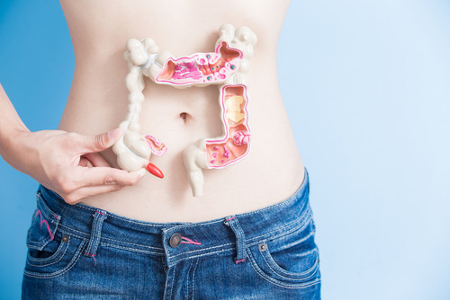
Colon Cancer Treatment in India

Colon cancer, also known as colorectal cancer, is a malignant tumor that develops in the colon or rectum. It is one of the most common types of cancer worldwide. Understanding the various aspects of colon cancer, including its treatment types, symptoms, diagnosis, treatment risks/complications, causes, procedure and recovery, treatment preparation, clinical trials, cost in India, side effects, success rate, and treatment diet, is crucial for patients and their loved ones.
Symptoms:
Symptoms of colon cancer include changes in bowel habits, abdominal discomfort, blood in the stool, unexplained weight loss, and fatigue.
Diagnosis:
Diagnosis involves tests such as colonoscopy, sigmoidoscopy, virtual colonoscopy, stool tests, and imaging scans to identify the presence and extent of the cancer.
Treatment Types:
Treatment options for colon cancer include surgery to remove the tumor, chemotherapy to kill cancer cells, radiation therapy to target the tumor with high-energy rays, targeted therapy to block specific cancer cell functions, and immunotherapy to boost the body's immune system to fight cancer.
Treatment Risks/Complications:
Risks and complications of colon cancer treatment can include infection, bleeding, damage to nearby organs during surgery, side effects like nausea and fatigue from chemotherapy, and potential long-term effects on bowel function.
Causes:
Colon cancer can be caused by a combination of genetic factors, environmental factors, and lifestyle choices. Risk factors include age, family history, certain genetic conditions, inflammatory bowel disease, obesity, a sedentary lifestyle, and a diet high in red and processed meats.
Procedure & Recovery:
Surgery is often the primary treatment for colon cancer, involving the removal of the tumor and nearby lymph nodes. Recovery after surgery may include pain management, changes in bowel habits, and a gradual return to normal activities. Other treatments have specific procedures and recovery timelines.
Treatment Preparation:
Treatment preparation involves discussing the treatment plan with healthcare professionals, understanding potential risks and benefits, and making necessary lifestyle adjustments to optimize overall health and well-being before starting treatment.
Clinical Trials:
Clinical trials offer access to innovative treatments and can contribute to advancements in colon cancer treatment. Participation in clinical trials allows patients to explore new treatment options and potentially benefit from cutting-edge therapies.
Cost in India:
The cost of colon cancer treatment in India can vary depending on factors such as the hospital, location, stage of cancer, and specific treatment options. It is advisable to consult with healthcare providers and insurance companies to get an accurate estimate of the expenses involved.
Side Effects:
Side effects of colon cancer treatment can vary depending on the specific modality used. They may include nausea, diarrhea, fatigue, hair loss, changes in taste, and other temporary or long-term effects. Supportive care is provided to manage these side effects and improve the patient's quality of life.
Success Rate:
The success rate of colon cancer treatment depends on factors such as the stage of cancer at diagnosis, overall health of the patient, and the effectiveness of the chosen treatment approach. Early detection and treatment tend to yield better outcomes. Advances in treatment options have improved survival rates and quality of life for patients with colon cancer.
Treatment Diet:
A healthy diet plays a crucial role in supporting colon cancer treatment. It is recommended to follow a well-balanced diet that includes plenty of fruits, vegetables, whole grains, lean proteins, and low in processed foods and red meats. Healthcare professionals and nutritionists can provide personalized dietary recommendations based on individual needs to support treatment and overall well-being.
#Colon Cancer Treatment in India#Colon Cancer Treatment#Colon Cancer#Cancer#Colon Cancer Symptoms#Colon Cancer Causes#Colon Cancer Diagnosis#Colon Cancer Risks#Colon Cancer Treatment Cost#Colon Cancer Side Effects
0 notes
Photo
The low cost, the high quality of care, and the ease of getting top-notch care are three important advantages of receiving the best medical treatment in Turkey. If anyone is looking for the affordable Colon Cancer Treatment in Turkey then do connect with Medsurge India now.
0 notes
Text
A Complete Guide to the Best Hospitals in India for Liver Cancer & Related Diseases
We have compiled a list of the best hospitals in India for liver cancer surgery and related diseases.
India is one of the most populated countries in the world. With such a large population, it is not surprising that with more and more people coming up with various diseases, hospitals are becoming overcrowded. In order to provide better medical care, it is essential to find solutions that would help reduce overcrowding. One solution is taking advantage of AI-assisted medical decision-making to improve patient outcomes at each step of care- from prevention through diagnosis and treatment all the way to rehabilitation.
Liver cancer surgery in India can be done at top-notch hospitals like CK Birla hospital, Apollo Hospitals, Fortis Escorts Heart Institute, Artemis Hospital & Research Centre, and others like these that offer world-class health care services across India.
What is the best hospital in India for liver cancer and related diseases?
India has not been known for its quality healthcare facilities. The country is seen to be a developing nation, with a huge population. But India has been making rapid progress in the medical field and a lot of things would change in the near future.
In India, patients have limited choices when it comes to hospitals that best provide treatment for liver cancer and related diseases. Nowadays, many medical treatments are made available online- which means more convenience for people who cannot afford to travel to Mumbai or Chennai.
In this article, we discuss the top 5 best hospitals in India that can treat liver cancer and pancreatic cancer surgeries, colorectal cancer surgeries, etc with high competence rates - AI hospitals!
How do you choose the best hospitals for surgery such as liver cancer surgery or pancreatic cancer surgery?
Choosing the best hospital for liver cancer surgery or pancreatic cancer surgery is not an easy task. The reasons behind this are many. Firstly, it depends on the country you are living in and your healthcare coverage status. Secondly, it is also dependent on the type of liver cancer or pancreatic cancer that you have and how advanced it is.
You will need to find out which hospitals around your country offer the highest quality of treatment for your condition and also provide test results after a given time period to ensure that they continue to provide a high-quality service.
A popular trend in the healthcare industry these days is finding out which hospitals provide cost-effective options while offering high-quality care for their patients. You will need to find out which hospitals around your city offer low rates as well as higher rates.
What are the most common types of surgeries performed in these hospitals?
Dr. Sundeep Jain is a renowned surgeon and a Professor in the Department of Surgery of the National University Hospital. He has performed more than 150,000 surgeries in a career spanning over 30 years.
Surgery is one of the most common types of procedures that doctors perform to treat different conditions or injuries. From routine to major surgeries, these are three of the most common surgeries performed in hospitals around India:
* Liver Cancer Surgery- A cancerous tumor can be removed from the liver by surgery
* Pancreatic Cancer Surgery in India- Pancreatic cancer can be treated by surgery, chemotherapy, and radiotherapy
* Colorectal Cancer Surgery- A polyp that grows out of the rectum or colon may be removed surgically by a doctor or it can be removed by your family physician, which means it will appear as if you have been diagnosed with cancer.
* Disfunctional Colon- This begins when there is a section of the colon that is all tight and has no opening in the center.
*Esophagus Cancer Treatment in India: Involves removal of the part of the esophagus that has become cancerous and re-establishing new healthy tissue that can be used to keep patients with this condition alive for an extended period
*Gallbladder Stone Surgery: This type of surgery involves removing stones from the gallbladder (a small organ about 4 inches long below the liver) and ensuring that there is no spillage or leakage after surgery.
1 note
·
View note
Photo

Лечение в Индии дает новую надежду пациентам с раком толстой кишки
Электронная почта: [email protected]
Телефон: + 91-9371136499
https://medicaltourfromrussia2india4health.blogspot.com/2019/08/colon-cancer-treatment-surgery-India-low-cost-advantages.html
1 note
·
View note
Photo

Carob Market by Type, Price, Competition, Forecast & Opportunities
Carob is a tree grown in the Mediterranean region known by its scientific name, Ceratonia siliqua L., and renowned worldwide as a substitute of chocolate. Seeds and pulp of the carob fruit are used for the manufacture of stabilizers and thickeners in the food sector. The global carob market report by Market Research Future (MRFR) contains a list of drivers, carob industry trends, and drawbacks for its producers and consumers alike with prospects forecasted for the period of 2019 to 2024 (forecast period).
Segmentation
The global carob market has been segmented based on category, form, application, and region.
By category, the global carob market has been divided into conventional and organic. The conventional segment has gained a major market share but may lose its position to the organic segment. On the other hand, the organic segment is likely to exhibit a strong growth rate in the coming years due to its use as an energy supplying feed to livestock animals and pets.
On the basis of form, the global carob market has been divided into powder, gum, and others. The gum segment is expected to be the topmost segment during the forecast period due to its use as a substitute for guar-guar gum. The carob bean gum, also known as locust bean gum, has various health benefits and can aid in the treatment of heart disease, colon cancer, and diabetes. But the powdered segment can gain in speed in the coming years due to the growing bakery industry and emergence of various bakeries experimenting with new fruits and flavors to appeal to an audience with modern tastes.
Major market applications include snacks, dairy products, bakery and confectionery, and others. The bakery and confectionery segment is likely to lead as the biggest application till 2024, however the snacks segment may overtake the segment due to massive demand for healthy snacks.
Market Outlook
The global carob market size is expected to rocket over the forecast period owing to the use of carob bean gum in stabilizers and thickeners. Need for chocolate substitutes among the general populace due to alarming levels of obesity, diabetes, and other lifestyle diseases can warrant the market demand. The use of carob in chocolate-free recipes as well as the support from the burgeoning food sector can push the market to new heights.
The use of carob pods in animal feeds to encourage the growth of farm animals can fuel the market growth. Rise of veganism and the inclusion of carob powder for the production of chocolate-free cakes and confectionery can support the rise of the global carob market. Its high-fiber and low-caffeine content has made it a favorite for food manufacturers looking to expand into alternative food sectors. The lactose-free ingredient can be used in various food items and appeal to a wider audience looking for modern tastes.
But limited availability of carob trees and skyrocketing costs of harvests can impede the market growth.
Regional Analysis
The global carob market has been studied with regard to four key regions—Europe, North America, Asia Pacific, and the Rest-of-the-World (RoW).
Europe is touted to be the region deemed to be highly profitable for the global market due to the easy growth of these trees in Italy, Spain, and Portugal. High consumption of the carob fruit as well as its use in powdered forms is driving the growth within the region.
On the other hand, the APAC region can exhibit a spectacular growth rate due to the large food industry and its plans to expand into new sectors. The rising economies of India and China and the rising purchasing power of the denizens are factors driving the regional carob market growth.
Competitive Landscape
DuPont, AEP Colloids, Savvy Carob Co Ltd, Altrafine Gums, Tate & Lyle, TIC Gums, Inc., Pedro Perez, Australian Carobs Pty Ltd., Stavros Parpis Foods Ltd, Carob S.A., and others are prime players profiled and discussed in the global carob market report.
Industry News
The consumption of collagen is gaining momentum and consumers are eating it in powdered form to increase their fiber intake. Maca powder is effective in enhancing the flavor of foods and beverages. This powder is being sold at local stores for making it to large masses. Carob powder is added to the mix to lower its bitter taste.
NOTE: Our Team of Researchers are Studying Covid19 and its Impact on Various Industry Verticals and wherever required we will be considering Covid19 Footprints for Better Analysis of Market and Industries. Cordially get in Touch for More Details.
1 note
·
View note
Text
Minimum cost of colon cancer treatment India is a very comforting for the patients because the surgical operation and treatment in India is far lower priced as compared to other growing countries and advanced international locations
#Colon Cancer Surgery in India#Low Cost of Colon Cancer Treatment India#Top Oncologist For Colon Cancer India#Top Colon Cancer Treatment Hospital of India#Top Surgeons for Colon Cancer Treatment India#Affordable Cost of Colon Cancer Treatment India#Minimum cost of colon cancer treatment India
1 note
·
View note
Text
Capsule Endoscopy Market Size, Key Players, SWOT, Revenue Growth Analysis Till 2027

Capsule endoscopy is the part of endoscopy in which a small capsule and pill shaped wireless camera used to examine the digestive system. It is the advanced step up of endoscopy. This method is used to identify diseases like gastrointestinal bleeding, oesophageal diseases, intestine diseases and colon related diseases. Increasing prevalence of these diseases are the major driving factor for the market. Continuous technological advancement in endoscopy and increasing healthcare spending has provided fuel for the growth of the market. But high cost for the diagnosis and limited reimbursement are the major hurdles for the growth of the Capsule endoscopy market.
Further an endoscopy is a procedure that allows physicians or surgeons to look inside interior parts of the organ. The technique is used for both diagnostic and therapeutic purpose. To perform endoscopy a specialized instrument is used named as endoscope. There are various type of endoscopes and adoption of endoscope depends upon the affected body organ such as, bronchoscopes are used for lungs related problem, arthroscopes are preferred for problems associated with joints, and many others. Other instruments required during an endoscopic procedure includes Biopsy forceps, Cytology brush, Flexible forceps, trocar sleeves, etc. For therapeutic purpose, endoscopes are either attached or passed through surgical instruments to perform particular surgery. Endoscopic Surgery is a type of minimally invasive surgery in which an endoscope is inserted in the body through small incisions to examine the internal organs. The major advantages associated with endoscopic surgery are small incisions, minimal blood loss, less scarring, low risk of infection, and less recovery time. As per the 2014 statistics suggested by Eurostat, it is observed that the Cataract surgery is the most common minimally invasive surgery in the European region as this surgery was performed 4.3 million times across the EU Member States and another most commonly used procedure was bronchoscopy for diagnosis with a frequency rate of 685 per 100 000 inhabitants in Croatia and more than 400 in Latvia and Germany.
Request Free Sample Copy at: https://www.marketresearchfuture.com/sample_request/1359
Rising prevalence rate of chronic diseases, technological advancement in endoscopy, and increasing funding and reimbursement towards screening and treatment with endoscopic techniques are promoting the growth of endoscopy devices market globally increasing popularity of minimally invasive surgery is also creating a huge scope for the growth of market.
Despite these drivers, there are some issues associated with endoscopy devices market. Lack of skilled physicians and endoscopists, and infections caused by endoscopes may hinder the growth of market to an extent.
Fujifilm Holding Corporation (Japan), Olympus Corporation (Japan), Given Imaging (Israel), RF System Lab (US), Capso Vision Inc. (US), IntroMedic Co., Ltd. (South Korea), Chongqing Jinshan Science & Technology Co. (China), Johnson and Johnson (US) and others are some of the major players in the Global Capsule endoscopy Market.
Global Capsule endoscopy Market – Regional Analysis
Geographically, Americas region is commanding the largest market share owing to the increasing adoption rate of endoscopic surgery. According to the Centers for Disease Control and Prevention, it is reported that in the United States due to increase in colonoscopy procedures the percentage of population detected with colon cancer has been increased. It is also estimated that on considering colonoscopy screening capacity of the United States now, 80% of the eligible population will be screened by 2023
Further Thus the market potential of nations such as India, Thailand, Vietnam etc. is huge which has been constrained by lack of healthcare professionals specifically endoscopists.
The market maturity of different nations also varies by an immense scale. China has the most market potential followed by India. India represents the next nation which will drive the future capsule endoscopy device market. Developed markets such as Japan, US etc. is likely to witness modest low single digit growth. The competition is also strong in the developed regions market. Thus developing region nations such as India, Argentina etc. are ideal for any new entrant.
There were approximately 15,000,000 endoscopy procedures in the US in 2016 alone. The population of US in 2016 is approximately 323.1 million. Thus there is one endoscopy procedure performed per 20 million population. The comparative rates for developing regions are far below taking the United States as reference base. Hence it is estimated that the market potential for global capsule endoscopy is expected to huge and will grow immensely in the coming future.
Browse Full Report @ https://www.marketresearchfuture.com/reports/capsule-endoscopy-market-1359
About Market Research Future:
At Market Research Future (MRFR), we enable our customers to unravel the complexity of various industries through our Cooked Research Report (CRR), Half-Cooked Research Reports (HCRR), & Consulting Services. MRFR team have supreme objective to provide the optimum quality market research and intelligence services to our clients.
Have a Look at Related Reports:
Telehealth Market
Implantable Cardioverter Defibrillator Market
Cardiomyopathy Medication Market
Onychomycosis Market
Liver Cirrhosis Treatment Market
Contact us:
Market Research Future (part of Wantstats Research and Media Private Limited),
99 Hudson Street, 5Th Floor,
New York, New York 10013
United States of America
+1 628 258 0071
Email: [email protected]
0 notes
Text
Best Cancer Hospital In Chandigarh
Why Grecian hospital is the best cancer hospital in Chandigarh?
If you live in the northern region of India, then you are definitely aware of Grecian hospital. The Grecian hospital is one of the best cancer hospitals in Chandigarh. We have a team of highly qualified and experienced cancer specialist doctors that can treat all forms of cancer. Surgery, radiation, and medicine are the different forms of treatment that our specialists use to treat all forms of cancer.
Our best cancer hospital in Chandigarh has the world-class therapy to cure cancer and at the same time, we ensure that it will never come back. We use different preventive measures to ensure it will never return. The whole staff of our hospital is very friendly and they will give you a feel like your home.
We have a capacity of 350 beds here. With this, we have multiple pharmacies and world-class diagnostic facilities that can give excellent healthcare to all types of cancer patients.
Know more about cancer
Cancer can be a fatal disease, especially if not diagnosed or treated on time. Cancer is simply an uncontrolled division of cells that can occur anywhere and any part of the body. Cancer has become most prevalent in India and is responsible for a large number of deaths. Besides this, it can put a lot of financial burden on any family.
In India, lung, oral, and throat cancers are most common in men. While in women, ovarian, breast, and cervix are the most common. For people of the old age group the colon, kidney, and prostate cancer is more prevalent.
The main reasons responsible for cancer are an unhealthy lifestyle. In men, smoking and tobacco use is the common cause of lung or oral cancer. The reason for other types of cancers may be obesity, pathogens, and genetics.
What are the treatments that the best cancer hospital in Chandigarh offers for its cancer patients?
Onco-Surgery
Laparoscopic surgery
• Genitourinary cancer (Radical Nephrectomy, Radical Cystectomy)
• Esophageal Cancer (Thoraco-Laparoscopic Esophagectomy)
•Colorectal Cancer (Abdominoperineal Resection, Colostomies, Low Anterior Resection)
•Gynecological Cancer (Extrafascial Hysterectomy, Pelvic Lymph Node Dissection, Radical Hysterectomy)
Head and Neck Surgery
•Oral Cancer (Composite Resection, Neck Dissection, Glossectomy for Carcinoma tongue, Flap Reconstruction)
•Thyroidectomy, Parotidectomy
•Larynx and Hypopharynx (Laryngopharyngeal esophagectomy, Laryngectomy)
Breast Surgery
•Breast Conservation Surgery
•Sentinel Node Biopsy
•Modified Radical Mastectomy
•LD Flap Reconstruction
Hepatopancreatico- Biliary Surgery
•Radical Cholecystectomy (for Carcinoma Gall Bladder)
•Whipples Operation (for Carcinoma Pancreas)
Chemotherapies
•All conventional chemotherapies are performed here
•We adhere to NCCN and ESMO guidelines
•We use the most advanced targeted therapies
•Intraperitoneal and Intrathecal Chemotherapy
•Immunotherapy
Conclusion
Grecian hospital's main aim is to provide world-class cancer treatment to our patients at a reasonable cost. Our doctors have a high success rate in surgeries related to cancer. If you have any confusion about our treatment you are free to contact us at any time. The best cancer hospital in Chandigarh is always ready to do your help.
0 notes
Link
0 notes
Link
Abstract
Turmeric has been used as a medicinal herb for thousands of years for treatment of various disorders. Although curcumin is the most studied active constituents of turmeric, accumulating evidence suggests that other components of turmeric have additional anti-inflammatory and anti-tumorigenic properties. Herein, we investigated anti-inflammatory efficacy and associated gene expression alterations of a specific, curcumin preparation containing essential turmeric oils (ETO-curcumin) in comparison to standard curcumin at three specific doses (0, 5, 25 or 50 mg/kg), in an animal model of dextran sodium sulfate (DSS)-induced colitis. The present study showed that both ETO and standard curcumin treatments provided protection against DSS-induced inflammation. However, ETO-curcumin improved disease activity index (DAI) dose-dependently, while the anti-inflammatory efficacy of standard curcumin remained constant, suggesting that ETO-curcumin may provide superior anti-inflammatory efficacy compared to standard curcumin. Gene expression analysis revealed that anti-inflammatory cytokines including IL-10 and IL-11 as well as FOXP3 were upregulated in the colon by ETO-curcumin. Collectively, these findings suggest that the combined treatment of curcumin and essential turmeric oils provides superior protection from DSS-induced colitis than curcumin alone, highlighting the anti-inflammatory potential of turmeric.
Go to:
Introduction
Turmeric has been used traditionally as a medicinal herb in India and South East Asia for thousands of years for various illnesses including biliary disorders, anorexia, coryza, cough, hepatic and rheumatic ailments and a variety of other chronic inflammatory diseases. In particular, curcumin, the active principle extracted from the dried rhizomes of Curcuma longa (or turmeric) is perhaps one of most studied natural compounds within the context of complementary medicine. In addition to its well-established anti-inflammatory properties, clinical studies in the recent years have highlighted its therapeutic efficacy in a variety of diseases including arthritis and depression1, 2, as well as its potent anti-tumorigenic potential3–6. Interestingly, growing body of data also indicate that in addition to curcumin, other constituents of turmeric, primarily essential turmeric oils (ETO) comprising of aromatic-tumerones (ar-tumerones), α-turmerones, β-turmerones, α-santalene and aromatic curcumene, also possess significant anti-inflammatory and anti-oxidant properties7–9. One of curcumin’s potential limitations is that it is poorly absorbed following ingestion; hence there has been a tremendous interest in developing strategies to enhance its absorption and systemic bioavailability. In this context, previous studies have demonstrated that administration of curcumin complexed with essential turmeric oils (ETO-curcumin) enhanced its bioavailability in circulation by 7–10 fold compared to standard curcumin, which subsequently lead to significantly improved bioactivity10, 11. The ETO-curcumin has been shown to exert superior anti-tumorigenic effects by permitting differentiation of cancer stem cells, reversing epithelial-to-mesenchymal transition and by enhancing the efficacy of chemotherapeutic agents such as 5-fluorouracil in in vitro and pre-clinical studies12–14. Although these data indicate that ETO-curcumin appears to have higher bioactivity, no studies have directly investigated the bioactivity of ETO-curcumin in comparison to standard curcumin and the underlying anti-inflammatory mechanisms.
Ulcerative colitis (UC) is an inflammatory disorder which affects the entire colorectum, and is one of the two major forms of inflammatory bowel diseases (IBD) along with Crohn’s disease (CD). Ulcerative colitis remains one of the most difficult gastrointestinal diseases to manage due to lack of definitive therapies15. While anti-tumor necrosis factor alpha (TNF-α) antibodies appear to be moderately effective in clinical management of CD and UC, not all patients respond to these antibodies, and these regimens often associate with severe side-effects16. Emerging evidence suggests that dysregulation of inflammatory transcription factors such as NF-κB and signal transducers and activators of transcription (STAT) could be involved in pathogenesis of UC17–19. Based upon these findings, several molecular inhibitors are currently being developed to target these inflammatory pathways. It is noteworthy to mention that the effectiveness of curcumin has been consistently demonstrated in preclinical models of colitis as well as in patients with UC20–22, through down-regulation of both NF-κB and STAT pathways23–26. Furthermore, a recent randomized, multinational clinical study demonstrated that combined treatment with curcumin and mesalamine resulted in remission in 54% of patients with colitis, while none of the patients achieved clinical remission in the control, untreated group27. Considering that curcumin is a readily available, safe and a cost-effective botanical, there is a growing interest in exploring its clinical efficacy individually or as an adjunctive treatment for managing and/or treating UC and subsequently improving the overall quality of life for patients with this inflammatory disease. Although curcumin is a well-established anti-inflammatory agent, the exact mechanism(s) by which it attenuates inflammatory burden in diseases such as UC remains unclear. Herein, we demonstrated that curcumin exhibited significant anti-inflammatory effects in a mouse model of dextran sodium sulfate (DSS)-induced colitis. In particular, a unique formulation of curcumin containing essential turmeric oils (ETO-curcumin) displayed greater anti-inflammatory efficacy compared to the standard curcumin, as evidenced by the reduced disease activity index (DAI) and changes in gene expression alterations of the inflammation-related genes, highlighting that ETO enhances the anti-inflammatory efficacy of curcumin.
Go to:
Results
Curcumin exerts anti-inflammatory effects in DSS-induced colitis
Since curcumin is a well-established botanical with anti-inflammatory properties, we wanted to determine whether presence of essential turmeric oils have any additional bioactivity, and hence, we sought to test the differences in anti-inflammatory efficacy between ETO- and standard curcumin. We first tested the efficacy of both types of curcumin at a relatively low treatment dose of 25 mg/kg body weight. In order for animals to be acclimatized to gavaging, we pretreated mice with curcumin for one week, followed by addition of 3% DSS in their drinking water to induce colitis (Fig. 1A). Based on the DAI values, both ETO-curcumin and standard curcumin-fed animals had less severe colitis from DSS treatment compared to the control animals, as early as day 6 (p < 0.001 and p < 0.01 respectively; Fig. 1B). Similarly, on day 7 of DSS treatment, both curcumin treatments showed reduction in DAI compared to DSS-control group, indicating their ability to attenuate inflammation-mediated colitis. Analyzing individual measures of DAI, ETO-curcumin treated mice maintained superior DAI parameters compared to the DSS control group, in particular the body weight loss category. None of the curcumin treatments altered stool consistency, but both were effective in reducing fecal bleeding compared to DSS-control mice (p < 0.01). Since colon length is another well-established indicator of DSS-induced colitis, we examined the length of colon after 7 days of DSS treatment. As expected, DSS treatment resulted in colon shortening, while ETO-curcumin, but not standard curcumin, significantly prevented such colon shortening (p < 0.01; Fig. 1C). Similarly, enlargement of spleen is another indicator of immune response to inflammation. Spleen weight in ETO-curcumin treated mice, but not standard curcumin, was lower compared to DSS-treated mice (p < 0.01), further confirming its anti-inflammatory effects (Fig. 1D). In addition, both ETO- and standard curcumin treated mice had lower histological scores than DSS-controls (p < 0.01 and p < 0.05 respectively; Fig. 1E). Taken together, while several DAI parameters showed an improved trend toward ETO-curcumin having superior anti-inflammatory efficacy than standard curcumin, our results demonstrated that both curcumin preparations were effective in mitigating DSS-induced colitis.
Open in a separate window
Figure 1
Curcumin attenuates DSS-induced inflammation at 25 mg/kg body weight. (A) Graphical representation of curcumin treatment strategy. (B) Changes in individual categories of disease activity index (DAI) (top): body weight changes, stool consistency, and stool bleeding (left-right). Changes in DAI (bottom). (C) Colon length, (D) Spleen weight and (E) Histology score on day 14. *p < 0.05, **p < 0.01, ***p < 0.001.
ETO-curcumin exhibited superior anti-inflammatory effects over standard-curcumin
In order to determine whether ETO-curcumin has a superior anti-inflammatory efficacy compared to standard curcumin, we doubled the treatment dose (50 mg/kg body weight) to re-evaluate the effects of these two curcumin types (Fig. 2A). Moreover, this dose has previously been established as an optimal dose for standard curcumin for DSS-induced rodent study20. During the first three days of treatment, no differences in DAI were observed between DSS-treated groups regardless of curcumin treatment (Fig. 2B). However, from day 4 of DSS treatment, ETO-curcumin treated mice displayed significant attenuation of DAI compared to DSS-control group, while animals treated with standard curcumin showed a delayed attenuation in DAI. Furthermore, ETO-curcumin showed lower DAI at day 7 of DSS treatment compared to both DSS control and standard curcumin treated groups (p < 0.001 and p < 0.05) indicating that ETO-curcumin was significantly more effective in attenuating DSS-induced colitis than standard curcumin. In order to determine which specific DAI parameters contributed to ETO-curcumin’s superior anti-inflammatory efficacy over standard curcumin, we further assessed each DAI criterion individually. While both ETO- and standard curcumin formulations were equally effective for maintaining consistency in feces (both p < 0.05 compared to DSS-control), ETO-curcumin-treated animals showed superior attenuation in DSS-induced body weight loss compared to standard curcumin (p < 0.05; Fig. 2B). In addition, we noted a statistical trend indicating that ETO-curcumin treatment resulted in less severe DSS-induced intestinal bleeding (p < 0.08) compared to standard curcumin treatment group. As expected, DSS treatment resulted in shrinkage of the colon, while both curcumin treatments attenuated shortening of the large intestine (both p < 0.05; Fig. 2C). Although ETO-curcumin treated mice demonstrated reduced spleen weight compared to DSS-treated group (p < 0.05), standard curcumin treated group only showed a trend in spleen weight reduction (p = 0.06; Fig. 2D). Consistent with our DAI measurements, histological evaluation showed that both ETO- and standard curcumin significantly attenuated DSS-induced colitis (p < 0.01 and p < 0.05 respectively; Fig. 2E and F). Collectively, these data indicate that ETO-curcumin resulted in superior anti-inflammatory effects compared to standard curcumin, suggesting that the presence of ETO may synergistically enhance the bioactivity of curcumin in the colon.
Open in a separate window
Figure 2
ETO-curcumin exerts superior anti-inflammatory effects compared to standard curcumin on DSS-induced inflammation at 50 mg/kg body weight. (A) Graphical representation of curcumin treatment strategy. (B) Changes in individual categories of disease activity index (DAI) (top), body weight changes, stool consistency and stool bleeding (left-right). Changes in DAI (bottom). (C) Representative image of colons (left) and average colon length (right). (D) Spleen weight and (E) Histology score on day 14. (F) Representative haematoxylin and easin (H&E) staining of large intestine on day 14. 100x magnification (left) and 400x magnification (right). *p < 0.05, **p < 0.01, ***p < 0.001.
Next, to determine whether the superior anti-inflammatory efficacy of ETO-curcumin was due to combined effects of ETO and curcumin or as a result of independent contribution of ETO, we repeated these experiments with the addition of an ETO alone group. Consistent with our initial experiments, once again we observed significant attenuation of DAI for both curcumin and ETO-curcumin treatment groups (Supplementary Fig. 1). Furthermore, to our surprise, ETO gavaged animals showed no difference in DAI compared to that of DSS-control group, suggesting that anti-inflammatory effects of ETO-alone are minimal (Supplementary Fig. 1). Therefore, our data indicates that ETO enhanced the anti-inflammatory efficacy of curcumin.
ETO-curcumin enhances anti-inflammatory effects in a dose-dependent manner
We next used a mathematical model, “linear mixed model”, to further evaluate the efficacy of ETO and standard curcumin. We assessed the dose-related effects over three doses - 5, 25 and 50 mg/kg body weight (5 mg/kg shown in Supplementary Fig. 2). The combined DAI across three doses for both types of curcumin are shown in Fig. 3A. Considering that all treatment doses for both curcumin extracts demonstrated significant attenuation in DAI, we first examined whether these protective effects are dose-dependent. To evaluate this hypothesis, we compared DAI and its associated sub-categories across all doses at day 5 and 7 of DSS treatment and graphically represented dose-associated effectiveness of each curcumin type (Fig. 3B and Supplementary Table 1). We color coded each cell according to its effectiveness with red representing the highest improvement in the effectiveness between doses, while blue representing no difference. Surprisingly, within the standard curcumin group, we observed no statistical difference between doses for DAI, weight change, stool consistency and fecal bleeding at both days 5 and 7. These data indicate that although standard curcumin clearly reduces DAI, the anti-inflammatory efficacy of standard curcumin was not dose-dependent (Fig. 3B and Supplementary Table 1). In contrast, ETO-curcumin demonstrated a dose-dependent improvement in efficacy at both days 5 and 7. Significant improvements were observed for DAI (p < 0.001), weight change (p < 0.001) and fecal bleeding (p = 0.001) when 50 mg/kg treatment group was compared to 5 mg/kg treatment group at both days 5 and 7 (Fig. 3Band Supplementary Table 1). Furthermore, the comparison between 25 mg/kg and 50 mg/kg showed improvement in DAI (p < 0.05) for ETO-curcumin in both days 5 and 7 compared to standard curcumin. Collectively these results suggest that ETO-curcumin enhanced anti-inflammatory efficacy in a dose-dependent manner in a DSS-induced colitis animal model, while standard curcumin appeared to show no dose-associated improvement in DAI, at least for the doses that we evaluated.
Open in a separate window
Figure 3
Dose-dependent comparison between standard curcumin and ETO-curcumin on disease activity index (DAI). (A) DAI of standard curcumin (left) and ETO-curcumin (right) according to their doses. (B) Graphical representation of comparison of doses within the treatment group at DSS treatment days 5 and 7 for DAI and DAI associated parameters. (C) Graphical representation of comparison between standard curcumin and ETO-curcumin at individual doses (5, 25, 50 mg/kg) on DSS treatment days 3, 5, 7 and cumulative comparsion. For both panel B and C red cells represent significant difference between groups, while blue represents similar values.
Next, we comprehensively analyzed the anti-inflammatory efficacies between ETO- and standard curcumin at each dose (5, 25 and 50 mg/kg) at specific time points (days 3, 5 and 7). Once again we represented our data graphically to illustrate time and dose-dependent dynamics of two curcumin types (Fig. 3C). Although differences between ETO and standard curcumin were small at DSS treatment day 5, the difference in DAI associated parameters between ETO- and standard curcumin treatments at day 7 increased substantially. In particular, we found statistically significant differences between ETO- and standard curcumin for body weight changes during days 5 and 7 for 25 and 50 mg/kg doses (p < 0.001). Within the group with highest treatment dose (50 mg/kg), ETO-curcumin demonstrated significant improvements in DAI, weight change, and fecal bleeding at day 7 compared to standard curcumin (all p < 0.05: Fig. 3C and Supplementary Table 2). Collectively, these data represent significant time-dependent shift in effectiveness between ETO- and standard curcumin for DAI suggesting that ETO-curcumin exerted superior anti-inflammatory effects compared to standard curcumin.
Curcumin alters gene expression profiles of inflammation-associated genes
To clarify the anti-inflammatory mechanism of curcumin, we investigated the gene expression profiles of inflammation-associated genes from tissues collected from colon in animals treated with 50 mg/kg dose using a Nanostring platform. As expected, there were significant alterations in the expression profiles of inflammatory genes in both curcumin treated vs. DSS-control groups (Fig. 4A). Since the overall gene expression profile of all DSS treatment groups appear to be relatively similar, we further analyzed our data using a mathematical estimation called “molecular distance to health” (MDTH) to determine whether there is a significant difference in overall gene expression profiles between DSS-control and curcumin groups. MDTH is a mathematical estimation which calculates overall shift in expression of genes between treatment groups according to their deviations in gene expression signature28. Intriguingly, MDTH analysis showed considerable shift in overall gene expression profile when animals treated with DSS were compared to non-DSS treatment control group (p < 0.001; Fig. 4B). However, both curcumin treatment groups significantly shifted MDTH towards non-DSS control group when compared to DSS-control group (ETO-curcumin, p < 0.01 and standard curcumin p < 0.05), indicating that curcumin treatments attenuated overall shift towards inflammatory molecular signature of control-DSS group (Fig. 4B). Due to relatively large variation in MDTH values within ETO-curcumin treated group, we were unable to detect difference between ETO- and standard curcumin treatments using MDTH.
Open in a separate window
Figure 4
Curcumin alters gene expression of inflammation associated genes (A) Treatment groups represented using molecular distance to health (MDTH) (B) Heat map of DSS-control, ETO-curcumin and curcumin normalized to untreated mice (C) Volcano plot comparison between control group and ETO-curcumin treated group (top) control group and curcumin treated group (bottom) (D) Expression levels of IL-10, IL-11, CCL17, CXCL5 determined by nanoString.
Next, we examined specific genes which were altered with curcumin treatment by first identifying differentially expressed genes between ETO- and standard curcumin against DSS-controls with greater than 1.5 fold change. A total of 50 differentially expressed genes were identified, suggesting that both ETO and standard curcumin altered common anti-inflammatory molecular pathways (Fig. 4C). We further interrogated the data to identify most differentially expressed genes for both ETO-curcumin and standard curcumin in comparison to DSS-controls and represented the data in volcano plots (Fig. 4D). We initially identified 10 such genes, which included 2 upregulated (IL10 and IL11) and 8 downregulated (CCL17, HSPB1, MEF2C, C7, HSPB2, PPP1R12B, H2-EA and CEBPB) genes for ETO-curcumin group, and one downregulated gene for the standard curcumin group, CXCL5, (Fig. 4D). Based on mechanistic findings of previous studies29–31, we decided to focus on two most highly upregulated genes for ETO-curcumin as well as a downregulated gene (CCL17) and the lone differentially expressed gene for standard-curcumin (CXCL5) (Fig. 4E).
ETO-curcumin attenuates inflammation through upregulation of anti-inflammatory cytokines
In order to validate the genes identified by Nanostring array, we used qRT-PCR to assess the gene expression of the identified genes from the colonic tissue samples (Fig. 5). Intriguingly, these two anti-inflammatory cytokines identified by Nanostring array, IL-10 and IL-11, are well-known anti-inflammatory cytokines. IL-10 is a well-established anti-inflammatory cytokine essential for the maintenance of the epithelial layer homeostasis and minimizes damages obtained from events such as infection29. Similarly IL-11 is another cytokine with anti-inflammatory properties manifested through suppression of TNF-α and IL-130. Confirming our Nanostring array data, the expression of anti-inflammatory cytokines, IL-10 and IL-11, was upregulated by ETO-curcumin compared to DSS-control (p < 0.01 and p < 0.05 respectively). We next determined whether upregulation of IL-10 is caused by upregulation of FOXP3, a master regulatory transcription factor of regulatory T-cells (Treg cells), in DSS-induced colitis32. Treg cells have been shown to suppress immune responses in humans and inhibit colitis through IL-10 production33. As expected DSS treatment resulted in significant downregulation of FOXP3 expression (p < 0.05), but ETO-curcumin was able to attenuate DSS-induced FOXP3 downregulation (p < 0.05). Our data suggest that ETO-curcumin may stimulate the Treg cell production and subsequently upregulate IL-10. In contrast, we confirmed that CCL17, a chemokine involved in induction of intestinal inflammation31, was suppressed by both standard curcumin and ETO-curcumin treatments. Interestingly, CXCL5 expression was downregulated in standard curcumin-treated mice, while the effect was not seen in the ETO-curcumin-treated group, suggesting that standard curcumin may exert anti-inflammatory effects through different mechanisms. Collectively, these data indicate that oral administration of curcumin protects against DSS-induced colitis through modulation of immune response.
Open in a separate window
Figure 5
Curcumin suppresses inflammation through modulation of inflammation associated genes. (A) Gene expression of IL-10, IL-11, CCL17, CXCL5 and FOXP3 normalized to 18S (B) Schematic representation of how ETO-curcumin exerts its anti-inflammatory effects.
Go to:
Discussion
In contrast to many south eastern Asian countries, consumption of turmeric as a dietary spice is generally uncommon in the western world. Nonetheless, in view of growing scientific evidence for the health benefits of curcumin, the active principle component present in the turmeric, curcumin supplements are becoming more widely accepted and popularized in the western nations. However, a question remains whether commercially-available curcumin extracts claiming enhanced bioavailability function comparably and whether curcumin supplementation is an equally effective approach as consuming this phytomedicine through dietary turmeric. The present study was undertaken to address some of these questions, and we deliberately chose to compare the anti-inflammatory effects of standard curcumin with a high-absorption curcumin, ETO-curcumin, in a well-established animal model of colitis. In this study we made several key observations. First, we confirmed anti-inflammatory properties of curcumin on the disease activity index in an animal model of DSS-induced colitis. Second, our data indicates that ETO-curcumin displayed a superior anti-inflammatory efficacy than that of standard curcumin and the subsequent mathematical analysis revealed that the ETO-curcumin associated anti-inflammatory effects were particularly pronounced at higher doses. Third, further investigation of the mechanisms by which ETO-curcumin exerted anti-inflammatory effects showed that ETO-curcumin upregulated anti-inflammatory cytokines including IL-10 and IL-11 and suppressed the chemokine CCL17.
Despite the majority of turmeric-related research has primarily focused on curcumin, less studied components of turmeric such as ETO, has been shown to have significant anti-inflammatory property. In particular, ar-turmerone has shown to possess potent anti-inflammatory, anti-oxidative and anti-platelet properties8, 9. Furthermore, clinical studies have demonstrated that regular ETO-curcumin consumption improved diseases including arthritis, depression and Alzheimer’s disease1, 2, 34. Although currently there is no study conducted on comparing anti-inflammatory properties of ETO and curcumin, ETO has been shown to exert more potent antifungal activity than curcumin35. Our data indicated that ETO-curcumin exerted superior anti-inflammatory effects than standard curcumin in DSS-induced mouse model of colitis and that the ETO enhances bioactivity of curcumin. Consistent with our data, combined treatment of curcumin and ar-turmerone has been shown to reduce adenoma multiplicity in DSS-dimethylhydrazine-induced inflammation associated model of CRC36. Although we showed that ETO-curcumin had overall superior anti-inflammatory efficacy than that of standard curcumin, we observed that ETO was less effective at lower doses. One potential reason for ETO-curcumin not being able to exert superior anti-inflammatory effects compared to the standard curcumin treatment group could be low ETO dose in these treatment groups. Furthermore, we demonstrate that mice gavaged with ETO alone showed no influence on DAI, suggesting that ETO contributed to enhanced activity of curcumin rather than acting as a potent anti-inflammatory agent on its own. Although our data indicates that the enhancement of bioactivity of ETO-curcumin is not exclusively related to bioavailability of curcumin, it is important to note that absorption of curcumin has been shown to increase significantly with the presence of lipophilic turmerones37.
In the present study, we investigated a range of curcumin doses which represented equivalent concentration of typical human consumption. Therefore, we chose our highest gavaging dose as 50 mg/kg body weight based on a previous study which showed that curcumin exerted anti-inflammatory effects at this dose in a DSS-induced colitis model20. According to the animal-to-human dose conversion formula using intestinal surface area, a mouse gavaged with 50 mg/kg body weight curcumin is equivalent to an individual with 70 kg weight consuming 283 mg curcumin38. While this calculation is relatively crude, this dose is still significantly less than a typical commercially available curcumin capsule which generally contains approximately 500 mg of curcumin. We confirmed that curcumin at 50 mg/kg resulted in significant attenuation of DSS-induced colitis and also demonstrated that curcumin at lower doses also attenuated DSS-induced colitis. This is of significant importance as several previous clinical studies have used doses exceeding 2 g per day which required multiple daily intakes39–41. While extrapolating animal data to human is difficult, it is important to maintain the doses used in the animal study as close as possible to clinical circumstances.
The present study also assessed gene expression changes of standard curcumin and ETO-curcumin in a panel of inflammation-associated genes. We identified anti-inflammatory cytokines, IL-10 and IL-11, were differentially expressed in ETO-curcumin compared to DSS-control. IL-10 is a well-established anti-inflammatory gene which is dysregulated in inflammatory disease and curcumin has been shown previously to stimulate promoter activity of IL-10 gene42. We have demonstrated that curcumin, ETO-curcumin in particular, elevated IL-10 expression in the colonic tissue indicating that ETO-curcumin exerts its anti-inflammatory effects through upregulation of anti-inflammatory cytokines. One of the IL-10 stimulatory mechanisms is Treg cells, which can be assessed through the expression of FOXP343. We demonstrated that FOXP3 expression was suppressed by DSS-induced colitis, but intriguingly curcumin treatments, ETO-curcumin in particular, reversed this suppression. While curcumin has been shown to stimulate Treg cells in an animal model of inflammation44, our study indicates that ETO-curcumin was more effective in exerting Treg cells mediated anti-inflammatory immune response. In addition, we identified inflammation-associated genes; CCL17 and CXCL5 were modulated by curcumin treatments. CCL17 is a chemokine which is required for induction of intestinal inflammation and counteracts Treg cells mediated protection from colitis31. While exact mechanism by which curcumin modulate this complex immune response requires further investigation, our data suggests that curcumin appears to have profound effects on immune response to inflammation. Although curcumin has been shown in modulate several anti-inflammatory pathways, the present study assessed the anti-inflammatory molecular profile at day 7 of DSS treatment. At this stage colitis was already severe for the majority of animals and this was reflected in the overall gene expression changes represented by the molecular distance to health (MDTH) assessment. Therefore, this could be the reason that only relatively small numbers of genes were differentially expressed in both standard curcumin and ETO-curcumin groups. Nevertheless, our data show that ETO-curcumin altered overall anti-inflammatory gene expression profile greater than that of standard curcumin reflecting its anti-inflammatory efficacy seen in DAI. Based on the findings of the present study, we illustrated potential mechanisms of ETO-curcumin in Fig. 5B. ETO-curcumin could exert more potent anti-inflammatory effect due to enhanced bioavailability, superior modulation of inflammatory cytokines and subsequent stimulation of Treg cells.
In conclusion, we demonstrated that both standard curcumin and ETO-curcumin attenuated DSS-induced colitis, while our data indicated that ETO-curcumin was significantly superior in reducing the DAI and inflammatory burden in DSS-induced colitis animal model. The combination of curcumin with essential turmeric oils appear to exert higher bioactivity than stand-alone curcumin highlighting the importance of other components of turmeric for treatment of large intestinal diseases.
Go to:
Methods
Animals and Experimental Protocol
Male C57BL/6 mice (5 weeks old) were purchased from Harlan Laboratories (Houston, TX) and kept under controlled conditions of light (12 hr light and dark cycles). All animals were fed Harlan Teklad Irradiated Global 19% Protein Extruded Diet (Harlan Laboratories) ad libitum and provided free access to water. The animal protocol was approved by the Institutional Animal Care and Use Committee of the Baylor Scott & White Research Institute and conducted strictly in accordance to the National Institute of Health Guide for the Care and Use of Laboratory Animals (8th Edition Institute for Laboratory Animal Research). After 1 week of acclimatization, mice were gavaged with curcumin (ETO-curcumin or standard curcumin) daily. ETO-curcumin (BCM-95) and the same batch of curcumin and ETO used to formulate ETO-curcumin (standard curcumin; n = 10 each group) were provided by Dolcas Biotech (Chester, NJ). Both ETO-curcumin and the standard curcumin were fed to animals at a dose range of 5, 25 and 50 mg/kg. 50 mg/kg ETO treatment group was gavaged with 7.5 mg/kg ETO (equivalent to 15% ETO which is equal to the composition of ETO in ETO-curcumin). All curcumin extracts were dissolved in 1% methyl cellulose (Sigma-Aldrich, St. Louis, MO) in water, while the non-treatment control groups were gavaged with water containing equivalent concentration of methyl cellulose (n = 5). Following 7 days of pre-treatment period, chronic colitis was induced by administration of DSS (3% w/v MW: 36,000~50,000 Colitis grade MP Biomedicals, Santa Ana, CA) dissolved in drinking water. All mice were sacrificed under anesthesia 7 days after DSS treatment and the sections of large intestine were collected in RNAlater and subsequently stored at −80 °C.
Evaluation of Colitis
The severity of colitis was assessed daily after DSS treatment using disease activity index (DAI) by evaluating changes in body weight, stool consistency and fecal blood as described previously45. For fecal blood analysis, presence of visible blood in feces and fecal bleeding were designated with a DAI classification score of 2 and 4 respectively. Colonic tissues including 2 cm long distal section of large intestine and 3 cm long section from the rectum were fixed in 10% buffered formalin, paraffin-embedded, sectioned and stained with haematoxylin and eosin (H&E). Histological scores of H&E-stained tissues were conducted blindly according to the criteria described previously46.
Analysis of Colonic Inflammatory Markers
Colon tissues, approximately 2 cm long, and 1 cm away from the rectum were rinsed thoroughly by PBS and stored in RNAlater (Sigma-Aldrich, St. Louis, MO) at −80 °C. Six randomly selected colonic tissues from 50 mg/kg standard and ETO- curcumin groups as well as their respective non-treatment and DSS-treatment control groups were used for the gene expression analysis. The nCounter GX mouse inflammation kit (Nanostring Technologies, Seattle, WA) was used to determine gene expression alterations in inflammation-associated genes. In brief, RNA extracts from colon tissues were subjected to nCounter RNA sample preparation. Thereafter, 100 ng of RNA was used to hybridize probes at 65 °C for 18 hours, followed by purification and analysis on an nCounter Prep Station and Digital Analyzer. The raw data were normalized using nSolver (Nanostring Technologies) using built-in positive controls and the housekeeping genes were used to normalize and calculate mRNA content. Data were then analyzed by multiple t-test with correction for multiple comparison using Sidak-Bonferroni methods using JMP genomics (SAS, Cary, NC).
Mathematical Analysis
To evaluate the dose associated-effects of standard curcumin and ETO-curcumin, we used linear mixed model approach to mathematically model correlation across time points within the same animal over a period of time. Response variables assessed by this model included DAI (disease activity index) as well as the three individual DAI parameters (fecal bleeding, stool consistency and body weight change). Explanatory variables incorporated in this model included curcumin type (ETO-curcumin v. standard curcumin), curcumin dosages (5, 25 and 50 mg/kg) and different time points (day 3, 5 and 7). We analyzed dose-dependent effects of each type of curcumin independent of time points, as well as at specific time points (e.g. treatment duration). Furthermore, statistical comparisons between ETO-curcumin and standard curcumin were evaluated at specific doses in both, time-dependent and independent fashion. All analyses were conducted using JMP Genomics (SAS, Cary, NC).
qRT-PCR Analysis
Total RNA was extracted from colonic tissue samples using the Oligotex Direct mRNA mini kit (Qiagen) following the manufacturer’s instructions. For analysis of the expression of mRNA, total RNA was reverse transcribed to complimentary DNA from 1 µg of total RNA using Advantage RT PCR-kit (Clontech Laboratories Inc., Mountain View, CA). Power SYBR Green (Applied Biosystems, Foster City, CA) real-time PCR was performed using StepOnePlus system (Applied Biosystems). For specific primer sequences refer to Supplementary Table (Supplementary Table 1). All qRT-PCR target gene expression was normalized to the expression of 18S and analyzed using the ΔΔCt method.
Statistical Methods
All analyses were performed using GraphPad Prism Ver. 6.0 (GraphPad Software Inc. San Diego, CA) and JMP Genomics (SAS, Cary, NC). All data were expressed as mean ± SEM with statistical significance indicated when p < 0.05. Statistical comparisons between control and treatment groups were determined using student’s t-test or one-way ANOVA with Tukey’s post-hoc correction.
Go to:
Electronic supplementary material
Supplementary methods and figures
(1.6M, pdf)
Go to:
Acknowledgements
Authors would like to thank Melissa Garcia for her technical assistance. The present work was supported by grants R01 CA72851, CA18172, CA184792 and U01 CA187956 from the National Cancer Institute, National Institutes of Health, funds from the Baylor Research Institute and a pilot grant from Charles A Sammons Cancer Center.
Go to:
Author Contributions
S.T. and A.G. conceived the study and wrote the manuscript. S.T., A.L.T. and A.G. designed the study. S.T. conducted the experiments. S.T. and X.W. analyzed the data. S.T., A.L.T., X.W. and A.G. interpreted data. A.G. supervised the study.
Go to:
Notes
Competing Interests
The authors declare that they have no competing interests.
Go to:
Footnotes
Electronic supplementary material
Supplementary information accompanies this paper at doi:10.1038/s41598-017-00812-6
Publisher's note: Springer Nature remains neutral with regard to jurisdictional claims in published maps and institutional affiliations.
Go to:
References
1. Chandran B, Goel A. A randomized, pilot study to assess the efficacy and safety of curcumin in patients with active rheumatoid arthritis. Phytother Res. 2012;26:1719–1725. doi: 10.1002/ptr.4639. [PubMed] [Cross Ref]
2. Sanmukhani, J. et al. Efficacy and Safety of Curcumin in Major Depressive Disorder: A Randomized Controlled Trial. Phytotherapy research: PTR, doi:10.1002/ptr.5025 (2013). [PubMed]
3. Rao CV, Simi B, Reddy BS. Inhibition by dietary curcumin of azoxymethane-induced ornithine decarboxylase, tyrosine protein kinase, arachidonic acid metabolism and aberrant crypt foci formation in the rat colon. Carcinogenesis. 1993;14:2219–2225. doi: 10.1093/carcin/14.11.2219. [PubMed] [Cross Ref]
4. Pereira MA, et al. Effects of the phytochemicals, curcumin and quercetin, upon azoxymethane-induced colon cancer and 7,12-dimethylbenz[a]anthracene-induced mammary cancer in rats. Carcinogenesis. 1996;17:1305–1311. doi: 10.1093/carcin/17.6.1305. [PubMed] [Cross Ref]
5. Huang MT, Wang ZY, Georgiadis CA, Laskin JD, Conney AH. Inhibitory effects of curcumin on tumor initiation by benzo[a]pyrene and 7,12-dimethylbenz[a]anthracene. Carcinogenesis. 1992;13:2183–2186. doi: 10.1093/carcin/13.11.2183. [PubMed] [Cross Ref]
6. Rao CV, Rivenson A, Simi B, Reddy BS. Chemoprevention of colon carcinogenesis by dietary curcumin, a naturally occurring plant phenolic compound. Cancer Res. 1995;55:259–266. [PubMed]
7. Singh G, et al. Comparative study of chemical composition and antioxidant activity of fresh and dry rhizomes of turmeric (Curcuma longa Linn.) Food Chem Toxicol. 2010;48:1026–1031. doi: 10.1016/j.fct.2010.01.015. [PubMed] [Cross Ref]
8. Park SY, Kim YH, Kim Y, Lee SJ. Aromatic-turmerone attenuates invasion and expression of MMP-9 and COX-2 through inhibition of NF-kappaB activation in TPA-induced breast cancer cells. J Cell Biochem. 2012;113:3653–3662. doi: 10.1002/jcb.24238. [PubMed] [Cross Ref]
9. Lee HS. Antiplatelet property of Curcuma longa L. rhizome-derived ar-turmerone. Bioresour Technol. 2006;97:1372–1376. doi: 10.1016/j.biortech.2005.07.006. [PubMed] [Cross Ref]
10. Shishu mm. Comparative bioavailability of curcumin, tumeric, and Biocurcumax in traditional vichles using non-everted rat intestinal sac model. J Functional Foods. 2010;2:60–65. doi: 10.1016/j.jff.2010.01.004. [Cross Ref]
11. Antony B, et al. A Pilot Cross-Over Study to Evaluate Human Oral Bioavailability of BCM-95CG (Biocurcumax), A Novel Bioenhanced Preparation of Curcumin. Indian J Pharm Sci. 2008;70:445–449. doi: 10.4103/0250-474X.44591. [PMC free article] [PubMed] [Cross Ref]
12. Buhrmann C, et al. Curcumin suppresses crosstalk between colon cancer stem cells and stromal fibroblasts in the tumor microenvironment: potential role of EMT. PLoS One. 2014;9:e107514. doi: 10.1371/journal.pone.0107514. [PMC free article] [PubMed] [Cross Ref]
13. Shakibaei M, et al. Curcumin chemosensitizes 5-fluorouracil resistant MMR-deficient human colon cancer cells in high density cultures. PLoS One. 2014;9:e85397. doi: 10.1371/journal.pone.0085397. [PMC free article] [PubMed] [Cross Ref]
14. Toden S, et al. Curcumin mediates chemosensitization to 5-fluorouracil through miRNA-induced suppression of epithelial-to-mesenchymal transition in chemoresistant colorectal cancer. Carcinogenesis. 2015;36:355–367. doi: 10.1093/carcin/bgv006. [PMC free article] [PubMed] [Cross Ref]
15. Katz JA. Advances in the medical therapy of inflammatory bowel disease. Curr Opin Gastroenterol. 2002;18:435–440. doi: 10.1097/00001574-200207000-00007. [PubMed] [Cross Ref]
16. Reddy JG, Loftus EV., Jr. Safety of infliximab and other biologic agents in the inflammatory bowel diseases. Gastroenterol Clin North Am. 2006;35:837–855. doi: 10.1016/j.gtc.2006.09.008. [PubMed] [Cross Ref]
17. Papadakis KA, Targan SR. Role of cytokines in the pathogenesis of inflammatory bowel disease. Annu Rev Med. 2000;51:289–298. doi: 10.1146/annurev.med.51.1.289. [PubMed] [Cross Ref]
18. Mudter J, et al. Activation pattern of signal transducers and activators of transcription (STAT) factors in inflammatory bowel diseases. Am J Gastroenterol. 2005;100:64–72. doi: 10.1111/j.1572-0241.2005.40615.x. [PubMed] [Cross Ref]
19. Stevens C, et al. Tumor necrosis factor-alpha, interleukin-1 beta, and interleukin-6 expression in inflammatory bowel disease. Dig Dis Sci. 1992;37:818–826. doi: 10.1007/BF01300378. [PubMed] [Cross Ref]
20. Liu L, et al. Curcumin ameliorates dextran sulfate sodium-induced experimental colitis by blocking STAT3 signaling pathway. Int Immunopharmacol. 2013;17:314–320. doi: 10.1016/j.intimp.2013.06.020. [PubMed] [Cross Ref]
21. Salh B, et al. Curcumin attenuates DNB-induced murine colitis. Am J Physiol Gastrointest Liver Physiol. 2003;285:G235–243. doi: 10.1152/ajpgi.00449.2002. [PubMed] [Cross Ref]
22. Singla V, et al. Induction with NCB-02 (curcumin) enema for mild-to-moderate distal ulcerative colitis - a randomized, placebo-controlled, pilot study. J Crohns Colitis. 2014;8:208–214. doi: 10.1016/j.crohns.2013.08.006. [PubMed] [Cross Ref]
23. Kim HY, Park EJ, Joe EH, Jou I. Curcumin suppresses Janus kinase-STAT inflammatory signaling through activation of Src homology 2 domain-containing tyrosine phosphatase 2 in brain microglia. J Immunol. 2003;171:6072–6079. doi: 10.4049/jimmunol.171.11.6072. [PubMed] [Cross Ref]
24. Bharti AC, Donato N, Aggarwal BB. Curcumin (diferuloylmethane) inhibits constitutive and IL-6-inducible STAT3 phosphorylation in human multiple myeloma cells. J Immunol. 2003;171:3863–3871. doi: 10.4049/jimmunol.171.7.3863. [PubMed] [Cross Ref]
25. Bharti AC, Donato N, Singh S, Aggarwal BB. Curcumin (diferuloylmethane) down-regulates the constitutive activation of nuclear factor-kappa B and IkappaBalpha kinase in human multiple myeloma cells, leading to suppression of proliferation and induction of apoptosis. Blood. 2003;101:1053–1062. doi: 10.1182/blood-2002-05-1320. [PubMed] [Cross Ref]
26. Singh S, Aggarwal BB. Activation of transcription factor NF-kappa B is suppressed by curcumin (diferuloylmethane) [corrected] J Biol Chem. 1995;270:24995–25000. doi: 10.1074/jbc.270.42.24995. [PubMed] [Cross Ref]
27. Lang A, et al. Curcumin in Combination With Mesalamine Induces Remission in Patients With Mild-to-Moderate Ulcerative Colitis in a Randomized Controlled Trial. Clin Gastroenterol Hepatol. 2015;13:1444–1449 e1441. doi: 10.1016/j.cgh.2015.02.019. [PubMed] [Cross Ref]
28. Berry MP, et al. An interferon-inducible neutrophil-driven blood transcriptional signature in human tuberculosis. Nature. 2010;466:973–977. doi: 10.1038/nature09247. [PMC free article] [PubMed] [Cross Ref]
29. Ouyang W, Rutz S, Crellin NK, Valdez PA, Hymowitz SG. Regulation and functions of the IL-10 family of cytokines in inflammation and disease. Annu Rev Immunol. 2011;29:71–109. doi: 10.1146/annurev-immunol-031210-101312. [PubMed] [Cross Ref]
30. Walmsley M, Butler DM, Marinova-Mutafchieva L, Feldmann M. An anti-inflammatory role for interleukin-11 in established murine collagen-induced arthritis. Immunology. 1998;95:31–37. doi: 10.1046/j.1365-2567.1998.00568.x. [PMC free article] [PubMed] [Cross Ref]
31. Heiseke AF, et al. CCL17 promotes intestinal inflammation in mice and counteracts regulatory T cell-mediated protection from colitis. Gastroenterology. 2012;142:335–345. doi: 10.1053/j.gastro.2011.10.027. [PubMed] [Cross Ref]
32. Zhang L, Zhao Y. The regulation of Foxp3 expression in regulatory CD4(+)CD25(+)T cells: multiple pathways on the road. J Cell Physiol. 2007;211:590–597. doi: 10.1002/jcp.21001. [PubMed] [Cross Ref]
33. Asseman C, Mauze S, Leach MW, Coffman RL, Powrie F. An essential role for interleukin 10 in the function of regulatory T cells that inhibit intestinal inflammation. J Exp Med. 1999;190:995–1004. doi: 10.1084/jem.190.7.995. [PMC free article] [PubMed] [Cross Ref]
34. Baum L, et al. Six-month randomized, placebo-controlled, double-blind, pilot clinical trial of curcumin in patients with Alzheimer disease. J Clin Psychopharmacol. 2008;28:110–113. doi: 10.1097/jcp.0b013e318160862c. [PubMed] [Cross Ref]
35. Ferreira FD, et al. Inhibitory effect of the essential oil of Curcuma longa L. and curcumin on aflatoxin production by Aspergillus flavus Link. Food Chem. 2013;136:789–793. doi: 10.1016/j.foodchem.2012.08.003. [PubMed] [Cross Ref]
36. Murakami A, Furukawa I, Miyamoto S, Tanaka T, Ohigashi H. Curcumin combined with turmerones, essential oil components of turmeric, abolishes inflammation-associated mouse colon carcinogenesis. BioFactors. 2013;39:221–232. doi: 10.1002/biof.1054. [PubMed] [Cross Ref]
37. Yue GG, et al. The role of turmerones on curcumin transportation and P-glycoprotein activities in intestinal Caco-2 cells. J Med Food. 2012;15:242–252. doi: 10.1089/jmf.2011.1845. [PMC free article] [PubMed] [Cross Ref]
38. Reagan-Shaw S, Nihal M, Ahmad N. Dose translation from animal to human studies revisited. FASEB J. 2008;22:659–661. doi: 10.1096/fj.07-9574LSF. [PubMed] [Cross Ref]
39. Irving GR, et al. Prolonged biologically active colonic tissue levels of curcumin achieved after oral administration–a clinical pilot study including assessment of patient acceptability. Cancer Prev Res (Phila) 2013;6:119–128. doi: 10.1158/1940-6207.CAPR-12-0281. [PMC free article] [PubMed] [Cross Ref]
40. Carroll RE, et al. Phase IIa clinical trial of curcumin for the prevention of colorectal neoplasia. Cancer Prev Res (Phila) 2011;4:354–364. doi: 10.1158/1940-6207.CAPR-10-0098. [PMC free article] [PubMed] [Cross Ref]
41. Golombick T, Diamond TH, Badmaev V, Manoharan A, Ramakrishna R. The potential role of curcumin in patients with monoclonal gammopathy of undefined significance–its effect on paraproteinemia and the urinary N-telopeptide of type I collagen bone turnover marker. Clin Cancer Res. 2009;15:5917–5922. doi: 10.1158/1078-0432.CCR-08-2217. [PubMed] [Cross Ref]
42. McCann MJ, et al. The effect of turmeric (Curcuma longa) extract on the functionality of the solute carrier protein 22 A4 (SLC22A4) and interleukin-10 (IL-10) variants associated with inflammatory bowel disease. Nutrients. 2014;6:4178–4190. doi: 10.3390/nu6104178. [PMC free article] [PubMed] [Cross Ref]
43. Murai M, et al. Interleukin 10 acts on regulatory T cells to maintain expression of the transcription factor Foxp3 and suppressive function in mice with colitis. Nat Immunol. 2009;10:1178–1184. doi: 10.1038/ni.1791. [PMC free article] [PubMed] [Cross Ref]
44. Bereswill S, et al. Anti-inflammatory effects of resveratrol, curcumin and simvastatin in acute small intestinal inflammation. PLoS One. 2010;5:e15099. doi: 10.1371/journal.pone.0015099. [PMC free article] [PubMed] [Cross Ref]
45. Murthy SN, et al. Treatment of dextran sulfate sodium-induced murine colitis by intracolonic cyclosporin. Dig Dis Sci. 1993;38:1722–1734. doi: 10.1007/BF01303184. [PubMed] [Cross Ref]
46. ten Hove T, et al. Dichotomal role of inhibition of p38 MAPK with SB 203580 in experimental colitis. Gut. 2002;50:507–512. doi: 10.1136/gut.50.4.507. [PMC free article] [PubMed] [Cross Ref]
#Resources#integrative medicine#curcumin#curcuma longa#BCM-95#family medicine#herbal medicine#gastroenterology#naturopathic medicine#medicinal herbs#herbal remedy#herbalism#herbs#turmeric#colitis
3 notes
·
View notes
Text
Ureteral Obstruction Market Landscape, Key Companies Profile and Solutions
Ureteral Obstruction Market Share, Growth And Trends Analysis By Type (Acute, Chronic) By Diagnosis (Ultrasound, Bladder Catheterization, Others) By Treatment (Surgery, Drugs) By Equipment (Devices, Consumables, Others) By End Users - Global Forecast Till 2023
Global Ureteral obstruction market Players:
Key players profiled in the Ureteral Obstruction Market report are C. R. Bard, Inc., Boston Scientific Corporation, Cook Medical Inc., Medline Industries, Inc., Coloplast A/S, DISA Vascular, Terumo Europe NV, Medtronic, Novartis AG, Johnson and Johnson and others.
Ureteral Obstruction Market Segmentation:
The global ureteral obstruction market is segmented based on type, diagnosis, treatment, equipment and end users.
Based on type, the market has been segmented as acute and chronic. Based on the diagnosis, the market has been segmented as ultrasound, bladder catheterization, computed tomography (CT), magnetic resonance imaging (MRI), voiding cystourethrography (VCUG), pyelography, endoscopy, blood and urine tests and others.
Based on the treatment, the Ureteral Obstruction Market has been segmented as surgery (ureteral stent, nephrostomy, open laparoscopy, catheterization, robot-assisted laparoscopy, others) and drugs (antibiotics, pain killers, others). Based on the equipment, the market has been segmented as devices (catheters, stents, others) and consumables (diagnostic reagents, urinary sample collectors, others) and others. Based on the end users, the market has been segmented as hospitals and clinics, research, others.
Request Free sample Copy at: https://www.marketresearchfuture.com/sample_request/4041
Ureteral Obstruction Market Overview
The market for ureteral obstruction is highly fragmented due to large number of players especially in the devices segment which has put pressure on the profit margin, rise of low quality and low-cost products especially catheters which reduces the market of high-quality products. Also, the market for ureteral obstruction is highly dependent on product development strategy such as miniaturization of ureter stents, development of coated ureter stents. The drug segment is growing at a slow pace due to saturation of the market. Considering all these factors, the ureteral obstruction market is expected to grow at a CAGR of 8.5 % during 2017-2023.
The global Ureteral Obstruction Market is growing at a rate of 8.5 % and is expected show a similar trend during the forecast period. Ureteral obstruction or urinary tract obstruction is a condition of blockage of urine through the urinary tract, which may be complete or partial. In Ureteral obstruction the urine may refluxes backward into your kidneys. Ureteral obstruction is curable, but if not treated, can lead to loss of kidney function, sepsis and eventually death. Other severe consequences are kidney stones, and infection of the urinary tract. Mild symptoms generally in partial blockage are pain in a side, abnormal urine flow and urinating at night, acute stinging pain etc. Ureteral obstruction is caused by injuries such as a pelvic fracture, ureter, kidneys, bladder or colon cancer, kidney stones in ureter etc.
Browse Detailed TOC with COVID-19 Impact Analysis at: https://www.marketresearchfuture.com/reports/ureteral-obstruction-market-4041
Brows More Healthcare Related Research Reports
Wearable Tracking Devices Market Trends, Growth | Size Analysis, 2023
Medical Billing Market Size, Trends | Growth Analysis, 2023
Cholesterol Management Devices Market Size, Growth | Share Analysis, 2023
About Market Research Future:
At Market Research Future (MRFR), we enable our customers to unravel the complexity of various industries through our Cooked Research Report (CRR), Half-Cooked Research Reports (HCRR), Raw Research Reports (3R), Continuous-Feed Research (CFR), and Market Research & Consulting Services.
MRFR team have supreme objective to provide the optimum quality market research and intelligence services to our clients. Our market research studies by products, services, technologies, applications, end users, and market players for global, regional, and country level market segments, enable our clients to see more, know more, and do more, which help to answer all their most important questions.
In order to stay updated with technology and work process of the industry, MRFR often plans & conducts meet with the industry experts and industrial visits for its research analyst members.
Contact:
Akash Anand,
Market Research Future
Office No. 528, Amanora Chambers
Magarpatta Road, Hadapsar,
Pune - 411028
Maharashtra, India
+1 646 845 9312
Email: [email protected]
0 notes

