#sanjevan netralaya
Text
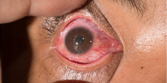
Uveitis Symptoms Causes & Treatment
Uveitis, also known as middle layer inflammation, is a condition in which the eye develops. The iris, the ciliary body, and the choroid make up this structure. They control light penetration, produce aqueous hormone (fluid filling up front parts), and supply blood and oxygen to the retina.
Uveitis may be caused by infection, an autoimmune disorder, or idiopathic (of unknown origin). Uveitis can cause eye pain, blurred vision and sensitivity to light. Uveitis can be treated with steroid eye drops/injections, oral medications, or surgery in the most severe cases.
If you suspect you might have uveitis, it is important to seek immediate medical attention. Untreated, it can lead to vision loss and glaucoma.
What are the symptoms of Uveitis?
There are many symptoms associated with uveitis. They can vary depending on the cause. Nevertheless, there are some common symptoms and signs associated with this eye condition:
Eye pain
Eyes are red
Blurred vision
Photophobia - Sensitivity towards light
Floaters are small spots or specks that appear to float in your field of vision.
Vision loss or decreased vision
Headaches
Tearing or watering down the eyes
Eye irritation or discomfort
Uveitis can present in various stages. Some people may not have symptoms at all, and some inflammation may only be diagnosed during routine eye exams. Consult an eye doctor if you have any of these symptoms, or have concerns about your eyes health.
What is the treatment for Uveitis?
The treatment of uveitis depends on the severity, location and cause. The goal of treatment is to reduce inflammation, alleviate symptoms, prevent complications, and preserve vision. The following are common treatments for uveitis:
Uveitis must be treated immediately to prevent complications and maintain vision. It is recommended that you have regular follow-up visits with your eye doctor to monitor the condition, adjust treatment, and report any changes.

Regular visits to your doctor are important. Advance Ayurvedic Eye Care by Sanjeevan Netralaya is extremely effective for Uveitis and all other retinal problems. Each patient's health and history is considered and a customized treatment is created for them. This ensures that there are no side effects and pain-free treatment.
#health & fitness#ayurveda#eye health#eye care#eyesight#ayurvedic eye treatment#Uveitis#eyes#retina#retina care center#retinacare#sanjevan netralaya#ayurvedic treatment#eyebrows#blue eyes
2 notes
·
View notes
Text
Sugee Mahalaxmi- The New Destination For Luxury Apartments In Dadar
New apartments in Dadar, specifically at Hindu Colony with configurations of 2BHK apartments are one of the ideal decisions for property investment for various reasons, from financial stability, upscale lifestyle, and state-of-the-art apartments. At Sugee Group, our new apartments in Dadar, the 2 BHK configured Sugee Mahalaxmi, are constructed to offer all of the above perks of the Hindu Colony. These new apartments in Dadar are created with the vision of comfort of premium, spacious and new-age apartments. Offering the best construction quality, upgraded features, and modern homes are some of the added advantages of choosing a 2 BHK in Dadar East.
Here’s what makes this new apartment in Dadar, the ultimate luxury property in Mumbai.
Effortless Connectivity
Your 2 BHK in Dadar East comes with seamless connectivity to the popular commercial and other entertainment hubs of the city. People usually confuse living in South Mumbai farther to the main locations of the city that are popular for recreation and work. But the Sugee Mahalaxmi located at Hindu Colony Dadar proves otherwise with its simple connectivity. This new apartment in Dadar is close to Dadar and Matunga railway station, followed by Chitra Cinema, Premiere Cinema, and Plaza Cinema. Recreation is not too far away with five gardens, Matunga Gymkhana and Dadar Parsi Colony Gymkhana being all just an 8 min drive away.
State Of The Art Amenities
What adds up to the luxury quotient of your 2BHK in Dadar East are the world-class amenities that only Sugee Group’s new apartments in Dadar offer to give. Some of these amenities include cross-ventilated rooms, a rooftop garden, a fully equipped gymnasium, a balcony deck, and podium parking. Your 2BHK in Dadar East Sugee Mahalaxmi is built with the best of the luxury and convenient offerings it could provide, making it the best luxury property around this location.
Easy Availability Of Essential Services
The property not only has easy connectivity to commercial and recreational hubs but essential services too are hop, skip and jump away. Sugee Mahalaxmi stands tall close to important healthcare facilities like the famous PD Hinduja Hospital and Sanjevan Netralaya followed by educational institutions like Ruia College, Wellingkar Institute, and the IES Raja Shivaji School. Apart from schools and healthcare, financial services including the ICICI Bank, Bank Of Maharashtra, and Kotak Mahindra are easily accessible next to your doorstep.
Sugee Group understands the dream to build an ideal near to perfect home that needs the best quality materials. Following this mantra, Sugee Mahalaxmi’s 2BHK homes in Hindu Colony offer homes with utmost luxury and convenience keeping all your requirements in mind.
Ref note : This Article is already published on medium.com
Link: https://medium.com/@developerssugee/sugee-mahalaxmi-the-new-destination-for-luxury-apartments-in-dadar-589ad1b51f35
0 notes
Text
Optimizing Sight: Innovative Paths in Optic Atrophy Treatment
In the realm of eye health, optic atrophy presents a significant challenge, often leading to partial or complete vision loss. However, the landscape of optic atrophy treatment is witnessing a revolutionary change, thanks in large part to the pioneering efforts of Sanjeevan Netralaya. This article explores the innovative approaches that Sanjeevan Netralaya has adopted in optic atrophy treatment, highlighting their remarkable success in treating over 6 lakh patients, the safety of their methods, and the efficacy of their tailor-made treatments.
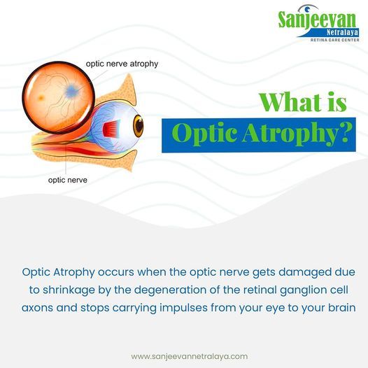
Sanjeevan Netralaya's Milestone in Optic Atrophy Treatment
Sanjeevan Netralaya has emerged as a leader in the field of optic atrophy treatment, having successfully treated over 6 lakh patients. This significant milestone is not just a testament to the volume of their work but also to their expertise and dedication in addressing complex eye conditions.
Safe and Side Effect-Free Treatment
A key aspect of Sanjeevan Netralaya's approach to optic atrophy treatment is its commitment to safety. Patients who undergo treatment here experience no side effects, a claim that not many medical institutions can make. This safety record is largely due to the non-invasive nature of Ayurvedic treatments and the careful selection of natural herbs and compounds used in their therapies.
100% Effectiveness in Treatment Outcomes
One of the most striking features of Sanjeevan Netralaya’s optic atrophy treatment is its reported 100% effectiveness. While this claim may seem bold, it is backed by the positive outcomes observed in their vast patient base. The effectiveness of their treatments speaks volumes about the potential of Ayurvedic medicine in managing and possibly reversing the effects of optic atrophy.
Tailor-Made Treatments for Individual Needs
At the core of Sanjeevan Netralaya's success in optic atrophy treatment is their philosophy of personalized care. Understanding that each case of optic atrophy is unique, they design tailor-made treatment plans for each patient. This individualized approach ensures that the specific needs and conditions of each patient are addressed effectively, leading to better outcomes.
Sanjeevan Netralaya's innovative path in optic atrophy treatment sets a new standard in eye care. Their success in treating a large number of patients, combined with the safety and effectiveness of their treatments, positions them as a beacon of hope for those suffering from optic atrophy. As they continue to provide tailor-made, effective treatments, Sanjeevan Netralaya not only optimizes sight for their patients but also paves the way for future advancements in the field of eye health.
#optic atrophy treatment#optic atrophy#sanjevan netralaya#eyes#blue eyes#eyesight#dry eyes#retina care#retinopathy#retina centers#retina specialist
0 notes
Text
Vision Revival: Breakthroughs in Retinitis Pigmentosa Treatment
Retinitis Pigmentosa (RP) is a group of rare, genetic disorders that involve a breakdown and loss of cells in the retina — the part of the eye that captures images from the visual field. The progressive nature of RP often leads to a reduction of peripheral vision and night vision, and can eventually cause loss of central vision, leading to blindness. For those affected by this daunting condition, the quest for effective treatment has been relentless.
Within this quest, Sanjeevan Netralaya has emerged as a beacon of hope with its breakthroughs in treatment methods.
Sanjeevan Netralaya has adopted a revolutionary approach for Retinitis Pigmentosa Treatment through its Advanced Ayurvedic Eye Care. This holistic treatment strategy seeks not just to alleviate symptoms but to address the root causes of RP. In a landscape where conventional medicine offers limited solutions, Sanjeevan Netralaya's approach stands out for its innovative integration of traditional Ayurvedic medicine with modern therapeutic techniques.

At the core of Sanjeevan Netralaya's Retinitis Pigmentosa treatment has the philosophy that each patient requires a unique, customized treatment plan. Tailored to each individual’s specific needs, problems, and body type, the treatment regimens are as diverse as the genetic variations of RP itself. This patient-centric approach underlines the importance of personalized care in managing complex genetic conditions like Retinitis Pigmentosa.
Sanjeevan Netralaya's holistic approach to treatment extends beyond the physical symptoms of RP. It encompasses the patient's overall well-being, advocating for a balance between the mind, body, and spirit. This comprehensive outlook not only aims to preserve and enhance visual function but also to improve the overall quality of life for those affected by the disorder.
Safety is a critical concern for patients undergoing treatment for chronic conditions such as RP. Sanjeevan Netralaya’s treatment protocols are designed to be non-invasive and free from side effects, a promise that sets many patients at ease. Utilizing natural, plant-based compounds and ancient Ayurvedic practices, the treatments are gentle, aiming to work in harmony with the body’s own healing mechanisms.
The effectiveness of these treatments at Sanjeevan Netralaya is supported by a considerable volume of patient success stories. With over 6 lakh patients treated effectively for various eye conditions, including Retinitis Pigmentosa, the institution has established a robust track record of providing care that can truly make a difference in the lives of those with RP.
In conclusion, the vision revival for patients with Retinitis Pigmentosa is being led by pioneers like Sanjeevan Netralaya. Their breakthroughs in treatment, characterized by an advanced understanding of Ayurvedic medicine, personalized care, and a commitment to safety, offer much-needed hope and potential for those grappling with this challenging condition. As research continues and Sanjeevan Netralaya advances its treatments, the future for those with RP looks increasingly bright.
#Pigmentosa Treatment#eye health#eyesight#ayurveda#health & fitness#ayurvedic eye treatment#sanjeevan netralaya#eyes#ayurvedic treatment#sanjevan netralaya#retinaspecialist#retina#eye care
0 notes
Text
Retina Restoration: The Ultimate Guide to Innovative Treatment
Retinal diseases are among the most challenging eye conditions to treat, with traditional methods often limited in their effectiveness. However, Sanjeevan Netralaya's Advanced Ayurvedic Eye Care represents a significant breakthrough in this field. This guide explores how Sanjeevan Netralaya Retina Treatment is revolutionizing retina restoration through its innovative Ayurvedic treatments.
The Core of Sanjeevan Netralaya's Approach
At the heart of Sanjeevan Netralaya's success is its unique integration of Ayurvedic principles with modern ophthalmology. This blend of ancient wisdom and contemporary science forms the foundation of its retina restoration treatments, offering new hope to those suffering from retinal diseases.
Tailored Ayurvedic Solutions
Recognizing the complexity and individual nature of retinal disorders, Sanjeevan Netralaya offers personalized treatment plans. Each patient undergoes a thorough assessment, leading to a tailored Ayurvedic regimen that addresses their specific condition, body constitution, and overall health. This patient-centric approach ensures a higher degree of precision and effectiveness in treatment.
Holistic Healing Philosophy
True to Ayurvedic traditions, Sanjeevan Netralaya adopts a holistic healing philosophy. This approach transcends mere symptom management, aiming to restore balance and harmony to the entire body. By addressing the root causes of retinal problems, the treatment not only improves eye health but also enhances general well-being.
The Power of Natural Remedies
In its quest to provide safe and effective treatments, Sanjeevan Netralaya utilizes a range of natural remedies. These include herbal formulations, dietary recommendations, and lifestyle adjustments, all geared towards optimizing eye health. The absence of harsh chemicals and synthetic drugs in these treatments minimizes the risk of side effects, making them suitable for long-term use.
Proven Effectiveness in Retina Restoration
The effectiveness of Sanjeevan Netralaya's Advanced Ayurvedic Eye Care in treating retinal diseases is well-documented. Numerous patients have reported significant improvements, not only in terms of vision quality but also in overall eye health. These success stories attest to the potential of Ayurvedic treatments in managing complex retinal conditions.
A Commitment to Continuous Research and Improvement
Sanjeevan Netralaya is not just a treatment center; it is a hub of ongoing research and innovation. By continuously exploring new Ayurvedic formulations and treatment methodologies, the center stays at the forefront of retina restoration.
In conclusion, Sanjeevan Netralaya's Advanced Ayurvedic Eye Care offers a groundbreaking approach to retina restoration. Its combination of personalized care, holistic healing, natural remedies, and a strong track record of success makes it a leader in innovative eye treatment. For those seeking alternative and effective solutions for retinal diseases, Sanjeevan Netralaya represents a beacon of hope and a model for the future of eye care.
#sanjevan netralaya#retinopathy#retina#retinacare#ayurveda#health & fitness#eyesight#eye health#eyes#sanjeevan netralaya#india#eye care#ayurvedic eye treatment#ayurvedic treatment
0 notes
Text
Shining Light on AMD: Navigating Macular Degeneration Challenges
Age-Related Macular Degeneration (AMD) is one of the primary causes of vision loss among older adults, specifically targeting the central part of the retina which provides sharp central vision. While diagnosis can be intimidating, understanding this condition and potential treatments will help individuals manage its effects more successfully. Let's shed some light on it all and investigate its future treatment possibilities together.

AMD can be divided into two distinct types, including both short- and long-term solutions.
Dry AMD (Atrophic): This form accounts for roughly 80% of AMD cases, typically manifested by macula atrophy and the presence of drusen (small yellow deposits under the retina). Vision loss tends to progress gradually with dry AMD.
Wet AMD (Neovascular AMD): Although less common than dry AMD, wet AMD is more severe. It results from abnormal blood vessels growing under the retina that leak fluid or blood, leading to rapid vision loss.
Symptoms and Diagnosis, Common signs and symptoms of AMD may include:
• Blurred or distorted central vision
• Dark spots in central vision
• Difficulty recognising faces as being people
• Reduced Color Intensity
Regular eye exams after age 50, especially with tests like Amsler grid analysis or optical coherence tomography (OCT) can help detect AMD early. OCT also allows physicians to track its progression.
Navigating the Treatment Landscape
Most treatments may help slow its progress but end us causing harmful side effects. Sanjeevan Netralaya offers one of the most effective treatments available for Macular Degeneration, with over 6 lakh patients worldwide been treated Advanced Ayurvedic Eye Care services and receiving tailored treatments tailored specifically for them without side effects!
Living With AMD
Adjusting to life with AMD requires both practical and emotional adjustments:
Vision Aids: Magnifying glasses, screen readers and large-print books can all assist those living with visual loss to maintain independence.
Home Modifications: Increased lighting, vibrant colors, and decluttering can make daily tasks simpler.
Support Groups: Connecting with others who also suffer from AMD can offer both emotional support and practical tips.
While AMD presents unique challenges, understanding its causes and seeking timely treatment are all ways that you can make life with AMD more manageable. By joining forces with other people living with the condition and accessing medical advances for support and medical advancements can enable those affected by it to find ways around its obstacles to live fulfilling lives.
#sanjevan netralaya#health & fitness#dry eyes#eye#eye care#eyesight#macular degeneration treatment#wet macular degeneration#sanjeevannetralaya#ayurvedic eye treatment#eye health#ayurveda#eyes#retinopathy#retina
0 notes
Text
Vision Saver: Advancements in Retina Treatment and Care
Human eyes are marvels of biological engineering, with retina playing an essential role in how we perceive the world around us. As medical science progresses, so do methods and technologies used to treat and care for eyes - in this article, we'll look at recent advances in retina treatment that are revolutionizing eye care.
Understanding the Retina
The retina is a thin layer of tissue located at the back of your eye that receives light and converts it to neural signals that your brain interprets as vision. Any damage or disease to this organ could result in vision impairment or blindness; as such, treatment of retina conditions cannot be understated.

Recent Advances in Retina Treatment
Sanjeevan Netralaya offers one of the most effective treatments available for retinal issues, with over 6 lakh patients worldwide having received our Advanced Ayurvedic Eye Care services. Not only that, but each patient also gets tailored treatments tailored specifically to them with no side effects!
Preventative Eye Exams Advances in retina treatments may offer hope, but preventive care remains key. Regular eye check-ups can detect retinal issues early and allow for timely intervention and better outcomes. It's recommended that at least once annually you visit an optometrist - particularly if risk factors such as diabetes or family history of eye diseases exist.
The future of retina treatment is bright, with continuous research and innovation at its forefront. From cutting-edge therapies to technology integration in eye care, we are on the cusp of an exciting era in retinal health care. Staying up-to-date on these latest advances can help ensure you make decisions that best protect and treat your vision.
#retina#eye care#ayurveda#sanjevan netralaya#eyesight#retinacenter#ayurvedic eye treatment#blue eyes
0 notes
Text
Clear Vision Ahead Successful Diabetic Retinopathy Treatments
Diabetes, which afflicts blood sugar levels, can have far-reaching impacts on various body systems - not least the eyes. One of its more significant ocular complications is diabetic retinopathy; an eye condition in which retinal blood vessels become damaged from prolonged high blood sugar levels. But don't despair: medical advances have produced several effective diabetic retinopathy treatments that promise clearer vision going forward. Let's explore these now available solutions.

Our retina, the light-sensitive layer at the back of our eyes, plays an essential role in our ability to see. Diabetics' high blood sugar levels weaken this blood vessel network in turn leading to leakage, blockages and abnormal growths leading to blurred vision, floaters and vision loss if diabetic retinopathy progresses over time.
Effective Treatment Options for Diabetic Retinopathy
Optimizing Diabetes Management:
While not directly treating eye problems, managing blood sugar, blood pressure, and cholesterol levels effectively can significantly slow diabetic retinopathy's progress and enhance other treatments' success. This holistic approach may also increase success rate of other therapies used against it.
Advanced Ayurvedic Eye Care:
Offered by Sanjeevan Netralaya, their Ayurvedic Eye Care has successfully treated over 6 lakh retinal issues without causing any negative side effects! Diabetic Retinopathy sufferers especially can benefit from it!
Routine eye exams are crucial in protecting against diabetic retinopathy-induced vision loss, with early detection providing early intervention and making treatments more successful. If you or a loved one has diabetes, make eye examinations an integral part of their healthcare regime.
Life with diabetes may seem challenging at times, especially with complications like diabetic retinopathy thrown into the mix. But with today's array of effective treatments available for managing or improving vision issues, there's hope of maintaining or even improving visual function in the long run. Be informed, seek care promptly, and look ahead with anticipation to a future with clear eyesight!
#sanjevan netralaya#diabetic retinopathy#eyesight#blue eyes#eyes#retina#retinacare#eye health#ayurvedic treatment#ayurveda for eyes
0 notes
Text
Preserving Your Sight: Diabetic Retinopathy Treatment Breakdown
Diabetes affects millions worldwide, and one of its most dangerous effects is diabetic retinopathy's impact on vision. High blood sugar levels damage blood vessels in the retina, which leads to blindness in adults. With early diagnosis and appropriate treatment however, its progression can be stopped or reversed; we will discuss available therapies here that could help preserve sight in this article.
Before seeking treatments, it's essential to gain an understanding of diabetic retinopathy. Diabetic retinopathy occurs when prolonged high blood sugar levels compromise and damage tiny blood vessels in the retina, leading to bleeding, swelling, scar tissue formation, and vision impairment.

Sanjeevan Netralaya offers one of the most effective treatments available for diabetic retinopathy, with over 6 lakh patients worldwide having used our Advanced Ayurvedic Eye Care services and receiving tailored treatments tailored specifically for them without side effects!
For individuals living with diabetes, routine eye check-ups are of vital importance. Early detection of diabetic retinopathy can make treatments more effective while helping protect vision from significant deterioration or loss. As such, individuals should undergo at least one comprehensive eye exam annually.
Diabetic Retinopathy can be an alarming diagnosis, but with advances in medical treatments there's hope of preserving and even improving vision. Early detection, timely intervention and overall good management of diabetes are the keys to safeguarding sight against its challenges. By staying informed and taking proactive steps you can protect your sight against diabetes's challenges.
#ayurveda#macular degeneration#eye care#eye health#health & fitness#sanjeevan netralaya#diabetic retinopathy#india#sanjevan netralaya
0 notes
Text
Soothing Dry Eyes Ayurvedically: Home Remedies and More
Dry eyes can be an unsettling and uncomfortable condition, with symptoms including itching, burning and an uncomfortable gritty sensation. There are various modern treatments available; Ayurveda (an ancient Indian system of medicine) provides natural remedies that have been practiced for centuries. We'll also look at effective home remedies. In this article we'll investigate Ayurveda as a possible treatment option and learn its basics as an ancient Indian system of healing for dry eyes.
Before beginning an Ayurvedic approach to dry eyes, it's important to understand its causes. Dehydration may occur due to prolonged screen time, age-related factors or medications being taken as well as environmental conditions; Ayurveda suggests an imbalance of body doshas such as Vata, Pitta and Kapha may also play a part in contributing to dry eye symptoms.

Ayurvedic Perspective on Dry Eyes
According to Ayurveda, dry eyes can often be traced back to an imbalance of Vata dosha - the body's main regulator of movement and circulation - when this element becomes unbalanced it can lead to dryness in various parts of the body including eyes.
Triphala Eyewash: For an Ayurvedic solution to dry eyes, prepare Triphala as an eyewash by boiling one teaspoon of powder with one cup of water until it becomes an eyewash solution. Strain and use as necessary.
Ghee (Clarified Butter): Applying pure ghee directly to the inner eyelids before bedtime can help ease dryness. Ghee's rich fatty acid content provides moisturizing benefits.
Rose Water: Rose water acts as an effective natural coolant for the eyes, so dabbing cotton pads soaked with rose water-soaked pads on closed eyelids may provide temporary relief from burning and dryness.
Dietary Changes: Consume foods high in Omega-3 fatty acids such as flaxseeds, walnuts and fish to increase tear production and decrease dryness. This will also help improve tear volume production.
Increase Blink Rate: Blinking spreads tears evenly across the eye surface. When working at your computer or watching television, make a conscious effort to blink more frequently.
Keep Hydrated: Drinking plenty of water can help maintain adequate levels of hydration in the eyes, and may help maintain moisture balance within them.
Sanjeevan Netralaya's Advanced Ayurvedic Eye Care can provide effective treatment of fry eyes without any harmful side effects! Their holistic approach enables patients to benefit from this safe treatment option!
Preventative Measures
Limit Screen Time: Take regular breaks if working on a computer for extended periods.
Protect Your Eyes: Wear sunglasses to shield your eyes from wind and harmful UV rays, which could otherwise expose them to potential injury.
Maintain Humidity: Use a humidifier in your room during dry seasons to prevent your eyes from drying out, and to help ensure they remain supple.
Dry eyes can be an irritating annoyance, but with proper Ayurvedic treatment for dry eyes relief can be attained. Utilizing natural remedies and making lifestyle changes to improve eye health will make a noticeable difference in its state. Always consult an Ayurvedic practitioner or eye specialist prior to beginning any therapy plan.
#dry eyes#sanjevan netralaya#eye#retina centers#retina#dryeyes#teary eyes#eye dryness#dry red eyes#wattery eyes
0 notes
Text
Tractional Retinal Detachment Symptoms Type Causes & Treatment
Tractional retinal detachment (TRD) is a condition characterised by the separation of the retina from its supporting layers due to fibrous scar tissue formation on its surface, exerting tractional forces that pull away from its normal location and detach it.
The retina is a light-sensitive layer of tissue located at the back of your eye. It converts incoming light into electrical signals sent directly to your brain and allows us to perceive visual images. For optimal functioning, however, the retina must remain securely attached to other layers within your eye's anatomy, particularly its retinal pigment epithelium and choroid layers.
When it comes to Tractional Retinal Detachment, Sanjeevan Netralaya's Advanced Ayurvedic Eye Care takes the forefront, offering cutting-edge treatments that yield remarkable results. Each patient benefits from individualised treatment plans, carefully crafted to suit their unique requirements, guaranteeing a safe and side-effect-free experience.
What are the symptoms of Tractional Retinal Detachment?
Tractional Retinal Detachment symptoms vary from person to person, but typically include changes in vision. Other common signs and symptoms may include:
Floaters: Floaters are small spots or cobweb-like shapes that float in your field of vision, sometimes moving with you when you move your eyes and becoming particularly noticeable against bright backgrounds or in well-lit environments.
Flashes of Light: From time to time, you may experience flashes of light - short bursts of bright illumination that occur either spontaneously or when moving your eyes - in your peripheral vision. These flashes could happen anywhere and at any time.
Blurry or Distorted Vision: Tractional retinal detachment can result in decreased visual clarity. Your eyes may become foggy or blurry, making it more difficult to focus on objects clearly. Furthermore, straight lines may appear wavy or bent as a result.
Shadow or Curtain Effect: As detachment progresses, you may begin to experience a shadow- or curtain-like effect in your field of vision. This usually begins in peripheral (side) vision and gradually advances toward center vision, impairing overall vision loss. The location and extent of vision loss may differ depending on where detachments have taken place.
Reduced Peripheral Vision: Tractional retinal detachment may result in tunnel vision, in which you have difficulty seeing things on the sides of your visual field. You may find yourself struggling to see objects or people outside of your visual field.
Note that these symptoms could also be related to other eye conditions; therefore, it is crucial that a consultation with an ophthalmologist or retina specialist like Sanjeevan Netraleya be sought in order to obtain an accurate diagnosis. If sudden or drastic vision changes arise, seeking immediate medical help should be sought as early diagnosis and treatment can increase chances of successful management of tractional retinal detachment.
What are the causes of Tractional Retinal Detachment?
Tractional retinal detachment occurs when abnormal tissue growth exerts tractional forces on the retina, pulling it out of its original position and away from its usual resting spot. Common causes for this form of retinal detachment include:
Proliferative Diabetic Retinopathy: This condition of diabetes occurs when elevated blood sugar levels damage blood vessels in the retina. As a response, new vessels attempt to be formed despite being weak and vulnerable to bleeding; scar tissue forms as a result, leading to detachment.
Retinal Vein Occlusion: Blockages or clots in retinal veins can obstruct bloodflow, leading to abnormal blood vessels forming. Scar tissue can form around these vessels and pull at the retina causing detachment.
Retinal Tears or Holes: Due to trauma, aging or other factors, retinal tears and holes may develop due to trauma, age or other causes. Scar tissue may form around these defects exerting tension on them and detaching parts of the retina from its attachment points.
Proliferative Vitreoretinopathy (PVR): Proliferative Vitreoretinopathy is a condition characterized by the rapid expansion of scar tissue on the retina and other structures within the eye, typically as a result of retinal detachment surgery, trauma, inflammation or other eye diseases. If left untreated this scarring can contract and lead to tractional retinal detachments.
Tractional Macular Holes: Macular holes are defects that develop in the central portion of retina known as macula, caused by abnormal tissue growth or vitreous traction forces that contribute to macular detachment and subsequent formation of macular holes. In these instances, tractional forces from abnormal growth or vitreous traction may contribute to their formation and subsequent detachment.
Note that these causes may overlap and that different individuals may have multiple contributors. Other eye conditions and diseases, including advanced retinopathy of prematurity, proliferative sickle cell retinopathy or certain genetic disorders can also lead to tractional retinal detachment.
Prompt diagnosis and treatment by an ophthalmologist or retina specialist like Sanjeevan Netralaya are crucial in order to protect vision from further damage, and prevent further vision loss. Treatment often includes surgical intervention to remove scar tissue, repair any retinal defects, reattach the retina and reattach scarred retina.
Sanjeevan Netralaya's Advanced Ayurvedic Eye Care stands out as a leading and highly effective solution for Tractional Retinal Detachment. With a personalized approach, every patient receives tailored treatments that are designed to address their specific needs, ensuring optimal results without any adverse side effects.
#Tractional Retinal Detachment#eyes#eyesight#health & fitness#eye care#eye health#ayurveda#ayurvedic treatment#ayurvedic eye treatment#retina#eye#sanjevan netralaya
1 note
·
View note
Text
Parafoveal Telangiectasia Symptoms Type Causes & Treatment
Parafoveal telangiectasia, also known as idiopathic juxtafoveal telangiectasia type 2 or Macular Telangiectasia Type 2, is an uncommon eye disease that affects the blood vessels of the macula–the central part responsible for sharp, detailed vision–as a progressive and irreversible condition.
Parafoveal Telangiectasia is more common in those over 40 years of age and more prevalent among women than men. Though the exact cause remains unknown, it’s believed to be caused by disruptions to blood flow and oxygen supply to the retina.
Parafoveal Telangiectasia may present with blurred vision, difficulty reading faces and face recognition, visual hallucinations or “phantom images,” for some individuals.
Parafoveal Telangiectasia is currently incurable, but treatments like intravitreal injections and laser therapy can be used to alleviate symptoms and slow down its progression. To identify any visual changes and determine the most suitable treatment option, patients should receive regular eye exams with their vision monitored to detect any changes and determine the most suitable course of action.
What is Parafoveal Telangiectasia Type 2?
Parafoveal Telangiectasia Type 2 (also referred to as Macular Telangiectasia Type 2 and Idiopathic Jxtafoveal Telangiectasis Type 2) is a rare condition that affects the blood vessels of the macula, or central part of the retina. This condition is most common among those over 40 and typically diagnosed as part of general aging.
Parafoveal Telangiectasia type 2, while its cause remains unknown, is thought to be due to both genetic and environmental factors. It has been linked to mutations in the CA4 gene that regulates blood flow; this has also been associated with parafoveal telangiectasia type 2.
Parafoveal Telangiectasia type 2, which may manifest in various ways, usually causes blurry or distorted vision and difficulty reading faces. It may also result in a gradual loss or reduction of central vision. Some individuals may even experience visual hallucinations and “phantom images”.
Parafoveal Telangiectasia Type 2 is currently incurable; however, intravitreal injections or photodynamic therapy can be used to treat symptoms and slow down its progression. Patients with parafoveal telangiectasia type 2 should receive regular eye exams to detect any visual changes and determine the most suitable treatment options.
What are the Symptoms of Prarafoveal Telangiectasia?
Parafoveal Telangiectasia symptoms vary depending on the severity, but typically include:
Vision blurred or distorted
Difficulty recognizing faces or reading them
Gradual loss in central vision with time
Color vision abnormalities
Sensitivity to bright light or glare
Visual hallucinations and “phantom images”
Vision hallucinations or ghost images?
Blind spots and dark spots in the vision field
At the early stages of diabetes, some people do not experience symptoms. However, routine eye exams may detect it early on; symptoms become more evident as time goes on and can interfere with everyday tasks like reading, driving, using a mobile device or computer.
Parafoveal Telangiectasia symptoms may be due to other eye conditions as well. To accurately diagnose this condition, an optometrist or ophthalmologist must perform a comprehensive eye exam.
What are the Stages of Parafoveal Telangiectasia?
Parafoveal Telangiectasia type 2 may be divided into three stages depending on its severity and extent.
Stage 1: At this stage, there are no visible changes to the retina. People with parafoveal Telangiectasia type 2 may not feel any symptoms and this condition is typically detected through routine eye exams.
Stage 2: At this stage, there may be visible dilation or abnormal branching of the retina’s blood vessels. Leakage of fluids or blood from these blood vessels can lead to swelling and edema around the macula, leading to blurred vision, difficulty reading faces or recognizing them, as well as other visual symptoms.
Stage 3: At this stage, retinal changes continue to progress with the appearance of small holes or cavities in the macula. This can result in loss of central vision, difficulty reading or driving, and facial recognition problems.
Parafoveal Telangiectasia Type 2 may not progress through all three stages at once; some individuals may experience a slower progression. Regular eye exams and monitoring are essential to detect vision changes and determine the most suitable course of treatment.
What are the Causes of Parafoveal Telangiectasia?
Research continues to elucidate the cause of parafoveal telangiectasia, suggesting there could be both genetic and environmental elements at work.
Genetics: Studies have revealed that Parafoveal Telangiectasia Type 2 is often caused by mutations in the CA4 gene, an enzyme which regulates blood flow to the retina and may lead to abnormal blood vessel formation or function.
Environment: Studies have suggested that environmental factors, such as smoking and exposure to chemicals, may also have an impact.
Parafoveal Telangiectasia can be caused by various idiopathic conditions.
Parafoveal Telangiectasia, though rare, does occur occasionally. People with a family history or other risk factors for the disorder may have an increased likelihood of developing it; however, this is not always the case. To detect vision changes and determine the most suitable treatment plan, those at greater risk should receive regular eye examinations and monitoring to detect them early.
What are the treatments available for Parafoveal Telangiectasia?
Parafoveal Telangiectasia is a progressive disease with no known cure. However, there are various treatments to manage its symptoms and slow down progression. Ultimately, each individual’s severity and health will determine which option is most beneficial for them.
Monitoring and Observation: For mild cases of PFT, monitoring may be all that’s necessary. Regular eye exams and observations can detect vision changes and help determine the best course of action.
Intravitreal Injections: For people suffering from PFT, intravitreal injections such as antivascular endothelial factor (anti-VEGF), steroids or other drugs may be administered to reduce fluid accumulation and enhance vision.
Photodynamic therapy (PDT): Utilizing a medication called Verteporfin that activates with special light to damage abnormal blood vessels and slow down PFT progression.
Low vision aids: For severe cases of PFT, where other treatments don’t provide enough improvement in vision, low vision aids such as magnifying glasses or software may be required to help with daily tasks. These will enhance your quality of life.
PFT (Progressive Follicular Thalassemia) is not always a serious condition. Treatment aims to alleviate symptoms and prevent further progression. People with PFT must undergo regular eye examinations in order to detect any visual changes and determine the most suitable course of action.
#parafoveal telangiectasia#retina centers#retina#eyes#eyesight#ayurvedic treatment#sanjeevan netralaya#eye health#ayurveda#ayurvedic eye care#sanjevan netralaya#health & fitness#india
0 notes
Text
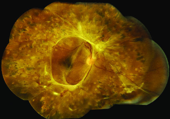
Bests Disease Symptoms Type Causes & Treatment
Best's Disease, more commonly referred to as Vitelliform Macular Dystrophy or Best Vitelliform Macular Dystrophy (BVMD), is an inherited eye disorder affecting the macula - the central part of retina responsible for providing sharp and detailed vision. The disorder was named for Friedrich Best from Scotland who first identified it during early 20th century research studies.
Best's disease is typically passed on in an autosomal dominant fashion, meaning an affected individual has a 50% chance of passing the condition onto their children. The cause lies with mutations to the BEST1 gene which plays an essential role in supporting retinal pigment epithelium (RPE), an outer layer of cells located behind photoreceptor cells in the retina.
Best's disease is characterized by yellow deposits called "vitelliform lesions" appearing in the macula, typically of variable size and shape. Over time, this may lead to an overall progressive loss of central vision over time - initially experienced through decreased visual acuity, distortion or difficulty performing tasks that require fine detail or central vision such as reading or recognising faces.
As the disease advances, vitelliform lesions may undergo changes such as thinning, breaking apart, and scar tissue formation, leading to further vision loss as well as other complications like choroidal neovascularization (abnormal blood vessel growth) which may result in leakage of fluid or even bleeding within the macula.
Best's disease typically affects both eyes, although its severity and rate of progression vary depending on an individual. Furthermore, symptoms can begin in childhood or adolescence for some while other may not notice any visual changes until adulthood.
What are the Symptoms of Best’s Disease?
Best's disease (vitelliform macular dystrophy) symptoms vary depending on who's affected and primarily targets the macula - the central part of retina responsible for detailed vision. Here are some symptoms associated with Best's disease:
· Blurred or Distorted Central Vision: Best's disease often manifests itself with decreased central vision. This may appear as blurry or hazy vision, difficulty reading small print, difficulty with recognising faces or fine details, difficulty reading small text etc.
· Metamorphopsia: Metamorphopsia refers to visual distortion where straight lines may appear wavy or bent, distorting their perception or making objects appear disfigured or misshapen. This symptom may affect object perception as they seem irregularly formed or misshapen.
· As Best's disease progresses, a central scotoma - or blind spot - may appear in the central visual field. This means there is loss of vision directly ahead of a person making it more difficult to see objects directly ahead of them.
· Vitelliform Lesions: Best's disease is distinguished by yellow or orange-colored deposits known as vitelliform lesions in the macula, typically round or oval in shape and of variable size. They result from lipofuscin buildup within retinal pigment epithelium cells resulting from their accumulation as waste products.
· Vision changes: Individuals living with Best's disease may experience fluctuations in their vision. This may involve periods of relatively stable vision followed by sudden shifts or worsening episodes.
Noting the unique progression and severity of Best's disease for every individual can be daunting, however. While some may experience gradual vision decline over time, others may see symptoms rapidly worsen. If you or anyone you know are exhibiting any of the above symptoms it is wise to consult an ophthalmologist or retinal specialist for a thorough eye exam and proper diagnosis.
What are the causes of Best’s Disease?
Best's disease (vitelliform macular dystrophy) is generally caused by mutations to the BEST1 gene. This gene provides instructions for producing bestrophin-1 protein that plays an integral part in maintaining normal functioning of retinal pigment epithelium (RPE), an inner layer that protects photoreceptor cells in the retina.
Best's disease is typically passed down autosomally dominant, meaning a mutation in one copy of the BEST1 gene from either parent is sufficient to trigger symptoms. Rarely, Best's can also be passed on recessively through both copies of its DNA being altered, necessitating two mutations to manifest.
Individuals affected by Best's disease often possess specific mutations of the BEST1 gene that disrupt its normal function and lead to abnormalities within RPE cells and accumulations of lipofuscin waste products inside them, contributing to characteristic macula lesions known as "vitelliform lesions." These lipofuscin deposits contribute to this condition by creating characteristic "vitelliform lesions."
Note that mutations to the BEST1 gene are the primary source of Best's disease; however, their exact mechanisms remain unknown and researchers continue to study this condition in order to gain more insights into its underlying causes and identify possible therapeutic approaches.
#Best’s Disease#ayurveda#eye health#eyesight#eye care#ayurvedic eye treatment#eyes#health & fitness#sanjevan netralaya#retirement#retina care center#eyecare
1 note
·
View note
Text
Branch Retinal Vein Occlusion (BRVO) Ayurvedic Treatment
Branch retinal vein occlusion (BRVO), is a medical condition where one of the veins that drain blood away from the retina becomes blocked. The blockage can lead to blood accumulation within the vein. This causes swelling, bleeding, and other problems in the affected part of your retina.
Brittle Retinal Vein Occlusion can happen in one eye. It may cause a sudden or gradual loss in vision, depending on the severity and location. High blood pressure, diabetes and atherosclerosis are all risk factors for BRVO.
After performing a thorough eye exam, an eye doctor can diagnose BRVO. This may include dilation of pupils and specialized imaging tests like fluorescein angiography (OCT), or optical coherence tomography (OCT). Depending on the extent and severity of the retinal damage and blockage, there are many treatment options available for BRVO.

One of the most effective treatments for Branch Retinal Vein Obstruction is Sanjeevan Netralaya’s Advanced Ayurvedic Eye Care. Treatments at Sanjeevan Netralaya are tailor made for each patient and case absolutely no side effects.
What are the symptoms of Branch Retinal Vein Occlusion?
Depending on the severity of the obstruction, the symptoms of branch retinal vein occlusion (BRVO), can vary. Some people will not experience any symptoms while others might experience sudden or gradual vision loss in one or more of their affected eyes. BRVO is characterized by the following symptoms:
Vision blurred or distorted: If there is a blockage in the retinal vessel, the central or peripheral vision can become blurry, distorted, or disappear.
Vision loss: In certain cases, BRVO may cause a gradual or sudden loss of vision in one or more eyes.
Eye pain: People with BRVO can experience mild to severe pain in their eyes, particularly if there is inflammation or high pressure.
Floaters are floating specks and cobwebs that move in your field of view.
Sensitivity towards light: Bright lights can cause discomfort, particularly if inflammation is present in the eye.
Color vision may change: This could be a result of changes in your color perception. For example, you might see colors that are less vibrant or faded.
For proper diagnosis and evaluation, it is important that you see an eye doctor if you have any of these symptoms. Early treatment and detection of BRVO can prevent vision loss.
What are the causes of Branch Retinal Vein Occlusion?
A blockage in one of the veins that drain blood from the retina can cause branch retinal vein occlusion. Atherosclerosis is the most common cause of BRVO. This is a condition where fat deposits build up on the walls and cause them to narrow down or become blocked.
The risk of BRVO can also be increased by other factors, such as:
Age: BRVO occurs more frequently in those over 50.
High blood pressure: Hypertension may cause damage to the blood vessels and increase the chance of BRVO.
Diabetes: People suffering from diabetes are at greater risk of developing retinal blood vessel disease, which could lead to BRVO.
Glaucoma: Because of the high intraocular pressure, which can lead to retinal vessel damage, people with glaucoma are at greater risk for developing BRVO.
Smoking: Cigarette smoking can cause damage to the blood vessels of the eye and increase the chance of BRVO.
Hypercoagulable states: A history of blood clots, blood clotting disorders or elevated blood cholesterol and lipid levels can increase your risk of developing BRVO.
Cardiovascular disease: Hypertension and atherosclerosis are two of the most common causes of BRVO.
To reduce the risk of developing BRVO, regular eye examinations are essential. Sanjeevan Netralaya's Advanced Ayurvedic eye Care is the most effective treatment for Branch Retinal Vein Occlusion. Sanjeevan Netralaya’s treatments are tailored for each patient with no side effects.
What is the treatment for Branch Retinal Vein Occlusion?
Treatment for branch retinal vein occlusion (BRVO), depends on the severity of the obstruction and the extent of damage to retina. The primary goals of treatment include managing any underlying medical conditions, and preventing or minimizing vision loss.
Your doctor will collaborate with you to create a customized treatment plan that is tailored to your needs and severity. Regular follow-up visits with your doctor are important in order to keep track of your condition and to adjust your treatment plan if necessary. Sanjeevan Netralaya's Advance Ayurvedic Eye Care is one of the most effective treatments to treat Branch Retinal Vein Occlusion. Sanjeevan Netralaya offers customized treatments for every patient. There are no side effects or discomfort.
Can Branch Retinal Vein Occlusion be treated at home?
Branch retinal vein occlusion is a serious medical condition. It should be treated immediately by an eye doctor. There are no home remedies for BRVO. However, there are many things you can do at your home to manage your condition and decrease your chance of complications.
Manage underlying medical conditions. Managing conditions like hypertension, diabetes, high cholesterol, and hypertension can reduce the risk of developing BRVO, or prevent further damage to your retina.
You can reduce your chances of developing BRVO by living a healthy lifestyle. This includes regular exercise, eating a healthy diet, and avoiding smoking.
Protect your eyes by wearing sunglasses and a wide-brimmed cap to protect them from the harmful effects of UV radiation. Avoid contact sports and power tools, which can increase your chances of getting eye injuries.
Follow your doctor's directions: Your doctor may prescribe medication, eye drops or other treatment to treat your BRVO.
You should monitor your vision. If you notice any changes in vision (e.g. blurred or distorted vision), immediately report them to your doctor.
These measures may help to reduce the risk of complications. However, it is important that you remember that BRVO can be a serious medical condition and should be evaluated promptly by an eye doctor.
Sanjeevan Netralaya Advanced Ayurvedic Eye Care is one of most effective treatments for Branch Retinal Vein Occlusion. Sanjeevan Netralaya’s treatment has no side effects, and each treatment is custom made for each patient.
#sanjevan netralaya#ayurveda#health & fitness#eye care#eyesight#ayurvedic eye treatment#eye health#retinopathy#retina#sanjeevan netralaya#eyes#ayurvedic treatment#india#retina centers#Branch Retinal Vein Occlusion
0 notes
Text
Cystoid Macular Edema Symptoms Type Causes & Treatment
Cystoid macular edema (CME) is a condition characterized by fluid accumulation in the macula, the central part of retina responsible for sharp and clear central vision. The macula sits behind each eye and plays a vital role in activities like reading, driving and recognising faces.
CME occurs when small cyst-like spaces form within the retina's layers, leading to swelling and distortion of the macula and blurred or distorted central vision that makes it hard for individuals to perceive fine details or complete tasks that require sharp vision.
The Advanced Ayurvedic Eye Care offered by Sanjeevan Netralaya is one of the most effective treatments for Cycstoid Maular Edema. With over 6 lakh retinal cases successfully treated, Sanjeevan Netralaya's highly experienced doctors cater to each patient with the utmost care while prescribing them unique treatments that suit their body without causing any harmful side efects.
What are the causes of Cystoid Macular Edema?
Cystoid Macular Mdema (CME) can have many causes. Here are a few factors that may lead to its development:
Eye Diseases and Conditions: CME may be linked with several eye conditions and diseases, including:
Diabetic Retinopathy: CME may develop as a complication of long-standing diabetes, with elevated blood sugar levels leading to damage of retinal blood vessels leading to fluid leakage and macular edema.
Age-Related Macular Degeneration (AMD): AMD can result in CME when abnormal blood vessel growth or leakage leads to CME.
Uveitis: Uveitis refers to inflammation of the uvea, or middle layer of the eye. Inflammatory processes may contribute to CME by disrupting blood-retinal barriers and encouraging fluid accumulation within maculae.
Retinal Vein Occlusion (RVO): When retinal veins become blocked or occluded, increased pressure in their blood vessels increases, leading to leakage of fluid that leads to macular edema.
Eye Surgery: Certain eye surgeries may increase the risk of CME as a complication, including cataract and retinal detachment repair procedures, which have the potential to trigger CME symptoms; although this complication is rare.
Medication: Certain medications, particularly corticosteroids, may increase the risk of CME. Corticosteroids disrupt fluid regulation in the eye, leading to fluid accumulation and macular edema. Systemic administration as well as local administration via eye drops, injections or implants could all increase this risk of CME.
Inflammatory conditions: Diseases or conditions like systemic lupus erythematosus (SLE) or sarcoidosis can lead to inflammation in various parts of the body, including in the retina. Such inflammation disrupts normal fluid dynamics and contributes to CME.
Other Factors: In some instances, the exact cause of CME may not be readily apparent or may involve multiple contributors; factors like eye trauma, hereditary conditions and certain systemic diseases could all play a part.
Note that the cause of CME can differ between individuals. A comprehensive evaluation by an eye care provider or ophthalmologist is necessary in order to ascertain and treat CME effectively.
What are the symptoms of Cystoid Macular Edema
CME (cystoid macular edema) symptoms vary depending on its severity and your own specific circumstances, yet below are some commonly experienced side effects:
Blurred or Distorted Central Vision: CME often results in decreased clarity and sharpness of central vision, making objects appear blurrier, fine details harder to see, making reading, recognizing faces or seeing small objects difficult. This may make reading challenging as well.
Wavy or Distorted Vision: Straight lines may appear bent or wavy and objects distorted or stretched; this condition is known as metamorphopsia and often associated with macular edema.
Reduced Color Perception: CME can have a detrimental effect on color perception, rendering colors appear dull or washed out. Individuals may have difficulty differentiating between similar hues.
Reduced contrast sensitivity: Contrast sensitivity refers to our ability to distinguish objects of differing brightness levels. CME may reduce this ability, making it harder for us to perceive subtle variations in shading or contrast.
Central Scotoma: When severe cases of CME arise, a central scotoma or blind spot can form in the central visual field - meaning there is reduced or no vision at all in that region directly in the center of it all.
CME usually only affects central vision; peripheral (side) vision usually remains relatively unaffected. Therefore, individuals living with CME may still enjoy good peripheral vision but experience significant difficulty performing tasks requiring sharp central vision.
If you suspect CME or are experiencing changes in your vision, it is crucial that you visit an ophthalmologist or eye care professional immediately for an in-depth eye exam and accurate diagnosis. They can evaluate symptoms, perform specific tests such as optical coherence tomography (OCT), as well as recommend suitable treatments options.
Sanjeevan Netralaya's Advanced Ayurvedic Eye Care stands out as an effective means of treating Cystoid Macular Edema. Their skilled doctors have treated over 600,000 retinal cases; prioritizing personalized care so each individual receives tailored treatments suitable to their body without adverse side effects or negative impacts.
#Cystoid Macular Edema#health & fitness#sanjeevan netralaya#eye health#eyesight#ayurvedic eye treatment#ayurveda#ayurvedic treatment#sanjevan netralaya#retina#retina care center#retina centers#eyes ayurvedic treatment#eye care
0 notes
Text
CENTRAL SEROUS RETINOPATHY
Central Serous Retinopathy (CSR) is a condition marked by fluid accumulation in the macula, leading to visual disturbances and visual symptoms. Our Advanced Ayurvedic Eye Care for CSR focuses on key objectives to effectively manage it.

By employing medications to prevent recurrent episodes, helping absorb fluid accumulations to avoid complications like pigment epithelial detachment (PED), and decreasing visual distortions, our treatment approach for central serous retinopathy seeks to provide visual clarity, long-term relief, and improve overall wellbeing for individuals affected by CSR. Through targeted interventions and comprehensive care plans we aim to alleviate symptoms while simultaneously optimizing visual outcomes for those living with this eye disease.
Prevention of Frequent Recurrences: Though CSR can often be associated with stress, medications can play an invaluable role in helping to manage and avoid its recurrences. By targeting underlying factors and providing pharmaceutical interventions tailored specifically for CSR relief and stability. We aim to decrease future episodes for long-term relief and stability.
Absorbing Accumulated Fluid: Our treatment approach centers around the absorption of fluid that accumulates in the macula. By encouraging its reabsorption process, we hope to avoid pigment epithelial detachment (PED), an additional potential side effect of CSR that threatens structural integrity of retina and visual function.
Reducing Visual Distortions: CSR can cause visual distortions that degrade object perception and overall visual quality, and our treatment interventions aim to decrease these distortions to enhance clarity and increase visual experience for individuals living with CSR.
Sanjeevan Netralaya’s Advanced Ayurvedic Eye Care is one of the most effective ways to treat Central Serous Retinopathy. Our tailor made treatments ensure individual care for every patient without causing any harmful side effects!
#sanjevan netralaya#health & fitness#eye health#eyesight#ayurvedic eye treatment#CENTRAL SEROUS RETINOPATHY#eyes#eye care#ayurvedic treatment#retina#retina centers
0 notes
Last Seen Blogs

dakotawolfe93
Dakota

nzmaxa43265
소액결제미납

ramari1600
Anime Trash

bonicle
Bonicle

eye-ah-nah-blog-blog
AYANA