#fetal echocardiogram
Text
0 notes
Text
Fetal echocardiogram in Bhubaneswar
TruScan Diagnostic Centre, which is the best diagnostic centre in Bhubaneswar is located near AIIMS Hospital, Bhubaneswar, Odisha.
In this diagnostic centre fetal echocardiogram test is likewise completed which helps to monitors the heartbeats of the child and also helps the doctors to predict the body functioning of the fetus and its growth.
The fetal echocardiogram test is done by Dr Rajeeb Jena who is one of the best doctors in the country who specialises in fetal remedy and prediction of pregnancy complications. He has conducted successful delivery of the child and mother's care when it comes to tests and surgeries. He is a renowned fetal medicine expert who shares the knowledge and skills to conduct experiments and tests to provide effective results.
To know more details visit : https://truscandiagnostics.com/fetal-medicine-expert-bhubaneswar.php
#diagnostic centre#truscan diagnostic centre#best diagnostic centre in bhubaneswar#truscan diagnostics#fetal echo#fetal echocardiogram in bhubaneswar#fetal echo test#echo#echocardiogram#echocardiography
0 notes
Text
ok tutoring was good and i got a bunch of work done in the late morning/early afternoon to make up for the unplanned nap. might do a little bit more tonight but i think it's also ok to have a relaxed week while most of the team is out of town, esp since we are about to dive into a very NOT relaxed two months. i release myself from guilt.
i did go to the dentist and receive the depressing news that I need about $250 in fillings done, plus they want me to get retainers/night guards for another $400. i said no to the retainer (can't afford it and my teeth aren't bothering me) but should probably say yes at some point down the line because they're like the third dentist to tell me i need it and i feel like they can't all be swindling me lol. i also had to pay off the $380 bill this week from the fetal echocardiogram which honestly just makes my blood boil because no one told me that it was going to be that expensive - it was just presented to me as the thing i had to do next because they couldn't get a couple views of the heart in the cheaper ultrasound, and no one was like oh btw this procedure costs $15k and even with good insurance you are going to have to pay a big chunk of money for it. i also have been calling every home daycare in the area and cannot find anyone providing infant care for under $1950/month which is just like. jesus! how do people DO this! i can aggressively pare back my day to day living expenses but the stuff i can't pare back is like, housing, childcare, and medical expenses, and i can already tell i'm just going to be drowning for the next five years. am very lucky to have a family who can act as a safety net so i know that i will never be in danger of losing my housing or not being able to buy food or whatever, but i am certainly probably going to dig myself into a very bad credit card debt hole and that sucks. but also just for my own sanity i think i have to choose not to stress about money - i will have many years to dig myself out of the debt hole and i will pick up as much tutoring & other work as i can and we will be fine. i did spiral pretty intensely at the dentist's today thinking oh my god what if this kid needs BRACES but you know what. we will deal with that when his adult teeth come in lol. it's years down the road. life is long and it will all be okay and it just sucks that it's so expensive to be a person, even a person with a stable job & good health insurance.
#money tag#bleh#it will be fine#i also looked at apartments to see if i can find anything smaller but#my place is a very good deal#and unless i downsize to a one bedroom moving will cost a lot without saving me much
13 notes
·
View notes
Text
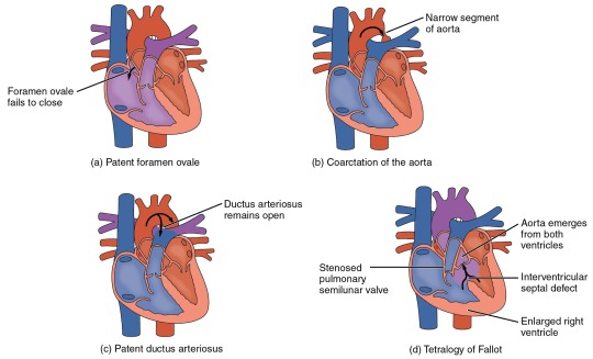
Heart Defects
Heart Defects
One very common form of interatrial septum pathology is patent foramen ovale, which occurs when the septum primum does not close at birth, and the fossa ovalis is unable to fuse. The word patent is from the Latin root patens for “open.” It may be benign or asymptomatic, perhaps never being diagnosed, or in extreme cases, it may require surgical repair to close the opening permanently. As much as 20–25 percent of the general population may have a patent foramen ovale, but fortunately, most have the benign, asymptomatic version. Patent foramen ovale is normally detected by auscultation of a heart murmur (an abnormal heart sound) and confirmed by imaging with an echocardiogram. Despite its prevalence in the general population, the causes of patent ovale are unknown, and there are no known risk factors. In nonlife-threatening cases, it is better to monitor the condition than to risk heart surgery to repair and seal the opening.
Coarctation of the aorta is a congenital abnormal narrowing of the aorta that is normally located at the insertion of the ligamentum arteriosum, the remnant of the fetal shunt called the ductus arteriosus. If severe, this condition drastically restricts blood flow through the primary systemic artery, which is life threatening. In some individuals, the condition may be fairly benign and not detected until later in life. Detectable symptoms in an infant include difficulty breathing, poor appetite, trouble feeding, or failure to thrive. In older individuals, symptoms include dizziness, fainting, shortness of breath, chest pain, fatigue, headache, and nosebleeds. Treatment involves surgery to resect (remove) the affected region or angioplasty to open the abnormally narrow passageway. Studies have shown that the earlier the surgery is performed, the better the chance of survival.
A patent ductus arteriosus is a congenital condition in which the ductus arteriosus fails to close. The condition may range from severe to benign. Failure of the ductus arteriosus to close results in blood flowing from the higher pressure aorta into the lower pressure pulmonary trunk. This additional fluid moving toward the lungs increases pulmonary pressure and makes respiration difficult. Symptoms include shortness of breath (dyspnea), tachycardia, enlarged heart, a widened pulse pressure, and poor weight gain in infants. Treatments include surgical closure (ligation), manual closure using platinum coils or specialized mesh inserted via the femoral artery or vein, or nonsteroidal anti-inflammatory drugs to block the synthesis of prostaglandin E2, which maintains the vessel in an open position. If untreated, the condition can result in congestive heart failure.
Septal defects are not uncommon in individuals and may be congenital or caused by various disease processes. Tetralogy of Fallot is a congenital condition that may also occur from exposure to unknown environmental factors; it occurs when there is an opening in the interventricular septum caused by blockage of the pulmonary trunk, normally at the pulmonary semilunar valve. This allows blood that is relatively low in oxygen from the right ventricle to flow into the left ventricle and mix with the blood that is relatively high in oxygen. Symptoms include a distinct heart murmur, low blood oxygen percent saturation, dyspnea or difficulty in breathing, polycythemia, broadening (clubbing) of the fingers and toes, and in children, difficulty in feeding or failure to grow and develop. It is the most common cause of cyanosis following birth. The term “tetralogy” is derived from the four components of the condition, although only three may be present in an individual patient: pulmonary infundibular stenosis (rigidity of the pulmonary valve), overriding aorta (the aorta is shifted above both ventricles), ventricular septal defect (opening), and right ventricular hypertrophy (enlargement of the right ventricle). Other heart defects may also accompany this condition, which is typically confirmed by echocardiography imaging. Tetralogy of Fallot occurs in approximately 400 out of one million live births. Normal treatment involves extensive surgical repair, including the use of stents to redirect blood flow and replacement of valves and patches to repair the septal defect, but the condition has a relatively high mortality. Survival rates are currently 75 percent during the first year of life; 60 percent by 4 years of age; 30 percent by 10 years; and 5 percent by 40 years.
In the case of severe septal defects, including both tetralogy of Fallot and patent foramen ovale, failure of the heart to develop properly can lead to a condition commonly known as a “blue baby.” Regardless of normal skin pigmentation, individuals with this condition have an insufficient supply of oxygenated blood, which leads to cyanosis, a blue or purple coloration of the skin, especially when active.
Septal defects are commonly first detected through auscultation, listening to the chest using a stethoscope. In this case, instead of hearing normal heart sounds attributed to the flow of blood and closing of heart valves, unusual heart sounds may be detected. This is often followed by medical imaging to confirm or rule out a diagnosis. In many cases, treatment may not be needed.
#atomic heart#science#biology#college#education#school#student#medicine#doctors#health#healthcare#nursing#physiology#pathology
3 notes
·
View notes
Text
Advancing Healthcare: Exploring Ultrasound Services in Perth
In the realm of modern medicine, ultrasound technology stands as a cornerstone in diagnostic imaging, offering invaluable insights into the inner workings of the human body. In Perth, Australia, ultrasound services play a pivotal role in healthcare delivery, providing patients and healthcare professionals with a non-invasive, safe, and effective tool for diagnosing and monitoring various medical conditions. In this article, we explore the significance of ultrasound services perth and their contributions to advancing healthcare in the region.
The Evolution of Ultrasound Technology:
Ultrasound technology has undergone remarkable advancements since its inception, revolutionizing the field of medical imaging and diagnostic medicine. Utilizing sound waves to create real-time images of internal structures, organs, and tissues, ultrasound imaging enables healthcare professionals to visualize anatomical abnormalities, assess organ function, and guide minimally invasive procedures with precision and accuracy.
Comprehensive Diagnostic Imaging Services:
In Perth, 4d Ultrasound Perth encompass a wide range of diagnostic imaging modalities, catering to the diverse needs of patients across various medical specialties. From obstetrics and gynecology to cardiology, oncology, and musculoskeletal imaging, ultrasound examinations provide valuable diagnostic information that aids in the detection, diagnosis, and management of medical conditions.
Obstetric Ultrasound:
Obstetric ultrasound Perth play a vital role in monitoring the health and development of unborn babies during pregnancy. Obstetric ultrasound examinations enable healthcare providers to assess fetal growth, detect congenital anomalies, evaluate placental function, and monitor maternal health, ensuring optimal pregnancy outcomes and providing expectant parents with reassurance and peace of mind.
Cardiac Ultrasound (Echocardiography):
Cardiac ultrasound, also known as echocardiography, is a non-invasive imaging technique used to assess the structure and function of the heart. Cardiac baby ultrasound Perth play a crucial role in diagnosing and managing various cardiac conditions, including coronary artery disease, valvular heart disease, cardiomyopathy, and congenital heart defects. Echocardiograms provide detailed images of the heart's chambers, valves, and blood flow, helping cardiologists evaluate cardiac function and formulate appropriate treatment plans.
Musculoskeletal Ultrasound:
Musculoskeletal ultrasound services offer valuable diagnostic insights into injuries, abnormalities, and degenerative conditions affecting the muscles, tendons, ligaments, joints, and soft tissues of the body. In Perth, musculoskeletal ultrasound examinations assist orthopedic specialists, sports medicine physicians, and physiotherapists in diagnosing conditions such as tendonitis, bursitis, arthritis, ligament tears, and muscle strains, facilitating targeted treatment and rehabilitation strategies.
Women's Health and Gynecological Ultrasound:
Gynecological ultrasound services in Perth are integral to the evaluation and management of various women's health issues, including reproductive disorders, pelvic pain, abnormal bleeding, and infertility. Transvaginal and transabdominal ultrasound examinations enable gynecologists to assess the uterus, ovaries, fallopian tubes, and surrounding structures, aiding in the diagnosis of conditions such as ovarian cysts, fibroids, endometriosis, and pelvic inflammatory disease.
Conclusion:
Ultrasound services in Perth represent a cornerstone in diagnostic imaging, providing patients and healthcare professionals with a versatile, non-invasive, and effective tool for evaluating and monitoring a wide range of medical conditions. From obstetrics and cardiology to musculoskeletal imaging and women's health, ultrasound examinations offer invaluable diagnostic insights that guide treatment decisions and improve patient outcomes. As technology continues to evolve and ultrasound imaging capabilities expand, ultrasound services in Perth will continue to play an essential role in advancing healthcare delivery and enhancing patient care across the region.
0 notes
Text
Exploring the Advancements and Applications of Ultrasound in Perth
Introduction: ultrasound Perth technology has revolutionized medical diagnostics and imaging, offering a non-invasive and safe method to visualize internal organs and tissues. In Perth, like in many other cities around the world, ultrasound technology is playing a pivotal role in healthcare, enabling healthcare professionals to diagnose and monitor various medical conditions with precision and efficiency.
Advancements in Ultrasound Technology: Over the years, ultrasound technology has seen significant advancements, making it more versatile and accurate than ever before. In Perth, healthcare facilities are equipped with state-of-the-art ultrasound machines that utilize advanced imaging techniques such as 3D and 4D ultrasound, Doppler imaging, and contrast-enhanced ultrasound. These technologies provide detailed and real-time images, allowing healthcare providers to make more informed decisions about patient care.
Applications of Ultrasound in Perth: Pregnancy Ultrasound Perth imaging has a wide range of applications across different medical specialties in Perth. In obstetrics and gynecology, ultrasound is commonly used for prenatal screenings, monitoring fetal development, and diagnosing gynecological conditions. In radiology, ultrasound assists in the detection and characterization of abnormalities in the abdomen, pelvis, thyroid, and musculoskeletal system. Additionally, ultrasound-guided procedures, such as biopsies and injections, are performed in Perth to ensure accuracy and safety.
Ultrasound in Cardiology: Cardiac ultrasound, also known as echocardiography, is indispensable in the field of cardiology. In Perth, echocardiograms are routinely performed to assess the structure and function of the heart, diagnose heart conditions such as valvular diseases and cardiomyopathies, and monitor the effectiveness of treatments. Advanced echocardiographic techniques like strain imaging and contrast echocardiography provide valuable insights into cardiac function, aiding in the management of cardiovascular diseases.
Point-of-Care Ultrasound (POCUS): Point-of-care 4d Ultrasound Perth has gained popularity in Perth and elsewhere as a bedside diagnostic tool that can be used by healthcare providers across various specialties. POCUS allows for rapid assessment of patients in emergency departments, intensive care units, and other clinical settings, aiding in the timely diagnosis and management of critical conditions such as trauma, cardiac arrest, and respiratory distress.
Ultrasound in Research and Education: In addition to clinical applications, ultrasound technology plays a crucial role in medical research and education in Perth. Researchers utilize ultrasound imaging to study disease mechanisms, develop new treatment modalities, and evaluate the efficacy of interventions. Moreover, ultrasound training programs are available for medical students, residents, and practicing healthcare professionals to acquire proficiency in ultrasound techniques and interpretation.
Conclusion: 3d Ultrasound Perth technology continues to be a cornerstone of modern medicine in Perth, offering invaluable diagnostic and therapeutic capabilities across various medical specialties. With ongoing advancements and innovations in ultrasound technology, the future holds even greater promise for improving patient outcomes and advancing medical knowledge in Western Australia's capital city.
Source Url:- https://sites.google.com/view/ohheybabycom1221212/home
0 notes
Text
Congenital Diaphragmatic Hernia
What Is A Congenital Diaphragmatic Hernia And How Does It Affect Infants?
Congenital diaphragmatic hernia is a condition in which the diaphragm is malformed and the opening between the lungs and stomach is not properly sealed. Congenital diaphragmatic hernia affects infants, usually before they are born. It can cause respiratory distress, feeding problems, and may lead to death or organ damage. If your infant has a congenital diaphragmatic hernia, it's important to be aware of what this means for them.
The diaphragm is a thin, dome-shaped muscle that separates the chest cavity from the abdomen. It is one of the most important layers of the lungs and plays a critical role in breathing and swallowing. The diaphragm also acts as a barrier between the interior of your baby's body and his or her environment - for example, protecting your baby from potential harmful substances in milk. People with congenital diaphragmatic hernia have an opening between their lungs and stomach that develops before birth. Without proper sealing, they can develop significant respiratory distress during infancy because their intestines are outside of the baby's body.
What are the symptoms of congenital diaphragmatic hernia?
Symptoms vary depending on the stage and severity of the hernia, but they can include: Difficulty eating or swallowing Upper airway obstruction, such as snoring and sleep apnea Difficulty breathing after an upper respiratory infection.
What causes congenital diaphragmatic hernia?
Congenital diaphragmatic hernia can be attributed to: Excessive pressure on the stomach and intestines during fetal development, known as gastric and duodenal compression. Softening of the ribs, which allows the stomach or intestines to protrude into a space between two ribs. Incomplete closure of one or both sides of the diaphragm during fetal development.
How is congenital diaphragmatic hernia diagnosed?
Congenital diaphragmatic hernia is usually diagnosed with a multi-phase CT scan. , which includes 2-D and 4-D scans, and a contrast study.
How to Reduce Your Risk of Having a Baby with Congenital Diaphragmatic Hernia?
Congenital diaphragmatic hernia is the most common congenital malformation of the diaphragm. It is characterized by a defect in the diaphragm that causes it to become displaced and protrude through the chest wall, which results in respiratory distress. The risk factors for having a baby with congenital diaphragmatic hernia include:
Multiple pregnancies
Long duration of labor
Premature rupture of membranes (PROM)
Low birth weight infants
Mother with previous history of fetal death or stillbirth
There are three types of congenital diaphragmatic hernias:
Type I: The fetal parts pass through the nares and into the mouth.
Type II: The fetal parts go through the trachea, then into either the esophagus or esophagus.
Type III: The infant has a normal diaphragm and no hernia is present. (Type II is more common than type I, but can occur in place of type III as well.)
How Diagnosis is Done for Someone With A Congenital Diaphragmatic Hernia?
Congenital diaphragmatic hernia is a rare type of congenital heart defect. It is present at birth and caused by an abnormal connection between the diaphragm and the esophagus. A diagnosis can be done through a number of tests that are typically done in the hospital setting. These tests include:
Chest x-ray
Electrocardiogram
Echocardiogram
Abdominal ultrasound
How to Prescribe the Right Nutrition for a Child With Congenital Diaphragmatic hernia?
The best way to get the right nutrition for a child with or without congenital diaphragmatic hernia is by consulting a dietitian. Sometimes children with this condition need extra calories, while others need to avoid certain foods. The most common types of nutrition for kids with diaphragmatic hernia are liquid meal replacement and nutritional supplements. Diets for children with congenital diaphragmatic hernia should include more calories than those without the condition, but they should also be low in fat, protein and cholesterol. .A dietitian can help you develop a meal plan that meets your child's needs.
Care for Your Newborn Baby with Congenital Diaphragmatic Hernia-
A newborn baby with congenital diaphragmatic hernia is a condition that can cause respiratory distress, breathing difficulties, and even death. Congenital diaphragmatic hernia is a rare condition that affects the lungs of a newborn baby. It occurs when the lower part of the intestines becomes lodged in the chest cavity and blocks the airway. This condition is often diagnosed during pregnancy or shortly after birth when it can be treated by surgical repair.
Neonatal care for the infant may include continuous positive airway pressure (CPAP) to help increase airflow into the lungs and improve oxygenation levels. Other treatments may include gastric decompression to relieve stomach distention and pain; intravenous fluids to correct dehydration; and antibiotics to prevent infection.
Long Term Care for Children with Congenital Diaphragmatic Hernia-
Congenital diaphragmatic hernia is a rare disorder that affects the diaphragm, which is the muscle used to breathe. It can cause respiratory problems, heart failure and even death. Long term care for children with congenital diaphragmatic hernia are challenging because of their physical limitations. The best option for these children includes ventilator support, which is necessary to maintain breathing during sleep. The long-term care process needs to be carefully planned before the child has any complications due to their condition.
It also requires a lot of patience and care from the family members who have to learn how to deal with this condition. and help their child with all the activities around them. Congenital diaphragmatic hernia can be treated surgically, through surgery. The two types of surgery are open and laparoscopic. The open repair is more complex and takes a significantly longer time to heal. A laparoscopic repair has been found to be a safe and effective option for most children with congenital diaphragmatic hernia, but it is not always possible due to poor surgical access or other reasons, depending on the case. Surgery requires a general anesthetic and overnight hospital stay while the scar tissue forms. Recovery is usually quick, but may take up to several months depending on the complexity of the repair.
The outcome for children with congenital diaphragmatic hernia after surgery depends on many factors, including the location and size of the hernia, whether it is associated with any other birth defects, and whether there has been any significant delay in diagnosis or treatment. Surgery often produces a "cure" for this condition but in some cases these children may need additional surgery later in life as their bodies grow or medical conditions change.
1 note
·
View note
Text
Echocardiography Centre In Ayapakkam
Echocardiography or Echo Test or Echocardiogram in Chennai
An echocardiography, also known as an echo test or echocardiogram, is a test that takes "moving pictures" of the heart using sound waves. This can be done at any time, no preparation is required. You do not need to stay in the hospital. This is not surgery and it is painless.
ECG (Electrocardiography)
It is based on the principle that the electrical impulses generated and propagated by the cardiac system are represented by peak waves. This is the most common test to detect the function of different parts of the heart and is the main test to detect cardiac abnormalities.
Echocardiography (ECHO) is available for pediatric, fetal (fetal), and adult patients. It allows the size and function of different types of valves and valves to be seen and with the advent of color Doppler, the function of the heart can be assessed correctly, especially if you suspect a heart problem. For high-quality test contact echocardiography centre in Ayapakkam Chennai!
Key Points about Echocardiography
Extremely high-frequency sound waves record 4,444 images of your heart and heart valves.
No X-rays will be used.
The movements of your heart can be seen on a video screen.
A videotape or photograph can be created from images.
You may occasionally view it during the test.
This usually takes an hour.
Painless and no side effects.
Characteristics of Echocardiography
An echocardiogram employs sound waves to show the blood flow through the heart and heart valves.
Sensors attached to the chest and legs check heart rate during the test.
This test can help healthcare providers diagnose heart disease.
There are 5 basic cardiac ultrasound views (Cardiac Windows) of the heart present include -
Parasternal Long Axis
Parasternal Short Axis
Apical 4 Chamber
Subxiphoid (Subcostal)
IVC Views
𝐌𝐨𝐝𝐞𝐫𝐧𝐋𝐚𝐛&𝐗-𝐑𝐚𝐲𝐬 – Best echocardiography centre in Chennai
Service is always the factor that makes 𝐌𝐨𝐝𝐞𝐫𝐧𝐋𝐚𝐛&𝐗-𝐑𝐚𝐲𝐬 stand out. From providing comfort, convenience, and courtesy in our centers to providing quick and accurate reporting, we are at the forefront of industry practices. Reputed echocardiography centre in Ayapakkam!
0 notes
Link
0 notes
Text
Fetal 2D Echo Test in Kothrud | Fetal 2D Echo Test in Baner
Advanced Fetal 2D Echo Test at DMS Diagnostic: Empowering Precise Insights in Kothrud and Baner Introduction: At DMS Diagnostic, we are committed to providing state-of-the-art diagnostic services that contribute to the well-being of expectant mothers and their unborn babies. As a leading healthcare facility in Kothrud and Baner, we offer advanced Fetal 2D Echo tests that play a crucial role in assessing fetal health and development. Our highly skilled team of medical professionals utilizes cutting-edge technology to perform accurate and comprehensive Fetal 2D Echo tests, ensuring the best possible care for both mother and baby. Accurate & compassionate Fetal 2D Echo Test in Kothrud & Baner by DMS Diagnostic. Early detection for your baby’s heart health. Trust in expert care.
What is a Fetal 2D Echo Test?
A Fetal 2D Echo test, also known as a fetal echocardiogram, is a specialized ultrasound examination that focuses on the fetal heart’s structure and function. This non-invasive and painless procedure employs high-frequency sound waves to create detailed images of the developing heart. The test helps identify any potential congenital heart defects or abnormalities, enabling early diagnosis and appropriate medical interventions.

Why Choose DMS Diagnostic for Fetal 2D Echo Test?
Expertise of Skilled Sonographers: At DMS Diagnostic, our team comprises certified and experienced sonographers who specialize in fetal imaging. They are well-versed in performing Fetal 2D Echo tests, and their expertise ensures accurate assessments of the fetal heart.
State-of-the-Art Equipment: We take pride in investing in advanced ultrasound equipment, specifically designed for fetal imaging. Our cutting-edge technology enables us to obtain high-resolution images, allowing for better visualization and analysis of the fetal heart.
Comprehensive Fetal Heart Evaluation: Our Fetal 2D Echo test provides a comprehensive evaluation of the fetal heart’s structure and function. We assess the heart’s chambers, valves, blood flow patterns, and cardiac activity to detect any anomalies that may require medical attention.
Early Detection of Congenital Heart Conditions: Detecting congenital heart defects early in pregnancy can significantly improve outcomes for both the baby and the mother. Our Fetal 2D Echo test aids in early diagnosis, allowing for timely consultations with pediatric cardiologists and appropriate management plans.
Compassionate Care and Support: We understand the importance of emotional support during pregnancy, especially when specialized tests are involved. Our caring team ensures a comforting environment for expectant mothers, answering any questions and alleviating concerns throughout the process.
The Fetal 2D Echo Test Procedure: Pre-Test Preparation: There is typically no special preparation required for the Fetal 2D Echo test. Expectant mothers are advised to wear loose and comfortable clothing, facilitating easy access to the abdominal area during the procedure.
Test Execution: During the test, the sonographer applies a gel to the mother’s abdomen and gently moves a transducer over the area. The transducer emits sound waves, which bounce off the fetal heart structures, creating real-time images displayed on a monitor.
Data Interpretation: Our skilled sonographers interpret the obtained images, looking for any structural or functional abnormalities in the fetal heart.
Consultation and Reporting: Once the test is complete, our team provides a detailed report of the findings. In cases where abnormalities are detected, we may recommend a consultation with a pediatric cardiologist for further evaluation and management.
Conclusion: At DMS Diagnostic, we prioritize the health and well-being of both expectant mothers and their unborn babies. Our Fetal 2D Echo test in Kothrud and Fetal 2D Echo test in Baner employs cutting-edge technology and expert medical guidance to ensure precise assessments of the fetal heart. With our compassionate care and commitment to excellence, you can trust us to provide the best possible insights into your baby’s cardiac health during this precious phase of life.
What are the various types of echocardiogram tests?
Transthoracic echocardiogram, Transesophageal echocardiogram, Doppler ultrasound, Three-dimensional echocardiogram, Two-dimensional echocardiogram
How much time does the 2D Echo test take?
2D Echo test takes around half an hour to 1 hour to complete the test.
Why is the 2d Echo test done?
Echocardiography or 2D Echo is mostly performed to discover the following: Any underlying heart disease or abnormalities. Congenital heart disease and blood clots or tumors.
#Fetal 2D Echo Test in Kothrud#Fetal 2D Echo Test in Baner#Fetal 2d Echo Test in Pune#Top Fetal 2d Echo in Pune#Fetal Echocardiography Testing Centres in Pune#Best Foetal 2D Echo Test in Pune
0 notes
Text
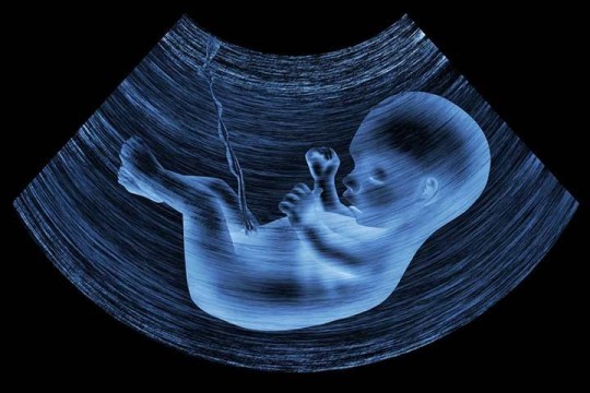
Fetal Echocardiography
What is fetal echocardiography?
A fetal echocardiogram is an ultrasound test performed during pregnancy to evaluate the fetal heart of your developing baby.
On average, it takes about 45-90 minutes to perform, depending on the complexity of the fetus’ heart.
Doctors usually perform fetal echocardiography between weeks 20 and 24 of the second trimester.
Fetal echo can help find heart defects before birth. If a heart problem can be found early, the more likely treatment will work. This is because:
Healthcare providers may be able to treat the problem before birth, in some cases.
Healthcare providers can get ready for problems that may happen during labor and delivery.
Once the baby is born, treatment may be done. This might be medicine or surgery.
Why is fetal echocardiography performed?
Fetal echocardiography can display the blood flow through the heart, the heart rhythm, and the structures of the baby’s heart. Doctors would perform this test if:
Fetal heart abnormalities suspected from a routine obstetric ultrasound
Family history (first degree relative) of CHD
Abnormal fetal heart rate or rhythm
Pregnancy through IVF
Twin-to-twin transfusion syndrome
Exposure to some drugs in early pregnancy, for example, some anti-epileptic drugs
Hydrops
Increased nuchal translucency on a first-trimester screening
Chromosomal abnormalities associated with CHD
What are the risks of fetal echo?
Fetal echo does not have any risks for either the fetus or mother. The lowest possible ultrasound settings are used.
What happens after fetal echo?
Your healthcare provider will look at the results. He or she may order more tests or procedures. They may include:
Treatment. This may be medicine or procedures to treat fetal heart problems.
Nonstress test. This checks fetal heart rate and movement.
Amniocentesis. This test can find chromosomal and genetic disorders and certain birth defects. The healthcare provider puts a needle through the abdominal and uterine wall and into the amniotic sac. He or she takes a sample of amniotic fluid.
Genetic counseling. A counselor helps you learn your risks of having a baby with genetic defects.
1 note
·
View note
Text
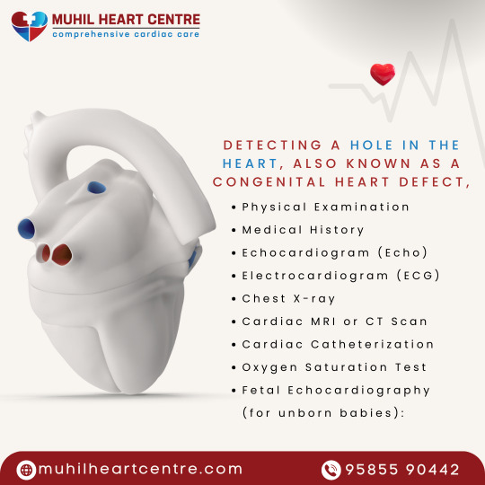
Detecting a hole in the heart, also known as a congenital heart defect,
Physical Examination
Medical History
Echocardiogram (Echo)
Electrocardiogram (ECG)
Chest X-ray
Cardiac MRI or CT Scan
Cardiac Catheterization
Oxygen Saturation Test
Fetal Echocardiography (for unborn babies):
Reach us @ +91 95855 90442
Visit@ muhilheartcentre.com
#muhilheartcentre #HeartHealth #HealthyHeart #CardiovascularHealth #HeartAwareness #HeartDiseasePrevention #HeartCare #HeartHealthyLifestyle #HeartWellness #HeartAware #HeartFitness #LoveYourHeart #HeartStrong #HeartDiseaseAwareness #HealthyLiving #HeartWellbeing #HeartSupport #HeartProtection #HeartCheck #HeartSmart #HeartStrong #HeartLife
0 notes
Text
Do fetal heart tests
Fetal echocardiography is a test similar to an ultrasound. This exam allows your doctor to better see the structure and function of your unborn child's heart. It's typically done in the second trimester, between weeks 18 to 24.
The exam uses sound waves that "echo" off the structures of the fetus's heart. A machine analyzes these sound waves and creates a picture, or echocardiogram, of their heart's interior. This image provides information on how your baby's heart is formed and whether it is working properly.
It also enables your doctor to see the blood flow through the fetus's heart. This in-depth look allows your doctor to find any abnormalities in the baby's blood flow or heartbeat.
Contact the University Hospital Sharjah now to perform fetal heart tests.
0 notes
Text
addon Health Care | Scan | Labs | Diagnostic Center
Improving The Quality Of Your Life Through Better Health add-on Health Care is the best diagnostic center in Bangalore offering affordable healthcare, ultrasound scanning, health checkups, digital X-ray, we promise to deliver our patients the ultimate medical diagnostics services. This includes delivering the highest quality Imaging, laboratory and comprehensive health check services with respect, courtesy and compassion add-on Scans & Labs exists to serve our clients with reliable and accurate diagnostics services, maintaining ethical standards coupled with patient safety measures, highest quality services with rapid turnaround time and ensuring a human touch to our services. We are committed to Delivering Reliable,Fast and Affordable Diagnostic services. Blood Test, COVID-19, Health Packages, Fetal Medicine, Consultations, Tele Consultation, Color Doppler Scan, Digital X-Ray, OPG (Dental X ray), ECG – Electrocardiogram, 2D Echocardiogram (Echo), Treadmill Test TMT, Physiotherapy
#addonscans #addonscanslabs #addonscansandlabs #pregnancy #pregnancytips #pregnancylife #pregnancyjourney
0 notes
Text
Q: Understanding of different types of ultrasound scan in pregnancy, purpose, risks and procedures.
John Korsah
1. Understanding of different types of ultrasound scan in pregnancy,
Ultrasound scans during pregnancy are used to monitor fetal development and ensure that the pregnancy is progressing normally. Here are some of the different types of ultrasound scans that may be performed during pregnancy:
▪️Transabdominal ultrasound: This is the most common type of ultrasound performed during pregnancy. It involves applying gel to the abdomen and using a handheld probe to produce images of the uterus and fetus. This type of ultrasound is typically performed in the first trimester to confirm the pregnancy and estimate the due date, and again in the second and third trimesters to monitor fetal growth and assess any abnormalities.
▪️Transvaginal ultrasound: This type of ultrasound involves inserting a small probe into the vagina to produce images of the uterus and fetus. It is typically performed early in the first trimester to confirm the pregnancy and estimate the due date, or in cases where a transabdominal ultrasound is not providing clear images.
▪️Doppler ultrasound: This type of ultrasound measures blood flow in the umbilical cord and other fetal blood vessels. It can help detect potential circulation problems or other abnormalities that may require medical intervention.
▪️3D/4D ultrasound: These types of ultrasound produce 3D or 4D images of the fetus, providing a more detailed look at the baby's features and movements. They are often used for entertainment purposes, but can also be helpful in identifying certain abnormalities.
▪️Fetal echocardiogram: This is a specialized ultrasound that is used to evaluate the structure and function of the fetal heart. It is typically performed when there is a concern about the baby's heart function, or in cases where the mother has a pre-existing heart condition.
Overall, ultrasound scans are an important tool for monitoring fetal development and ensuring a healthy pregnancy. Your healthcare provider will recommend the appropriate types of ultrasound scans based on your individual needs and the stage of your pregnancy.
2. Purpose
Ultrasound scans are an important tool used during pregnancy to monitor fetal development and ensure the health of the mother and baby. Some of the specific purposes of ultrasound scans during pregnancy include:
▪️Confirming the pregnancy: Ultrasound scans can confirm the presence of a pregnancy, estimate the due date, and detect any potential problems early on.
▪️Monitoring fetal growth and development: Ultrasound scans are used to monitor the baby's growth and development throughout pregnancy, ensuring that the baby is developing properly and identifying any potential problems early on.
▪️Assessing the health of the placenta and umbilical cord: Ultrasound scans can detect problems with the placenta or umbilical cord, which can affect the baby's growth and development.
▪️Screening for genetic abnormalities: Some ultrasound scans, such as the nuchal translucency scan, can screen for genetic abnormalities that may affect the baby's development.
▪️Evaluating the health of the mother: Ultrasound scans can also be used to evaluate the health of the mother's uterus, ovaries, and other pelvic organs, and to monitor for potential complications such as pre-eclampsia or placenta previa.
Overall, the purpose of ultrasound scans during pregnancy is to ensure that the baby is developing properly, to detect any potential problems early on, and to monitor the health of the mother and baby throughout pregnancy.
3. Risk
Ultrasound scans during pregnancy are considered safe and have no known risks to the mother or baby. Unlike some other imaging tests, such as X-rays, ultrasound does not involve ionizing radiation, which can be harmful to the developing fetus.
However, there is a small risk that the ultrasound probe can cause slight heating of the tissue being scanned, which could potentially affect fetal development. To minimize this risk, ultrasound technicians are trained to use the lowest possible power settings and to limit the duration of the scan.
Additionally, there is a small risk of false positive or false negative results from ultrasound scans. In some cases, abnormalities or problems may not be detected on the scan, while in other cases, abnormalities may be detected that turn out to be benign.
It's important to note that while ultrasound scans are generally considered safe, they should only be performed when medically necessary. Unnecessary exposure to ultrasound during pregnancy should be avoided. Your healthcare provider can help you determine when ultrasound scans are necessary and what risks, if any, may be associated with the procedure.
4. Procedures
Ultrasound scans during pregnancy are non-invasive and typically painless procedures. Here is what you can expect during an ultrasound scan:
▪️Preparation: You will be asked to drink water before the scan to fill your bladder, which helps to provide clearer images of the uterus and fetus. You may also be asked to change into a hospital gown.
▪️Positioning: You will lie down on an examination table and the technician will apply a warm, gel-like substance to your abdomen or insert a small probe into your vagina, depending on the type of ultrasound being performed.
▪️Scanning: The technician will move the ultrasound probe over your abdomen or insert it into your vagina to produce images of the uterus and fetus. You may be asked to hold your breath or change positions to help provide clearer images.
▪️Interpretation: The technician will interpret the images and may point out different parts of the fetus, such as the head, heart, or limbs. In some cases, the technician may be able to tell you the sex of the baby, but this is not always possible.
▪️Follow-up: Your healthcare provider will review the results of the ultrasound scan and discuss any findings with you. Additional scans may be recommended if there are concerns or abnormalities detected.
Overall, ultrasound scans during pregnancy are a routine part of prenatal care and are generally safe and non-invasive procedures. Your healthcare provider can help you prepare for the scan and answer any questions you may have about the procedure.
0 notes
Last Seen Blogs

vampsickle
jackpot! ⭐️

digitalbeachrave
OUT OF THE OOZE AND BORN TO CRUISE

black-diamond-cabo
∆ Irwinkabo☠

cellerbration
Riley Farrell

untitled42566
Untitled