#NIH_NIAMS
Text
National Institute of Arthritis and Musculoskeletal and Skin Diseases SCIENCE TRANSLATIONAL MEDICINE The mechanosensitive ion channel PIEZO1 is expressed in tendons and regulates physical performance
National Institute of Arthritis and Musculoskeletal and Skin Diseases SCIENCE TRANSLATIONAL MEDICINE The mechanosensitive ion channel PIEZO1 is expressed in tendons and regulates physical performance
https://blog.naver.com/artnouveau19/222803608570
National Institute of Arthritis and Musculoskeletal and Skin Diseases
“The mechanosensitive ion channel PIEZO1 is expressed in tendons and regulates physical performance”
Read the abstract at https://www.science.org/doi/10.1126/scitranslmed.abj5557
Full access requires subscription or payment. Science Translational Medicine
#MouseModels #NIAMSFunded Scripps Research
NIAMS/NIH/DHHS
@NIH_NIAMS
“The mechanosensitive ion channel PIEZO1 is expressed in tendons and regulates physical performance”
Read the abstract at
https://www.science.org/doi/10.1126/scitranslmed.abj5557?fbclid=IwAR24o2XSU-AUrTFLjHQdlcQzkna6MivRraURamS2vTUq2FVFWIuOQ_SfU2c
@ScienceTM
#MouseModels #NIAMSFunded
@ScrippsResearch
https://twitter.com/NIH_NIAMS/status/1544697693007593475
RESEARCH ARTICLE
TENDON
The mechanosensitive ion channel PIEZO1 is expressed in tendons and regulates physical performance
RYO NAKAMICHI HTTPS://ORCID.ORG/0000-0002-0145-2135SHANG MATAKAYUKI NONOYAMA HTTPS://ORCID.ORG/0000-0001-8554-0636TOMOKI CHIBA HTTPS://ORCID.ORG/0000-0001-5472-9030RYOTA KURIMOTO HTTPS://ORCID.ORG/0000-0003-3029-5287HIROKI OHZONO HTTPS://ORCID.ORG/0000-0001-7170-5401MERISSA OLMER HTTPS://ORCID.ORG/0000-0002-2483-4216CHISA SHUKUNAMI HTTPS://ORCID.ORG/0000-0001-5616-6761NORIYUKI FUKU HTTPS://ORCID.ORG/0000-0001-7792-5835[...]HIROSHI ASAHARA HTTPS://ORCID.ORG/0000-0002-5215-8745 +8 authors Authors Info & Affiliations
SCIENCE TRANSLATIONAL MEDICINE
1 Jun 2022
Vol 14, Issue 647
DOI: 10.1126/scitranslmed.abj5557
Potentiating physical performance
Tendons connect muscle to bone and are important for musculoskeletal function, including athletic performance. Here, Nakamichi et al. used a mouse model to study the effects of a gain-of-function variant in Piezo1, a mechanosensitive ion channel, in tendon. Mutant tendons were wider, with larger collagen fibril cross-sectional diameter, and had increased compliance and greater storage of elastic energy. Tendon-specific mutant mice demonstrated greater jumping ability and faster running. The authors also observed increased frequency of a gain-of-function PIEZO1 deletion in a cohort of Jamaican sprinters, supporting a role for tendon expression of PIEZO1 in potentiating physical performance.
Abstract
How mechanical stress affects physical performance via tendons is not fully understood. Piezo1 is a mechanosensitive ion channel, and E756del PIEZO1 was recently found as a gain-of-function variant that is common in individuals of African descent. We generated tendon-specific knock-in mice using R2482H Piezo1, a mouse gain-of-function variant, and found that they had higher jumping abilities and faster running speeds than wild-type or muscle-specific knock-in mice. These phenotypes were associated with enhanced tendon anabolism via an increase in tendon-specific transcription factors, Mohawk and Scleraxis, but there was no evidence of changes in muscle. Biomechanical analysis showed that the tendons of R2482H Piezo1 mice were more compliant and stored more elastic energy, consistent with the enhancement of jumping ability. These phenotypes were replicated in mice with tendon-specific R2482H Piezo1 replacement after tendon maturation, indicating that PIEZO1 could be a target for promoting physical performance by enhancing function in mature tendon. The frequency of E756del PIEZO1 was higher in sprinters than in population-matched nonathletic controls in a small Jamaican cohort, suggesting a similar function in humans. Together, this human and mouse genetic and physiological evidence revealed a critical function of tendons in physical performance, which is tightly and robustly regulated by PIEZO1 in tenocytes.
https://www.science.org/doi/10.1126/scitranslmed.abj5557?fbclid=IwAR24o2XSU-AUrTFLjHQdlcQzkna6MivRraURamS2vTUq2FVFWIuOQ_SfU2c
H. Langberg, H. Ellingsgaard, T. Madsen, J. Jansson, S. P. Magnusson, P. Aagaard, M. Kjaer, Eccentric rehabilitation exercise increases peritendinous type I collagen synthesis in humans with Achilles tendinosis. Scand. J. Med. Sci. Sports 17, 61–66 (2007).
T. Wang, Z. Lin, M. Ni, C. Thien, R. E. Day, B. Gardiner, J. Rubenson, T. B. Kirk, D. W. Smith, A. Wang, D. G. Lloyd, Y. Wang, Q. Zheng, M. H. Zheng, Cyclic mechanical stimulation rescues achilles tendon from degeneration in a bioreactor system. J. Orthop. Res. 33, 1888–1896 (2015).
B. Coste, J. Mathur, M. Schmidt, T. J. Earley, S. Ranade, M. J. Petrus, A. E. Dubin, A. Patapoutian, Piezo1 and Piezo2 are essential components of distinct mechanically activated cation channels. Science 330, 55–60 (2010).
K. Nonomura, V. Lukacs, D. T. Sweet, L. M. Goddard, A. Kanie, T. Whitwam, S. S. Ranade, T. Fujimori, M. L. Kahn, A. Patapoutian, Mechanically activated ion channel PIEZO1 is required for lymphatic valve formation. Proc. Natl. Acad. Sci. U.S.A. 115, 12817–12822 (2018).
W. Sun, S. Chi, Y. Li, S. Ling, Y. Tan, Y. Xu, F. Jiang, J. Li, C. Liu, G. Zhong, D. Cao, X. Jin, D. Zhao, X. Gao, Z. Liu, B. Xiao, Y. Li, The mechanosensitive Piezo1 channel is required for bone formation. eLife 8, e47454 (2019).
M. R. Servin-Vences, M. Moroni, G. R. Lewin, K. Poole, Direct measurement of TRPV4 and PIEZO1 activity reveals multiple mechanotransduction pathways in chondrocytes. eLife 6, e21074 (2017).
F. S. Passini, P. K. Jaeger, A. S. Saab, S. Hanlon, N. A. Chittim, M. J. Arlt, K. D. Ferrari, D. Haenni, S. Caprara, M. Bollhalder, B. Niederöst, A. N. Horvath, T. Götschi, S. Ma, B. Passini-Tall, S. F. Fucentese, U. Blache, U. Silván, B. Weber, K. G. Silbernagel, J. G. Snedeker, Shear-stress sensing by PIEZO1 regulates tendon stiffness in rodents and influences jumping performance in humans. Nat. Biomed. Eng. 5, 1457–1471 (2021).
R. Schweitzer, J. H. Chyung, L. C. Murtaugh, A. E. Brent, V. Rosen, E. N. Olson, A. Lassar, C. J. Tabin, Analysis of the tendon cell fate using Scleraxis, a specific marker for tendons and ligaments. Development 128, 3855–3866 (2001).
H. Liu, C. Zhang, S. Zhu, P. Lu, T. Zhu, X. Gong, Z. Zhang, J. Hu, Z. Yin, B. C. Heng, X. Chen, H. W. Ouyang, Mohawk promotes the tenogenesis of mesenchymal stem cells through activation of the TGFβ signaling pathway. Stem Cells 33, 443–455 (2015).
A. J. Bayliss, A. M. Weatherholt, T. T. Crandall, D. L. Farmer, J. C. McConnell, K. M. Crossley, S. J. Warden, Achilles tendon material properties are greater in the jump leg of jumping athletes. J. Musculoskelet. Neuronal Interact. 16, 105–112 (2016).
G. J. Ettema, P. A. Huijing, A. de Haan, The potentiating effect of prestretch on the contractile performance of rat gastrocnemius medialis muscle during subsequent shortening and isometric contractions. J. Exp. Biol. 165, 121–136 (1992).
J. M. McBride, G. O. McCaulley, P. Cormie, Influence of preactivity and eccentric muscle activity on concentric performance during vertical jumping. J. Strength Cond. Res. 22, 750–757 (2008).
C. T. Thorpe, K. J. Karunaseelan, J. N. C. Hin, G. P. Riley, H. L. Birch, P. D. Clegg, H. R. Screen, Distribution of proteins within different compartments of tendon varies according to tendon type. J. Anat. 229, 450–458 (2016).
K. D. Smith, A. Vaughan-Thomas, D. G. Spiller, J. F. Innes, P. D. Clegg, E. J. Comerford, The organisation of elastin and fibrillins 1 and 2 in the cruciate ligament complex. J. Anat. 218, 600–607 (2011).
R. K. Smith, M. Gerard, B. Dowling, A. J. Dart, H. L. Birch, A. E. Goodship, Correlation of cartilage oligomeric matrix protein (COMP) levels in equine tendon with mechanical properties: A proposed role for COMP in determining function-specific mechanical characteristics of locomotor tendons. Equine Vet. J. Suppl. 241–244 (2002).
H. Nakahara, A. Hasegawa, K. Otabe, F. Ayabe, T. Matsukawa, N. Onizuka, Y. Ito, T. Ozaki, M. K. Lotz, H. Asahara, Transcription factor Mohawk and the pathogenesis of human anterior cruciate ligament degradation. Arthritis Rheum. 65, 2081–2089 (2013).
C. Spang, J. Chen, L. J. Backman, The tenocyte phenotype of human primary tendon cells in vitro is reduced by glucocorticoids. BMC Musculoskelet. Disord. 17, 467 (2016).
Nuclear factor of activated T-cells (NFAT) is a family of transcription factors shown to be important in immune response. One or more members of the NFAT family is expressed in most cells of the immune system. NFAT is also involved in the development of cardiac, skeletal muscle, and nervous systems. NFAT was first discovered as an activator for the transcription of IL-2 in T cells (as a regulator of T cell immune response) but has since been found to play an important role in regulating many more body systems.[1] NFAT transcription factors are involved in many normal body processes as well as in development of several diseases, such as inflammatory bowel diseases and several types of cancer. NFAT is also being investigated as a drug target for several different disorders.
https://en.wikipedia.org/wiki/NFAT
The gastrocnemius muscle (plural gastrocnemii) is a superficial two-headed muscle that is in the back part of the lower leg of humans. It runs from its two heads just above the knee to the heel, a three joint muscle (knee, ankle and subtalar joints). The muscle is named via Latin, from Greek γαστήρ (gaster) 'belly' or 'stomach' and κνήμη (knḗmē) 'leg', meaning 'stomach of the leg' (referring to the bulging shape of the calf).
https://en.wikipedia.org/wiki/Gastrocnemius_muscle
Proteoglycan 4 or lubricin is a proteoglycan that in humans is encoded by the PRG4 gene.[3][4][5] It acts as a joint/boundary lubricant.
https://en.wikipedia.org/wiki/Proteoglycan_4
List of flexors of the human body
A flexor is a muscle that flexes a joint. In anatomy, flexion (from the Latin verb flectere, to bend)[1] is a joint movement that decreases the angle between the bones that converge at the joint. For example, one’s elbow joint flexes when one brings their hand closer to the shoulder. Flexion is typically instigated by muscle contraction of a flexor.
https://en.wikipedia.org/wiki/List_of_flexors_of_the_human_body
Actinin is a microfilament protein. Alpha-actinin-1 is necessary for the attachment of actin myofilaments to the Z-lines in skeletal muscle cells,[1] and to the dense bodies in smooth muscle cells.[2] The functional protein is an anti-parallel dimer, which cross-links the thin filaments in adjacent sarcomeres, and therefore coordinates contractions between sarcomeres in the horizontal axis.
The non-sarcomeric alpha-actinins, encoded by ACTN1 and ACTN4, are widely expressed. ACTN2 expression is found in both cardiac and skeletal muscle, whereas ACTN3 is limited to the latter. Both ends of the rod-shaped alpha-actinin dimer contain actin-binding domains.
Mutations in ACTN4 can cause the kidney disease focal segmental glomerulosclerosis (FSGS).
https://en.wikipedia.org/wiki/Actinin
Actin is a family of globular multi-functional proteins that form microfilaments in the cytoskeleton, and the thin filaments in muscle fibrils. It is found in essentially all eukaryotic cells, where it may be present at a concentration of over 100 μM; its mass is roughly 42 kDa, with a diameter of 4 to 7 nm.
An actin protein is the monomeric subunit of two types of filaments in cells: microfilaments, one of the three major components of the cytoskeleton, and thin filaments, part of the contractile apparatus in muscle cells. It can be present as either a free monomer called G-actin (globular) or as part of a linear polymer microfilament called F-actin (filamentous), both of which are essential for such important cellular functions as the mobility and contraction of cells during cell division.
https://en.wikipedia.org/wiki/Actin
The scleraxis protein is a member of the basic helix-loop-helix (bHLH) superfamily of transcription factors.[1] Currently two genes (SCXA and SCXB respectively) have been identified to code for identical scleraxis proteins.
https://en.wikipedia.org/wiki/Scleraxis
1 note
·
View note
Photo
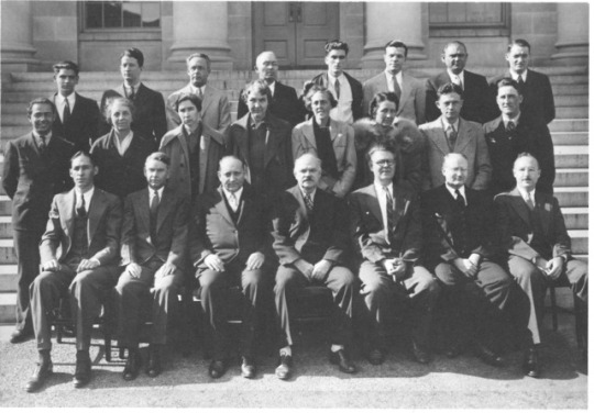
This cheerful group of people posing on the steps of the NIH administration building 80 years ago worked in the NIH Division of Chemistry. They were getting ready to move from their labs at 25th and E Streets, NW in Washington, DC, to their new building on the current Bethesda campus in 1939. Dr. Claude Hudson, in the middle of the front row, was a founder of carbohydrate chemistry and the Division director; many of his staff went on to greater glory, such as Floyd Daft, who became director of the National Institute of Arthritis and Metabolic Diseases. The Division included four women Ph.D. chemists. Learn more about their work on page 54 and 74 https://history.nih.gov/research/downloads/70acresofscience.pdf
Front row: Dr. W. Dayton MacLay, Dr. Ernest L. Jackson, Dr. Elias Elvove, Dr. Claude S. Hudson, Dr. Raymond M. Hann, Dr. Havelock F. Fraser, and Dr. Nelson K. Richtmyer. Middle row: Mr. Samuel Dove, Dr. Alice T. Merill, Dr. Allene Jeanes, Dr. Evelyn B. Tilden, Dr. Mildred Adams, Miss Edna M. Montgomery, Dr. Willard T. Haskins, and Mr. Harry W. Diehl. Back row: Mr. Richard Maggenti, Dr. William S. McClenaham, Dr. Frank J. McClure, Mr. Charles G. Remsburg, Mr. Thomas Collins, Dr. Floyd S. Daft, Mr. John T. Sipes, and Dr. Albert E. Knauf.
2 notes
·
View notes
Text
NIAMS/NIH/DHHS (@NIH_NIAMS) Tweeted:
NIAMS is recruiting volunteers for a clinical study on the natural history, genetics, and pathophysiology of systemic juvenile idiopathic #arthritis, adult-onset Still’s disease & related inflammatory conditions: https://t.co/63tYKXpWgT #JuvenileArthritis https://t.co/wXIdszDeoK https://twitter.com/NIH_NIAMS/status/1172195668036747264?s=17
3 notes
·
View notes
Text
0 notes
Text
@NIH_NIAMS: Image: Skin and Fat Cells. Credit: Richard Gallo, M.D., Ph.D., UC San Diego Health, and Ling-juan Zhang, Ph.D., Xiamen University, Xiamen, China. @UCSDHealth
from http://twitter.com/NIH_NIAMS
via IFTTT
0 notes
Link
via Twitter https://twitter.com/Funding_Grants
0 notes
Text
#UBI Victim "Mary" in @CityofPerris. Homeless, painful poverty. This young american suffers daily from the pain of being ignored. #BREAKING @CBSLA #ABC7Eyewitness @NIH_NIAMS @RivCoDPSS #HelptheHomeless January 29th 2020 Wednesday 7:42 P.M. California USA.

1 note
·
View note
Last Seen Blogs

exgo
Sin título

valt-the-wonder-deer
Valt the Wonder Deer (Unofficial Page)

trending-info
Megha Verma

kennysaysthings
Kenny
milwaukeephotographer
Untitled