#rare autoimmune liver disease
Text
Exploring the Interplay Between Diseases and Liver Transplant
Understanding the Complexities and Implications
Liver transplant surgery is a life-saving procedure that offers hope to patients with end-stage liver disease and certain liver-related conditions. However, the decision to undergo a liver transplant is often influenced by the underlying disease or condition that necessitates the procedure. In this article, we delve into the intricate connection between diseases and liver transplants, exploring the complexities, implications, and considerations involved.

Liver Diseases Leading to Transplant: Liver transplant is commonly indicated for patients with end-stage liver disease (ESLD), a condition characterized by irreversible liver damage and loss of function. Chronic liver diseases such as cirrhosis, hepatitis B and C, alcoholic liver disease, non-alcoholic fatty liver disease (NAFLD), and autoimmune hepatitis are among the primary causes of ESLD. These diseases progress over time, leading to liver failure and ultimately necessitating a transplant to restore liver function and prolong survival.
Hepatobiliary Cancers: Hepatocellular carcinoma (HCC), the most common type of liver cancer, often develops in the setting of chronic liver disease or cirrhosis. In cases where the cancer is confined to the liver and has not spread beyond, liver transplant may be considered as a curative treatment option. However, stringent criteria and careful patient selection are essential to ensure favorable outcomes and prevent cancer recurrence post-transplant.
Metabolic Liver Diseases: Inherited metabolic disorders such as Wilson's disease, hemochromatosis, and alpha-1 antitrypsin deficiency can affect liver function and lead to progressive liver damage. For patients with severe and unmanageable symptoms, liver transplant may offer a chance for improved quality of life and long-term survival. However, the presence of underlying metabolic abnormalities may pose challenges during the transplant process and require specialized pre-transplant evaluation and management.
Acute Liver Failure: Acute liver failure (ALF) is a rare but life-threatening condition characterized by rapid onset of liver dysfunction and hepatic encephalopathy. Causes of ALF include viral hepatitis, drug-induced liver injury, autoimmune hepatitis, and acute fatty liver of pregnancy, among others. Liver transplant may be considered for select patients with ALF who fail to respond to medical therapy or develop complications such as hepatic coma. Timely referral and evaluation are crucial in optimizing outcomes for these patients.
Autoimmune Liver Diseases: Autoimmune liver diseases, including autoimmune hepatitis, primary biliary cholangitis (formerly known as primary biliary cirrhosis), and primary sclerosing cholangitis, are characterized by immune-mediated damage to the liver and biliary tract. While medical therapy is the mainstay of treatment for most patients, those with advanced disease and progressive liver failure may require liver transplant as a definitive treatment option. Careful management of post-transplant immunosuppression is essential to prevent disease recurrence and graft rejection.
Challenges and Considerations: Despite the potential benefits, liver transplant poses inherent challenges and considerations, particularly in the context of underlying diseases. Patient selection, pre-transplant evaluation, and post-transplant management require a multidisciplinary approach involving hepatologists, transplant surgeons, oncologists, and other specialists. Additionally, the shortage of donor organs, immunosuppression-related complications, and the risk of disease recurrence post-transplant are important factors to consider when weighing the risks and benefits of liver transplant in patients with underlying diseases.
The interplay between diseases and liver transplant is multifaceted and complex, with diverse implications for patient management and outcomes. While liver disease treatment in Bangalore, offers a lifeline to patients with end-stage liver disease, hepatobiliary cancers, metabolic disorders, acute liver failure, and autoimmune liver diseases, careful consideration of the underlying disease, patient characteristics, and transplant-related factors is essential in optimizing outcomes and ensuring long-term success. Through continued research, innovation, and collaboration, clinicians and researchers strive to advance our understanding of this intricate connection and improve the care and outcomes of patients undergoing liver transplant for various diseases.
Let’s know what are approaches taken to Liver transplant
Deceased Donor Liver Transplantation (DDLT) and Living Donor Liver Transplantation (LDLT) are two approaches to liver transplantation, each with its own distinct characteristics and considerations.

Donor Source:
Deceased Donor Liver Transplantation: Deceased Donor Liver Transplantation in Bangalore, the liver is procured from a deceased donor who has been declared brain-dead and has consented to organ donation either during their lifetime or by their family after death.
Living Donor Liver Transplantation: In Living Donor Liver Transplantation, the liver is donated by a living donor, typically a family member or close relative of the recipient. The donor undergoes a thorough evaluation process to assess their suitability for donation, including medical, psychological, and ethical considerations.
Timing of Transplantation:
Deceased Donor Liver Transplantation: The timing of Deceased Donor Liver Transplantation depends on the availability of deceased donor organs, which can vary depending on factors such as organ donation rates, waitlist prioritization, and organ allocation policies.
Living Donor Liver Transplantation: Living Donor Liver Transplantation offers the advantage of scheduling the transplant at a time that is convenient for both the recipient and the donor, minimizing the risk of disease progression and optimizing outcomes for both parties.
Graft Size and Compatibility:
Deceased Donor Liver Transplantation: The size and compatibility of the deceased donor liver may not always match the recipient's requirements, leading to potential mismatches in size or blood type. This can sometimes result in a longer wait time on the transplant waitlist or the need for additional surgical techniques to adapt the donor liver to the recipient's anatomy.
Living Donor Liver Transplantation: Living Donor Liver Transplantation allows for a more tailored approach to graft selection, as the donor liver can be selected based on size, blood type compatibility, and other factors specific to the recipient's needs. This often results in a better match and reduces the risk of graft rejection or complications post-transplant.
Waiting Time and Urgency:
Deceased Donor Liver Transplantation: Due to the limited availability of deceased donor organs, patients awaiting Deceased Donor Liver Transplantation may experience longer wait times on the transplant waitlist, particularly for those with less severe illness or lower priority status.
Living Donor Liver Transplantation: Living Donor Liver Transplantation offers the advantage of shorter waiting times, as the transplant can be scheduled based on the recipient's clinical status and urgency of need. This can be particularly beneficial for patients with rapidly progressive liver disease or acute liver failure who may not have the luxury of waiting for a deceased donor organ.
Risk to Donor:
Deceased Donor Liver Transplantation: There is no risk to the donor in Deceased Donor Liver Transplantation, as the liver is procured from a deceased donor who has already passed away.
Living Donor Liver Transplantation: Living Donor Liver Transplantation carries inherent risks to the living donor, including potential complications related to the surgical procedure, anesthesia, and recovery process. However, advances in surgical techniques and donor selection criteria have significantly reduced the risk of complications for living donors in recent years.
while both Deceased Donor Liver Transplantation and Living Donor Liver Transplantation in Bangalore, offer life-saving options for patients with end-stage liver disease, each approach has its own unique characteristics, considerations, and advantages. The choice between Deceased Donor Liver Transplantation and Living Donor Liver Transplantation depends on various factors, including the recipient's clinical status, urgency of need, compatibility, and availability of suitable donor organs. Ultimately, the goal of liver transplantation is to provide the best possible outcome for the recipient while ensuring the safety and well-being of the donor.
For details get the specialists for the Best Liver Transplant Center in Bangalore
#fatty liver treatment in Bangalore#Liver inflammation treatment in Bangalore#Liver disease treatment in Bangalore#Liver transplantation surgery in Bangalore#Deceased Donor Liver Transplantation in Bangalore#Living Donor Liver Transplantation in Bangalore#best liver transplant center in Bangalore#fatty liver#liver treatment center#liver treatment#fastrecoveryoffattyliver#liver care center Bangalore
0 notes
Text
Track 22: Liver Biopsy
A liver biopsy is a medical procedure in which a small piece of liver tissue is removed for examination under a microscope. This procedure is typically performed to diagnose various liver conditions and diseases, assess the severity of liver damage, and determine the best course of treatment.
There are different methods of performing a liver biopsy:
Percutaneous biopsy: This is the most common method, where a thin needle is inserted through the skin and into the liver to collect a small sample of tissue. This procedure is usually done under local anesthesia, and the patient may be asked to hold their breath to reduce the risk of injury to surrounding organs.
Transvenous biopsy: In some cases, particularly when the liver tissue needs to be examined more closely or if there is ascites (fluid buildup in the abdomen), a biopsy may be performed through a vein in the neck or groin, using a special needle and imaging guidance.
Laparoscopic biopsy: In this method, a small incision is made in the abdomen, and a laparoscope (a thin, flexible tube with a camera) is inserted to guide the biopsy needle to the liver. This method may be used if other biopsy techniques are not possible or if a larger sample of liver tissue is needed.
After the biopsy, the liver tissue sample is sent to a laboratory for analysis by a pathologist. The pathologist examines the tissue under a microscope to look for signs of liver disease, inflammation, scarring (fibrosis), cirrhosis, infections, or tumors.
Understanding Liver Biopsy: Uses, and Risks
Liver biopsy is a diagnostic procedure used to assess liver health and diagnose various liver conditions. It involves the removal of a small sample of liver tissue for examination under a microscope. In this blog post, we will explore the liver biopsy procedure, its uses, and associated risks.
Uses:
Liver biopsy is used to diagnose a variety of liver conditions, including:
Hepatitis (both viral and autoimmune)
Cirrhosis
Nonalcoholic fatty liver disease (NAFLD)
Liver cancer
Biliary tract diseases
Genetic liver disorders
The results of a liver biopsy can help healthcare providers determine the severity of liver disease, guide treatment decisions, and monitor disease progression over time.
Risks:
While liver biopsy is generally considered safe, it does carry some risks, including:
Bleeding at the biopsy site
Pain or discomfort
Injury to nearby organs, such as the gallbladder or lungs
In rare cases, infection or allergic reaction to anesthesia
Conclusion
In conclusion, liver biopsy serves as a crucial diagnostic tool in the assessment and management of liver diseases. Through the extraction of a small tissue sample, healthcare providers gain invaluable insights into the health of the liver, enabling accurate diagnosis, treatment planning, and disease monitoring.
Despite its importance, it's essential to acknowledge the potential risks associated with liver biopsy, including bleeding, pain, and the rare possibility of complications such as injury to nearby organs or infection. However, with proper preparation, skilled medical professionals, and diligent post-procedural care, these risks can be minimized.
Ultimately, the decision to undergo a liver biopsy should be made collaboratively between patients and their healthcare providers, taking into account individual medical history, the suspected condition, and the potential benefits and risks of the procedure.
By providing valuable diagnostic information, liver biopsy plays a vital role in the comprehensive care and management of liver diseases, contributing to improved patient outcomes and quality of life.
Important Information:
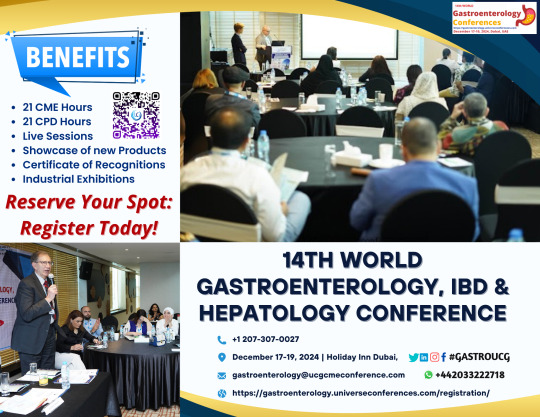
Conference Name: 14th World Gastroenterology, IBD & Hepatology Conference
Short Name: 14GHUCG2024
Dates: December 17-19, 2024
Venue: Dubai, UAE
Email: [email protected]
Visit: https://gastroenterology.universeconferences.com/
Call for Papers: https://gastroenterology.universeconferences.com/submit-abstract/
Register here: https://gastroenterology.universeconferences.com/registration/
Exhibitor/Sponsor: https://gastroenterology.universeconferences.com/exhibit-sponsor-opportunities/
Call Us: +12073070027
WhatsApp Us: +442033222718
0 notes
Text
Navigating the Landscape of the Immunosuppressant Drugs Market: Trends, Challenges, and Future Prospects
Introduction:
The global immunosuppressant drugs market has witnessed significant growth in recent years, driven by an increasing prevalence of autoimmune diseases, organ transplant procedures, and advancements in medical research. Immunosuppressant drugs play a pivotal role in managing conditions where the immune system attacks the body's own tissues, as well as in preventing organ rejection post-transplantation. This article explores key trends, challenges, and future prospects shaping the immunosuppressant drugs market.
Market Overview:
The immunosuppressant drugs market has experienced substantial expansion, with a diverse range of pharmaceutical companies contributing to the development and commercialization of novel therapies. The market encompasses a broad spectrum of drugs, including corticosteroids, calcineurin inhibitors, and monoclonal antibodies, catering to various autoimmune disorders and transplant-related needs.
Key Trends:
1. Rising Incidence of Autoimmune Diseases:
The increasing prevalence of autoimmune diseases such as rheumatoid arthritis, lupus, and multiple sclerosis has fueled the demand for immunosuppressant drugs. These medications help manage symptoms by modulating the immune response, providing relief to patients and improving their quality of life.
2. Organ Transplantation Surge:
Organ transplantation has become a mainstream medical procedure, with a growing number of patients benefiting from kidney, liver, heart, and lung transplants. Immunosuppressant drugs are crucial in preventing graft rejection, ensuring the success of transplantation and enhancing patient survival rates.
3. Advancements in Biotechnology:
The field of biotechnology has witnessed remarkable strides in the development of targeted immunosuppressant therapies. Monoclonal antibodies, in particular, have gained prominence for their specificity in targeting immune cells involved in autoimmune responses, offering more effective and tailored treatment options.
Challenges:
1. Side Effects and Safety Concerns:
The use of immunosuppressant drugs is associated with side effects, ranging from mild to severe. Balancing the need for immune suppression with the risk of infections and other adverse reactions poses a constant challenge for healthcare providers. Striking the right balance is crucial to ensuring patient well-being.
2. High Cost of Treatment:
The cost of immunosuppressant drugs can be a significant barrier to access for many patients, especially in developing regions. As these medications are often required for extended periods, the financial burden on patients and healthcare systems remains a concern.
3. Emergence of Biosimilars:
The impending expiration of patents for some established immunosuppressant drugs has paved the way for the development and approval of biosimilars. While offering cost-effective alternatives, the entry of biosimilars brings challenges in terms of ensuring equivalent efficacy and safety.
Future Prospects:
1. Personalized Medicine:
The future of immunosuppressant therapy lies in personalized medicine, tailoring treatments based on an individual's genetic makeup and immune response. This approach aims to optimize therapeutic outcomes while minimizing adverse effects, marking a paradigm shift in the field.
2. Innovations in Drug Delivery:
Ongoing research focuses on developing innovative drug delivery systems to enhance the efficiency and precision of immunosuppressant drug administration. Nanotechnology and targeted drug delivery methods hold promise in improving therapeutic efficacy and reducing side effects.
3. Focus on Rare Diseases:
As the understanding of rare autoimmune disorders grows, there is an increasing emphasis on developing specialized immunosuppressant drugs to address unmet medical needs in these niche markets.
Conclusion:
The immunosuppressant drugs market continues to evolve, driven by the intersection of scientific advancements, increasing disease prevalence, and the pursuit of more targeted and personalized treatment approaches. Despite challenges, the market's trajectory points towards a future where patients can benefit from safer, more effective, and accessible immunosuppressant therapies, ultimately improving the management of autoimmune diseases and enhancing the success of organ transplantation.
0 notes
Text
Liver function tests can help identify a wide range of liver-related conditions and provide valuable information about the health and function of the liver. Some of the conditions that can be identified or assessed using liver function tests include:
Hepatitis: Liver function tests can detect inflammation of the liver caused by various forms of hepatitis, including hepatitis A, B, C, and others.
Cirrhosis: Elevated liver enzymes (such as ALT and AST) and low levels of albumin may indicate chronic liver damage and cirrhosis, which is scarring of the liver tissue.
Fatty liver disease: Elevated ALT and AST levels can be indicative of non-alcoholic fatty liver disease (NAFLD) or alcoholic liver disease.
Alcohol-induced liver damage: Chronic alcohol consumption can lead to elevated liver enzymes and other abnormalities in liver function tests.
Biliary obstruction: High levels of alkaline phosphatase (ALP) and total bilirubin can suggest problems with the bile ducts, such as blockages or gallstones.
Liver tumors: Liver function tests may show abnormalities in cases of liver tumors, such as hepatocellular carcinoma.
Drug-induced liver injury: Some medications can lead to liver damage, which may be detected through changes in liver function tests.
Autoimmune liver diseases: Conditions like autoimmune hepatitis and primary biliary cholangitis may be identified through liver function tests, along with additional specific tests.
Wilson's disease: Elevated liver enzymes and low serum ceruloplasmin levels can be indicators of Wilson's disease, a rare genetic disorder that causes copper buildup in the liver.
Hemochromatosis: Elevated ferritin levels may suggest hemochromatosis, a condition characterized by excessive iron absorption and accumulation in the liver.
Gilbert's syndrome: This is a benign genetic condition that may lead to mild increases in bilirubin levels without causing any serious liver damage.
Liver injury from other medical conditions: Liver function tests can provide information about the impact of other medical conditions, such as heart failure, sepsis, or certain infections, on the liver.
Get full body checkups at Saifee Hospital Mumbai done.
#health#liver transplant#liver cancer#liver disease#liver damage#liver#cancer#hepatica#hepatitis a#hepatitis c#hepatitis#hepatitis b#wilson disease
0 notes
Text
Acute Liver Failure and its Management
Acute liver failure, also known as fulminant hepatic failure, is a rare and life-threatening condition in which the liver rapidly loses its ability to function. This can occur over a period of days to weeks and is often associated with the sudden onset of severe liver dysfunction. There are many potential causes of acute liver failure, including viral infections (such as hepatitis), drug toxicity, autoimmune disorders, and metabolic diseases.
Symptoms of acute liver failure may include jaundice (yellowing of the skin and eyes), confusion, abdominal pain, bleeding tendencies, and changes in mental status. It is crucial to seek immediate medical attention if acute liver failure is suspected, as it can be fatal if not treated promptly.
The management of acute liver failure involves several key aspects:
Hospitalization: Patients with acute liver failure need to be admitted to the hospital, typically in an intensive care unit (ICU), to receive close monitoring and supportive care.
Identifying and treating the underlying cause: The first step is to identify and address the cause of acute liver failure. Depending on the specific cause, this may involve antiviral medications (for viral hepatitis), discontinuation of offending drugs or toxins, or treatments for autoimmune or metabolic conditions.
Supportive care: Supportive care is essential for patients with acute liver failure. This includes maintaining adequate nutrition, managing complications, and addressing the symptoms. Patients may require nutritional support via a feeding tube if they are unable to eat.
Medications: In some cases, medications such as N-acetylcysteine may be used to treat certain types of drug-induced liver injuries. Other medications may be used to manage complications and symptoms.
Monitoring and management of complications:
Coagulation and bleeding issues: Patients often have blood clotting abnormalities, which may require blood products or medications to correct.
Cerebral edema: Acute liver failure can lead to swelling in the brain, which is a life-threatening complication. Intracranial pressure is monitored, and therapies such as mannitol or hypertonic saline may be used to reduce brain swelling.
Infections: Patients with acute liver failure are at an increased risk of infections, so vigilance for and prompt treatment of infections is crucial.
Liver transplant evaluation: In cases of severe acute liver failure, a liver transplant may be the only life-saving option. A patient's eligibility for a transplant is determined based on various factors, including the severity of liver damage, overall health, and availability of suitable donor organs.
Dialysis: If kidney function is also affected, hemodialysis or continuous renal replacement therapy (CRRT) may be necessary to support kidney function.
The prognosis for acute liver failure can vary depending on the cause, the patient's overall health, and the speed at which treatment is initiated. Timely diagnosis and appropriate management are critical for improving the chances of survival. It's essential for patients and their healthcare providers to work closely together to determine the best course of action based on the individual circumstances.
0 notes
Link
0 notes
Text
Unraveling the Mystery: Diagnosis and Treatment of aTTP - Conquering a Rare Blood Disorder
In the vast world of medical conditions, some rare and lesser-known disorders can significantly impact the lives of those affected. One such condition is acquired Thrombotic Thrombocytopenic Purpura, commonly known as aTTP. This rare blood disorder is characterized by the formation of blood clots in small blood vessels throughout the body, leading to a low platelet count and potential organ damage. Despite its rarity, aTTP demands urgent attention and specialized care. This blog explores the diagnosis and treatment approaches for this challenging and potentially life-threatening condition.
Write to us at [email protected] Learn how GRG Health is helping clients gather more in-depth market-level information on such topics.
Understanding aTTP
Thrombotic Thrombocytopenic Purpura (TTP) is a group of disorders, with aTTP being an acquired form of the disease. It primarily affects the microvasculature, leading to clot formation within small blood vessels, especially in the brain, heart, kidneys, and other vital organs. The formation of these clots can obstruct blood flow and cause damage to the affected tissues.
The exact cause of aTTP is not always clear. Still, it often involves an autoimmune response, where the body's immune system mistakenly targets and damages its own blood vessels and platelets. Certain triggers, such as infections, medications, or pregnancy, can potentially set off the disease in predisposed individuals.
Diagnosis of aTTP
Diagnosing aTTP can be challenging due to its rarity and similarity in symptoms with other conditions. A prompt and accurate diagnosis is crucial to initiate appropriate treatment promptly.
Healthcare providers may consider the following steps in diagnosing aTTP:
Clinical Assessment: The initial step involves a comprehensive review of the patient's medical history, including any recent infections or potential triggers, and a thorough physical examination.
Blood Tests: Blood tests are critical in diagnosing aTTP. The analysis may include a complete blood count to check for a low platelet count and a peripheral blood smear to look for abnormal red blood cells and platelet clumps.
ADAMTS13 Activity Test: This specialized test measures the activity of ADAMTS13, a specific enzyme responsible for breaking down large von Willebrand factor multimers in the blood. Reduced activity of ADAMTS13 is a hallmark feature of aTTP.
Other Tests: Additional tests, such as kidney and liver function tests, coagulation profiles, and assessment of organ damage, may be performed to evaluate the severity of the disease and its impact on vital organs.
Treatment of aTTP:
Treating aTTP requires a multidisciplinary approach involving hematologists, nephrologists, and other specialists. The main goals of treatment include stopping the formation of blood clots, increasing platelet count, and preserving organ function.
Depending on the severity of the disease, the following treatments may be employed:
Plasma Exchange (PE): PE is considered the first-line treatment for aTTP. During this procedure, the patient's plasma containing faulty antibodies and other pro-clotting factors is removed and replaced with fresh frozen plasma or other plasma products. This helps to remove the pathogenic components and restore the balance of clotting factors.
Corticosteroids: Corticosteroids, such as prednisone, are often used in combination with PE to suppress the immune response and reduce the production of autoantibodies that attack ADAMTS13.
Rituximab: In cases where PE and corticosteroids are insufficient, the monoclonal antibody rituximab may be used to target and deplete specific immune cells involved in the autoimmune response.
Immunosuppressants: Drugs such as cyclosporine or azathioprine can dampen the immune system and prevent further attacks on ADAMTS13.
Splenectomy: In some refractory cases, surgical removal of the spleen (splenectomy) may be considered to reduce the destruction of platelets and enhance treatment efficacy.
Supportive Care: Patients with aTTP often require supportive care, including close monitoring, pain management, and treatment for any associated organ damage.
Conclusion:
aTTP is a rare but serious blood disorder that demands timely diagnosis and appropriate treatment. The collaboration between medical professionals, advanced diagnostic tools, and novel therapies plays a vital role in improving outcomes for those affected by this condition. While aTTP presents significant challenges, ongoing research, and medical advancements offer hope for better management and potentially a cure in the future. Early recognition, proper intervention, and continuous support are essential in the journey towards bettering the lives of individuals battling this rare blood disorder.
Visit our website now: https://www.grgonline.com/
0 notes
Text
How is peripheral neuropathy diagnosed?
The diagnosis of peripheral neuropathy typically involves a combination of medical history, physical examination, and various diagnostic tests to determine the underlying cause and assess the extent of nerve damage. Here is an overview of how peripheral neuropathy is diagnosed:
1. Medical History and Physical Examination:
- The healthcare provider will begin by taking a detailed medical history, which includes asking about the onset and progression of symptoms, any underlying medical conditions, medications, family history, and exposure to toxins or risk factors.
- A thorough physical examination is conducted to assess muscle strength, reflexes, coordination, and sensitivity to touch, temperature, and vibration. The provider may also check for signs of other medical conditions that could be contributing to neuropathy.
2.Neurological Examination:
- A neurological examination focuses on evaluating the peripheral nerves, which includes testing muscle strength, reflexes, sensation, and coordination. The doctor may use tools such as a monofilament or tuning fork to assess sensory perception.
3. Nerve Conduction Studies (NCS) and Electromyography (EMG):
- NCS and EMG are common tests used to diagnose peripheral neuropathy and assess nerve function. During NCS, small electrical shocks are applied to specific nerves, and the speed and strength of nerve signal conduction are measured. EMG involves the insertion of tiny needles into muscles to evaluate their electrical activity.
- These tests can help determine the location and severity of nerve damage.
4. Blood Tests:
- Blood tests may be ordered to identify underlying medical conditions that can cause or contribute to peripheral neuropathy. Common tests include those to measure blood sugar levels (for diabetes), vitamin B12 levels, kidney and liver function, thyroid function, and markers of autoimmune diseases.
5. Imaging Studies:
- In some cases, imaging studies such as magnetic resonance imaging (MRI) or computed tomography (CT) scans may be recommended to visualize the nerves, spinal cord, or any structural abnormalities that could be affecting nerve function.
6. Nerve Biopsy:
- In rare cases where the diagnosis is challenging or when a specific cause is suspected, a nerve biopsy may be performed. This involves removing a small piece of nerve tissue for examination under a microscope.
7. Skin Biopsy:
- In certain neuropathies, such as small fiber neuropathy, a skin biopsy may be performed to evaluate nerve endings in the skin.
8. Lumbar Puncture (Spinal Tap):
- In cases where inflammation of the nervous system is suspected, a lumbar puncture may be performed to analyze cerebrospinal fluid for signs of infection or autoimmune disorders.
Diagnosing peripheral neuropathy can be complex, as there are numerous potential causes and types of neuropathy. A comprehensive evaluation, including the above diagnostic tests, helps identify the underlying cause and determine the most appropriate treatment plan.
Once a diagnosis is established, treatment can be tailored to manage symptoms, slow progression, and address the underlying condition if possible. It's essential to consult with Dr. Amit Shah, a healthcare professional or Neurologist in Kandivali practicing at Dr. Amit Shah Neurology Clinic for a proper diagnosis and individualized care.
0 notes
Text
Elevated Liver Enzymes: A Possible Sign of Pancreatic Cancer
Elevated liver enzymes are a common sign of liver damage, irritation, or inflammation. However, they can also be a sign of other serious conditions, including pancreatic cancer.
Pancreatic cancer is a rare but deadly cancer that begins in the pancreas, an organ that produces enzymes that help with digestion and hormones that regulate blood sugar. Pancreatic cancer is often difficult to diagnose in its early stages, but elevated liver enzymes can be an early sign of the disease.
Other possible causes of elevated liver enzymes include:
Hepatitis
Cirrhosis
Fatty liver disease
Alcohol abuse
Medications
Liver infections
Autoimmune diseases
If you have elevated liver enzymes, it is important to see a doctor to determine the cause. If pancreatic cancer is suspected, your doctor may order additional tests, such as a CT scan, MRI, or PET scan.
There is no cure for pancreatic cancer, but there are treatments that can help prolong life. If you are diagnosed with pancreatic cancer, it is important to work with a team of doctors who specialize in this type of cancer.
Blood Tests for Pancreatic Cancer
There are a few blood tests that can be used to screen for pancreatic cancer. These tests include:
CA 19-9: This is a tumor marker that is often elevated in people with pancreatic cancer. However, it can also be elevated in people with other conditions, such as pancreatitis.
CEA: This is another tumor marker that can be elevated in people with pancreatic cancer. However, it is not as specific as CA 19-9.
Lipid profile: This test measures levels of cholesterol and other fats in the blood. High levels of triglycerides and low levels of HDL cholesterol have been linked to an increased risk of pancreatic cancer.
If you have elevated liver enzymes or other symptoms of pancreatic cancer, your doctor may order a pancreatic cancer blood test to help diagnose the condition.
If you are concerned about what cancers cause elevated liver enzymes, please talk to your doctor. They can help you determine the cause of your symptoms and recommend the best course of treatment.
0 notes
Text
What are the disease conditions associated with carbohydrates metabolism?

Carbohydrates are one of the three macronutrients that provide energy to the body. They are found in a variety of foods, including fruits, vegetables, grains, and dairy products. Carbohydrate metabolism is the process by which the body breaks down carbohydrates into glucose, which is used as energy by the cells.However, when carbohydrate metabolism is disrupted, it can lead to several disease conditions. Some of these conditions include:1. Diabetes: Diabetes is a chronic condition in which the body is unable to produce or properly use insulin, a hormone that regulates blood sugar levels. This results in high blood sugar levels, which can damage the blood vessels and organs over time. Type 1 diabetes is caused by an autoimmune response that destroys the insulin-producing cells in the pancreas, while type 2 diabetes is caused by a combination of genetic and lifestyle factors.2. Metabolic syndrome: Metabolic syndrome is a cluster of conditions that increase the risk of heart disease, stroke, and diabetes. It is characterized by high blood pressure, high blood sugar levels, excess body fat around the waist, and abnormal cholesterol levels.3. Hypoglycemia: Hypoglycemia is a condition in which blood sugar levels drop too low, causing symptoms such as dizziness, confusion, and fainting. It can be caused by medications, excessive alcohol consumption, and certain medical conditions.4. Glycogen storage disease: Glycogen storage disease is a rare genetic disorder in which the body is unable to break down glycogen, a form of stored glucose. This results in an accumulation of glycogen in the liver and muscles, leading to symptoms such as muscle weakness and fatigue.5. Lactose intolerance: Lactose intolerance is a condition in which the body is unable to digest lactose, a sugar found in milk and dairy products. This can cause symptoms such as bloating, gas, and diarrhea.To prevent these disease conditions, it is important to maintain a balanced and healthy diet that includes a variety of macronutrients, including carbohydrates. It is also important to exercise regularly, maintain a healthy weight, and manage any underlying medical conditions. Consult with a healthcare provider or registered dietitian for personalized recommendations
1 note
·
View note
Text
Expert Medical Oncologist in Jaipur: Specialized Cancer Care for Patients
Gallbladder cancer, though relatively rare, is a formidable disease that often presents at an advanced stage, making it challenging to treat effectively. However, the good news is that understanding and awareness of the risk factors associated with gallbladder cancer can significantly contribute to its prevention and early detection.
Understanding Gallbladder Cancer
Gallbladder cancer is a malignancy that originates in the gallbladder, a small organ located beneath the liver. Its function is to store bile produced by the liver, aiding in digestion. Unfortunately, gallbladder cancer is often asymptomatic in its early stages, leading to delayed diagnosis and limited treatment options. By recognizing the risk factors, individuals can take steps to reduce their chances of developing this aggressive disease.
Risk Factors for Gallbladder Cancer
Gender and Age: Gallbladder cancer is more common in women and typically occurs in individuals over the age of 65. Hormonal factors, including estrogen exposure, are believed to play a role in this gender disparity.
Ethnicity: Certain ethnic groups, such as Native Americans, Hispanics, and South Asians, have a higher incidence of gallbladder cancer. Genetic and lifestyle factors within these populations contribute to the increased risk.
Obesity: Obesity has been linked to an elevated risk of gallbladder cancer. Excess body weight is associated with changes in bile composition and increased inflammation, which can contribute to cancer development.
Gallstones: Gallstones are a common condition where hardened deposits of digestive fluid form in the gallbladder. Long-standing gallstones can lead to chronic inflammation, a known precursor to gallbladder cancer.
Chronic Gallbladder Inflammation: Chronic inflammation of the gallbladder, known as chronic cholecystitis, can increase the risk of cancer over time. This inflammation may result from conditions like gallstones, infections, or autoimmune disorders.
Family History and Genetics: A family history of gallbladder cancer or certain genetic mutations can predispose individuals to the disease. Genetic counseling and screening may be recommended for those with a strong family history.
Diabetes: Diabetes is associated with an increased risk of gallbladder cancer. The underlying metabolic changes in diabetes may contribute to the development of cancerous cells.
Porcelain Gallbladder: In this rare condition, the gallbladder becomes covered with calcium deposits, resembling porcelain. It is associated with an elevated risk of cancer and often requires surgical removal.
Preventing Gallbladder Cancer
Maintain a Healthy Weight: Engaging in regular physical activity and adopting a balanced diet can help prevent obesity, reducing the risk of gallbladder cancer.
Healthy Eating Habits: A diet rich in fruits, vegetables, whole grains, lean proteins, and low in saturated fats can support overall health and reduce the risk of gallbladder cancer.
Manage Gallstones: If diagnosed with gallstones, follow medical advice for their management, which may include lifestyle changes, medications, or surgery.
Stay Hydrated: Drinking an adequate amount of water can help maintain the proper composition of bile and reduce the risk of gallstones.
Regular Medical Check-ups: Routine health check-ups can aid in the early detection and management of any underlying conditions that may increase the risk of gallbladder cancer.
Understand Genetic Predisposition: If there is a family history of gallbladder cancer, consider genetic counseling to assess the risk and discuss appropriate screening options.
With over 8 years of experience in Oncology, Dr. Tara Chand Gupta is the best medical oncologist in Jaipur excelled in Tata Memorial Hospital's top 5 entrance candidates nationally. Beginning at S.M.S. Medical College in Jaipur, he ranked 30th in Rajasthan's Pre-Medical Examination. After their M.B.B.S., he pursued an M.D. in General Medicine at S.N. Medical College, Agra, where his interest shifted towards cancer treatment.
Conclusion
Gallbladder cancer, while relatively rare, can be a devastating diagnosis due to its advanced stage at presentation. However, by recognizing the risk factors associated with this disease and taking proactive steps to mitigate them, individuals can significantly reduce their chances of developing gallbladder cancer. Education, regular medical check-ups, and a healthy lifestyle are vital components of prevention and early detection. As we continue to advance our understanding of gallbladder cancer, raising awareness about its risk factors remains a crucial strategy in the fight against this formidable disease.
#Medical Oncologist in Jaipur#cancer specialist#cancer doctor#best cancer doctor in jaipur#top cancer doctor in jaipur
0 notes
Text
Unraveling the Mystery: Diagnosis and Treatment of aTTP - Conquering a Rare Blood Disorder
In the vast world of medical conditions, some rare and lesser-known disorders can significantly impact the lives of those affected. One such condition is acquired Thrombotic Thrombocytopenic Purpura, commonly known as aTTP. This rare blood disorder is characterized by the formation of blood clots in small blood vessels throughout the body, leading to a low platelet count and potential organ damage. Despite its rarity, aTTP demands urgent attention and specialized care. This blog explores the diagnosis and treatment approaches for this challenging and potentially life-threatening condition.
Read more: https://www.grgonline.com/post/unraveling-the-mystery-diagnosis-and-treatment-of-attp-conquering-a-rare-blood-disorder
Understanding aTTP
Thrombotic Thrombocytopenic Purpura (TTP) is a group of disorders, with aTTP being an acquired form of the disease. It primarily affects the microvasculature, leading to clot formation within small blood vessels, especially in the brain, heart, kidneys, and other vital organs. The formation of these clots can obstruct blood flow and cause damage to the affected tissues.
The exact cause of aTTP is not always clear. Still, it often involves an autoimmune response, where the body's immune system mistakenly targets and damages its own blood vessels and platelets. Certain triggers, such as infections, medications, or pregnancy, can potentially set off the disease in predisposed individuals.
Diagnosis of aTTP
Diagnosing aTTP can be challenging due to its rarity and similarity in symptoms with other conditions. A prompt and accurate diagnosis is crucial to initiate appropriate treatment promptly.
Healthcare providers may consider the following steps in diagnosing aTTP:
Clinical Assessment:The initial step involves a comprehensive review of the patient's medical history, including any recent infections or potential triggers, and a thorough physical examination.
Blood Tests:Blood tests are critical in diagnosing aTTP. The analysis may include a complete blood count to check for a low platelet count and a peripheral blood smear to look for abnormal red blood cells and platelet clumps.
ADAMTS13 Activity Test:This specialized test measures the activity of ADAMTS13, a specific enzyme responsible for breaking down large von Willebrand factor multimers in the blood. Reduced activity of ADAMTS13 is a hallmark feature of aTTP.
Other Tests:Additional tests, such as kidney and liver function tests, coagulation profiles, and assessment of organ damage, may be performed to evaluate the severity of the disease and its impact on vital organs.
Treatment of aTTP:
Treating aTTP requires a multidisciplinary approach involving hematologists, nephrologists, and other specialists. The main goals of treatment include stopping the formation of blood clots, increasing platelet count, and preserving organ function.
Depending on the severity of the disease, the following treatments may be employed:
Plasma Exchange (PE):PE is considered the first-line treatment for aTTP. During this procedure, the patient's plasma containing faulty antibodies and other pro-clotting factors is removed and replaced with fresh frozen plasma or other plasma products. This helps to remove the pathogenic components and restore the balance of clotting factors.
Corticosteroids:Corticosteroids, such as prednisone, are often used in combination with PE to suppress the immune response and reduce the production of autoantibodies that attack ADAMTS13.
Rituximab:In cases where PE and corticosteroids are insufficient, the monoclonal antibody rituximab may be used to target and deplete specific immune cells involved in the autoimmune response.
Immunosuppressants:Drugs such as cyclosporine or azathioprine can dampen the immune system and prevent further attacks on ADAMTS13.
Splenectomy:In some refractory cases, surgical removal of the spleen (splenectomy) may be considered to reduce the destruction of platelets and enhance treatment efficacy.
Supportive Care:Patients with aTTP often require supportive care, including close monitoring, pain management, and treatment for any associated organ damage.
Conclusion:
aTTP is a rare but serious blood disorder that demands timely diagnosis and appropriate treatment. The collaboration between medical professionals, advanced diagnostic tools, and novel therapies plays a vital role in improving outcomes for those affected by this condition. While aTTP presents significant challenges, ongoing research, and medical advancements offer hope for better management and potentially a cure in the future. Early recognition, proper intervention, and continuous support are essential in the journey towards bettering the lives of individuals battling this rare blood disorder.
0 notes
Text

Complications associated with Crohn's disease include the following:
1. Intestinal Complications:
a) Bowel Obstruction: Chronic inflammation causes narrowed passages, leading to severe abdominal pain, constipation, and vomiting. b) Fistulas: Abnormal passageways form, causing pain, recurrent infections, and sometimes requiring surgery. c) Abscesses: Pus-filled pockets form, causing localized pain, swelling, and potential infections. d) Perforation: Rare cases involve the formation of holes, requiring immediate medical attention to avoid life-threatening complications.
2. Nutritional Deficiencies and Malabsorption: a) Malnutrition: Reduced nutrient absorption leads to weight loss, fatigue, and impaired immune function. b) Iron Deficiency Anemia: Chronic bleeding and reduced iron absorption cause symptoms like fatigue and pale skin. c) Vitamin B12 Deficiency: Inflammation hampers vitamin B12 absorption, leading to fatigue and anemia. d) Calcium and Vitamin D Deficiency: Malabsorption results in weakened bones and increased fracture risk. e) Electrolyte Imbalance: Inflammation and diarrhea disrupt electrolyte balance, potentially causing dehydration and muscle cramps.
3. Extraintestinal Manifestations: a) Joint Problems: Inflammation affects joints, causing pain, stiffness, and limited mobility. b) Skin Conditions: Crohn's disease leads to skin conditions like painful ulcers and skin tags. c) Eye Complications: Inflammation affects the eyes, causing conditions like uveitis and dry eyes. d) Liver and Gallbladder Disorders: Inflammation in the liver can cause autoimmune hepatitis and primary sclerosing cholangitis. e) Kidney Problems: Inflammation and medications contribute to kidney complications. f) Osteoporosis: Chronic inflammation and malnutrition increase the risk of reduced bone density.
4. Increased Risk of Colorectal Cancer: Long-standing Crohn's disease in the colon slightly raises the risk of colorectal cancer, requiring regular surveillance.
5. Psychological and Emotional Impact: Living with Crohn's disease can lead to anxiety, depression, and reduced quality of life, necessitating psychological support.
6. Medication-Related Complications: Medications used to manage Crohn's disease carry complications such as increased infection risk, side effects, and potential toxicity.
7. Surgical Complications: Surgery may be necessary and can involve complications like leaks, adhesions, short bowel syndrome, and surgical recurrence.
For reducing your risk of developing complications due to Crohn's disease and manage the condition well, you can contact Dr. Amit Maydeo, who is one of the best senior gastroenterologists based in Mumbai.
0 notes
Text
What is Portal hypertension and what causes it ?
Portal hypertension is a condition characterized by increased blood pressure within the portal venous system, which carries blood from the digestive organs to the liver. Normally, blood flows through the portal vein into the liver, where it is filtered and detoxified. However, when there is obstruction or increased resistance to blood flow within the portal vein or its branches, it leads to increased pressure in the portal venous system, resulting in portal hypertension.
Dr. Ronak Ajmera says the most common causes of portal hypertension include:
Liver cirrhosis: This is the most frequent cause of portal hypertension. Cirrhosis is a condition in which healthy liver tissue is replaced by scar tissue due to chronic liver diseases such as chronic hepatitis, alcohol abuse, non-alcoholic fatty liver disease, or autoimmune hepatitis.
Portal vein thrombosis: Blood clots within the portal vein or its branches can obstruct blood flow and lead to increased pressure.
Budd-Chiari syndrome: This rare condition occurs when there is a blockage or narrowing of the hepatic veins, which carry blood from the liver to the heart. It can result from blood clots, tumors, or other anatomical abnormalities.
Schistosomiasis: This parasitic infection, caused by certain species of Schistosoma, can lead to portal hypertension by causing fibrosis and obstruction in the portal venous system.
Congenital abnormalities: Rarely, portal hypertension can be caused by congenital malformations or genetic disorders affecting the liver or blood vessels.
For more information visit https://arihantgastroenterologycentre.com/portal-hypertension/
Contact 7666806080
Email [email protected]

0 notes
Text
A paediatric liver transplant is a surgical procedure performed on children who have end-stage liver disease or liver failure. It involves replacing the diseased or damaged liver with a healthy liver from a deceased or living donor. This procedure is typically reserved for children whose liver cannot function properly or is at risk of life-threatening complications.
Pediatric liver transplantation may be necessary due to various conditions, such as:
Biliary Atresia: A rare condition where the bile ducts are absent or blocked, leading to liver damage and failure.
Metabolic Disorders: Inherited disorders like Wilson's disease, alpha-1 antitrypsin deficiency, and tyrosinemia, where the liver cannot process certain substances properly.
Liver Tumors: Certain types of liver cancer or benign tumors that cannot be treated with other methods.
Acute Liver Failure: Sudden and severe liver damage, often caused by infections, drug reactions, or metabolic disorders.
Chronic Liver Disease: Conditions like hepatitis, cirrhosis, and autoimmune liver diseases that progress over time, leading to liver failure.
Pediatric liver transplantation involves several steps:
Evaluation: The child undergoes a thorough evaluation to determine their suitability for transplantation, including medical tests, imaging, and assessments by a multidisciplinary team.
Donor Selection: If a living donor is considered, typically a family member, they are evaluated for compatibility and overall health.
Surgery: The transplant surgery involves removing the diseased liver and replacing it with the healthy donor liver. The new liver's blood vessels and bile ducts are carefully connected.
Postoperative Care: After the surgery, the child is closely monitored in the intensive care unit (ICU) to ensure proper recovery. Immunosuppressant medications are administered to prevent organ rejection.
Follow-up Care: Long-term follow-up care is essential to monitor the child's progress, manage medication, and address any complications that may arise.
Pediatric liver transplantation can significantly improve the child's quality of life and survival rates. However, it is a complex procedure with potential risks and challenges. The success of the transplant depends on factors such as the child's overall health, donor compatibility, surgical expertise, and postoperative care.
#health
#liver transplant
0 notes
Text
Who Undergoes a Gall Bladder Surgery
Gallstones are usually formed when there is an excess of cholesterol and bile in your belly. The main reason for gallbladder removal is to treat gallstone-related problems. There are several different types of gallstone surgeries, but the most common way to remove a gallbladder is through laparoscopic gallbladder surgery, which uses tiny incisions instead of one large cut.
Gallbladder surgery is performed on people who have gallstones or inflammation of the gallbladder.
Gallbladder surgery is performed on people who have gallstones or inflammation of the gallbladder. Gallstones are usually formed when there is an excess of cholesterol and bile in your belly.
Gallstones can be caused by eating a diet high in fat, cholesterol and animal protein. Sometimes they form because you don't produce enough bile or your body absorbs too much fat from what you eat. gallbladder surgery is a procedure to remove the gallbladder, which is located just below your liver on the right side of your abdomen (stomach).
Gallstones are usually formed when there is an excess of cholesterol and bile in your belly.
Gallstones are usually formed when there is an excess of cholesterol and bile in your belly. This can result in small, hard deposits that form in the gallbladder. Gallstones can be as small as a grain of sand or as large as a golf ball, but most people with gallstones don't need surgery unless they experience symptoms such as pain, nausea and vomiting.
The main reason for gallbladder removal is to treat gallstone-related problems.
If you have gallstones, your doctor may recommend surgery. Gallstones are tiny stones that form in the gallbladder and can cause pain and inflammation of the gallbladder. They can also cause biliary colic, which is a sharp pain in the upper right side of your abdomen.
Gallstone-related problems include:
Gallbladder disease (cholecystitis)
Gallstone pancreatitis (gallstones in pancreas)
Biliary colic
There are several different types of gallstone surgeries
There are several different types of gallstone surgeries, each with its own pros and cons.
Laparoscopic surgery: The surgeon operates through a small incision in the abdomen, using tiny instruments and a camera to view the inside of your body on a monitor. This method is typically less painful than open surgery but may take longer to recover from because it requires more time for your wounds to heal.
Open surgery: An incision is made in your abdomen so that the surgeon can access your gallbladder through an open wound (the same way laparoscopic surgery works). Because there's no need for sutures after this type of procedure, you might be able to go home sooner than if you had laparoscopic or robotic cholecystectomy performed instead
Some people may need to have their gallbladder removed if they have familial pancreatitis
If you have a rare condition called familial pancreatitits, which causes inflammation of both the pancreas and biliary tree, then your doctor may recommend removing your gallbladder.
This condition is more common in people who have family members with diabetes or other autoimmune diseases like celiac disease or Crohn's disease. It can also be triggered by trauma to the abdomen, surgery on other organs in the body (such as an appendectomy), infection with bacteria called Escherichia coli (E. coli) and alcoholism. Symptoms include:
Fatigue
Nausea or vomiting after eating fatty foods such as pizza or fast food burgers
Weight loss despite eating large amounts of food because it goes right through you without being digested properly
You should talk to your doctor if you're experiencing any symptoms related to your gallbladder before deciding whether or not to undergo surgery
If you're experiencing any of the following symptoms, it's important to talk to your doctor before deciding whether or not to undergo gallbladder surgery:
Nausea and vomiting that isn't related to eating
Stomach pain that doesn't go away with medication or rest
Jaundice
Conclusion
If you are experiencing gallbladder problems, you should talk to your doctor. He or she will be able to determine if gallbladder surgery is right for you and help make sure that the procedure goes as smoothly as possible.
Here you can find our reference post: https://gallstoneremovalsingapore.blogspot.com/2023/03/who-undergoes-gall-bladder-surgery.html
0 notes
Last Seen Blogs