#Cardiac MR (CMR)
Text
A Case of Multiple Embolic Strokes Caused by a Congenital Left Ventricle Diverticulum Undetected on Echocardiography
Rovere G¹; Perazzolo A²; Moliterno E²; Giarletta L²; Brancasi A²; Marano R²*
1Department of Diagnostic Imaging, Oncological Radiotherapy and Haematology, Agostino Gemelli University Polyclinic Foundation IRCCS, Rome, Italy
2Department of Radiological and Haematological Sciences; Section of Radiology, Catholic University, Rome, Italy
*Corresponding author: Marano R Agostino Gemelli University Polyclinic Foundation IRCCS, Department of Diagnostic Imaging, Oncological Radiotherapy and Haematology, Rome, Italy. Catholic University, Department of Radiological and Haematological Sciences; Section of Radiology, Rome, Italy. Email: [email protected]
Received: April 26, 2023 Accepted: May 18, 2023 Published: May 25, 2023
Abstract
Congenital Left Ventricular Diverticulum (CLVD) is a rare cardiac malformation caused by a localized protrusion of the left ventricular wall and usually diagnosed with routine echocardiography. CLVD is often associated with other cardiac and noncardiac abnormalities, but it can also occur alone. When echocardiography is non diagnostic, other noninvasive techniques such as cardiac CT (CCT) and Cardiac MR (CMR) can help to rule in/out the diagnosis by providing additional information on myocardial structure, morphology, and kinetics of both ventricles and of the diverticulum itself. We report the case of a patient who arrived in the Emergency Department for an acute cerebrovascular ischemic event with embolic pattern, who underwent non invasive diagnostic tests in order to identify the etiology.
Keywords: Cardiac malformation; Left ventricle wall abnormalities; Left ventricle diverticulum; Stroke; Echocardiography; Cardiac MR (CMR)
Abbreviation: CVLD: Congenital Left Ventricular Diverticulum; CCT: Cardiac CT; CMR: Cardiac Magnetic Resonance; CAD: Coronary Artery Disease; RV: Right Ventricle; LV: Left Ventricle; CLVC: Congenital Left Ventricle Cleft; LVA: Left Ventricle Aneurysm
#Cardiac malformation#Left ventricle wall abnormalities#Left ventricle diverticulum#Stroke#Echocardiography#Cardiac MR (CMR)
0 notes
Text
Nuclear Imaging Equipment Market Adopt Next-Generation Tech 2018- 2026

The Global Nuclear Imaging Equipment Market size was valued at US$ 2,220.5 million in 2017 and is expected to exhibit a CAGR of 4.4% over the forecast period (2018 – 2026).Nclear imaging allows physicians to diagnose and treat various diseases with the help of radiopharmaceuticals and gamma cameras. It offers valuable medical information to help diagnose a disease in its early stages. Nuclear imaging equipment provides promising prognosis, thereby allowing patients for early treatment to save additional therapy cost.
Market Dynamics
Increasing adoption of nuclear imaging equipment in healthcare settings is attributed to benefits of nuclear imaging equipment in improving selection therapy and monitoring patient response for a specific treatment. Furthermore, Medicare and private insurance companies cover the cost of various nuclear imaging procedures, thereby increasing the adoption rate of the equipment in emerging economies. Increasing prevalence of cardiovascular diseases also propels demand for nuclear imaging. Cardiovascular diseases require diagnostic tests to identify heart disease, severity of prior heart attacks, and the risk of future heart attacks. According to a report by the Centers for Disease Control and Prevention (CDC), 2015, around 800,000 people in the U.S. die due to stroke and other cardiovascular diseases.
The global nuclear imaging equipment market is expected to be driven by increasing prevalence of chronic diseases including cardiac diseases and cancer
Increasing prevalence of chronic diseases is expected to propel growth of the nuclear imaging equipment market. According to a report published by Center for Chronic Disease Prevention and Health Promotion 2012, around 117 million people were suffering from at least one chronic health condition and one in four adults had two or more chronic health conditions in the U.S. Moreover, according World Health Organization (WHO), chronic diseases are expected to account for around three-quarters of all deaths worldwide by 2020. WHO further stated that around 71% of these deaths would occur due to Ischaemic Heart Disease (IHD) and 75% due to stroke.
Furthermore, increasing funding for development of advanced nuclear imaging equipment is expected to propel the market growth. For instance, the National Institute of Biomedical Imaging and Bioengineering is focusing on offering funding to develop new radio tracers and technologies in order to enhance image quality of nuclear imaging equipment. Moreover, in November 2018, the Radiological Society of North America (RSNA) Research & Education (R&E) Foundation granted US$ 40,000 for its joint research grants with Canon Medical Systems.
Ask For Sample Copy Of This Business Research Report : https://www.coherentmarketinsights.com/insight/request-sample/2366
Key Players
Key players in the global nuclear imaging equipment market are focused on new product launch to enhance their market share. For instance, in May 2018, the Food and Drug Administration (FDA) cleared— Biograph Vision— a positron emission tomography/computed tomography system developed by Siemens Healthineers. The system helps to reduce scan time to improve patient throughput and also reduces patient radiation exposure and tracer cost.
Major players operating in the global nuclear imaging equipment market include, Siemens Healthineers, Koninklijke Philips N.V.,GE Healthcare, Toshiba Medical Systems Corporation, Neusoft Medical Systems Co., Ltd., Mediso Medical Imaging Systems Co., Ltd., CMR Naviscan Corporation, Digirad Corporation, SurgiEye GmbH, and Positron Corporation
Increasing technological innovations in nuclear imaging equipment and research grants are expected to bolster the market growth
North America is expected to hold a dominant position in the global nuclear imaging equipment market. This is owing to increasing adoption of new imaging equipment, which in turn is expected to improve cancer treatment with enhanced image quality. For instance, in October 2018, the Center for Quantitative Cancer Imaging at Huntsman Cancer Institute (HCI), University of Utah, installed— nanoScan PET/MRI 3T preclinical in vivo imaging system for screening rodent tumor models. Furthermore, increasing prevalence of cancer in North America is expected to propel demand for nuclear imaging equipment over the forecast period. A report by the National Cancer Institute projected around 1,735,350 new cases of cancer and 609,640 deaths due to the disease in the U.S in 2018.
About Coherent Market Insights:
Coherent Market Insights is a prominent market research and consulting firm offering action-ready syndicated research reports, custom market analysis, consulting services, and competitive analysis through various recommendations related to emerging market trends, technologies, and potential absolute dollar opportunity.
Contact Us:
Mr. Shah
Coherent Market Insights
1001 4th Ave,
#3200
Seattle, WA 98154
Tel: +1-206-701-6702
Email: [email protected]
#Nuclear Imaging Equipment Market#Nuclear Imaging Equipment Market Size#Nuclear Imaging Equipment Market Share#Nuclear Imaging Equipment Market Outlook
0 notes
Video
Mild aortic regurgitation in cardiac MRI - Shared by Dr. Daniel Lorenzatti
1 note
·
View note
Text
Old MI Found on CMR Common, Same Prognosis as Known MI
Newly disclosed prior MIs in 15% of referrals to stress cardiac MR imaging were as prognostically dire as old known MIs in a large cohort study, whether or not there was ischemia.
Medscape Medical News
from Medscape Medical News Headlines https://ift.tt/2CYv7cV
via IFTTT
0 notes
Text
The Cardio Kinesiograph System

Authored by *
Abstract
The Cardio Kinesiograph System (CKinG) is a novel computerized diagnostic system incorporating a computer model of cardiac kinesis. Cardiac Kinesis (CK) is the interpretation of heart movement or electrical activity of specialized cardiac muscle cells in response to biochemical reactions. The mechanical events occurring during the cardiac cycle consist of changes in pressure in the ventricular chamber which cause(s) blood to move in and out of the ventricle [1]. The events of the cardiac cycle start with an electrical signal and proceed through excitation-contraction coupling (which involves chemical and mechanical events) to contraction of the ventricle (pressure generation) and ejection of blood (flow) into the pulmonary and systemic circulations [2]. Thus, we can characterize the cardiac cycle by tracking changes in ventricular volume (LVV), ventricular pressure (LVP), left atrial pressure (LAP) and aortic pressure (AoP) [3]. In the other hand, because electrical events always precede mechanical events in the cardiac cycle, distortions of a part or parts of the electrical signal have been used as diagnostic indicators of both electrical and mechanical dysfunctions of the heart muscle [4]. There are numerous methods and technical systems available for diagnosis or predict heart disease. However, medical errors and undesirable results [5] in these systems are reasons for a need for conventional computer-based diagnosis of heart diseases systems. The purpose of this study is to design and develop a system that can observe and analyze Kinesiographical Cardiac basted on statistical models and identifies some fundamental characteristic of heart motions.
Keywords: Cardiovascular biomechanics; Statistical analysis methods; Medical heart imaging modalities; DNA modelization in biomedical image
Introduction
Since 1950 cardiologists have studied the functions of heart moments to employ them in the diagnostics of ischemic heart disease (IHD). Indeed, changes of the movements have found their diagnostic application in this field. If blood supply to a certain area of ventricular myocardium is insufficient the con-tractions in this area diminish and even ceases. After systolic increase in ventricular pressure this area dilates and forces intercostal tissues out, causing a “bulge” wave on the record.
Cardiokymography was one of several noninvasive techniques able to detect coronary artery disease. It can qualitatively determine abnormal left ventricular motion, and, based on animal models, this can be directly related to abnormalities in the left coronary artery [7]. Nevertheless sensitivity of cardiokymography in detecting patients with ischaemic left ventricular wall motion abnormalities depended on the extent of left ventricular ischaemia [8]. Cardiokymography is no longer valid [9]. Today Computer Aided Diagnosis (CAD) is one of the trusted methods in the field of medicine [10]. Advances in medical imaging and image processing techniques have greatly enhanced interpretation of medical images. Computer aided diagnosis (CAD) systems based on these techniques play a vital role [11] in the early detection of cardiovascular diseases and hence reduce death rate. CAD is the most preferable method for the initial diagnosis of heart disease. The combination of Digital and Medical Image Pro-cessing, Cardiac Electrophysiology, Ventricular Pressure-Volume Technique and Phonocardiogram etc makes the CAD system more reliable and efficient. One example of the medical applications in computer aided diagnosis is the detection system for heart disease based on Cardiovascular Magnetic Resonance (CMR). Cardiovascular magnetic resonance of the heart provides a potentially useful way to assess cardiac mechanical function. Besides CMR, positron emission tomography (PET), and cardiac CT are able to illustrate kinesis of cardiac muscles. Perfusion imaging with cardiac PET is used clinically to produce images of myocardial blood flow, aiding the diagnosis of coronary artery disease and the monitoring of condition of coronary circulation in response to treatment [12].
Clinical imaging in positron emission tomography (PET) is often performed using single-time-point estimates of tracer uptake or static imaging that provides a spatial map of regional tracer concentration [13]. However, dynamic cardiac techniques (e.g. PET, Myocardial perfusion imaging and Dynamic cardiac SPECT [14]) are used to estimate rate parameters activity of myocardial blood flow, and there are limited studies evaluating the role of Cardiovascular Magnetic Resonance and cardiac PET and cardiac CT for the assessment of cardiac kinesis. Therefore, scientific communities in computational cardiovascular science [15] have contributed to developing mathematical models and algorithms to improve efficiency of cardiac safety data management in clinical trials. In this way in 2014 proposed a new method based on a mathematical model, “Fourier Transform” which calculates an amplitude parametric image for the assessment of cardiac kinetics. This image, calculated from the Cine MR images, allows the localization and quantification of abnormalities related to difference in contraction and their extent [16]. In 2015 Zakynthinaki [17] has also presented effective mathematical model of heart rate kinetics in response to movement. She made conclusion that the new model is able not only to provide important information regarding an individual’s cardiovascular condition but to also simulate and predict heart rate kinetics for any given exercise intensities. The existing models of cardiac kinetics focus mainly on amplitude images or are limited to simulation of biological transformation. The present study provides a novel mathematical model of cardiac kinesis based on visual presentation of numerical data (obtained through the cardiac kinesis) in the form of graph, with a particular focus on Human Cardio Kinesiograph Analysis. The purpose of this study was to de-sign, develop and evaluate a novel method for Kinesiograph of Cardiac for the clinical assessment of cardiac and vascular function.
Methods and Materials
The system includes the following steps:
Data collection: In order to obtain an accurate data, the Cardiovascular Magnetic Resonance imaging (CMR), Cardiovascular Ultrasound or Cardiac Computed Tomography are produced better performances for detection of cardiac mobility. However, CMR is provided the most comprehensive anatomic picture for patient selection [18]. The system therefore is obtained relevant information from the CMR.
Video quality assessment: Different medical imaging methods may introduce common artifacts include image distortion, signal pileup (bright regions), and image dropout (area without signal) [19], therefore the quality assessment is an important factor at the operational level. The assessment of quality of video depends upon the type of distortion [20]. Numerous video quality assessment methods and metrics have been proposed over the past years with varying computational complexity and accuracy [21]. We utilize different forms of quality assessment methods, however the merit of these methods is often judged by assessing the quality of a set of results through lengthy user studies [22].
Motion estimation and inertial measurements: Motion estimation is the process of determining the movement of blocks between adjacent video frames (MathWorks). Efficient and accurate motion estimation is an essential component in the domains of image sequence analysis [23] and medical video processing. The estimation of motion is also important from the viewpoint of matching metric technique, which is computed the context similarity between two images. There exist several methods for motion estimation image, and video processing (e.g. pixel-based motion estimation, block-based motion estimation, optical flow method). In this study in order to increase the computational accuracy and improve efficiency in solving problem, DNA Modeling (Dawoudi, 2017) method in biomedical image matching has been proposed. The method is based on the linear mapping and the one-to-one correspondences between point features extracted from the frames and on calculating similarities in pixel values. This correspondence is determined by comparing two strings constructed from pixel values of the frames. The method uses a table called the Quarter Code table, which is the set of characters and numbers. In this table every number between 0 and 255 is translated into a unique string of four letter alphabet. Letters A,C,G,T are chosen, since they are the same as used in DNA sequences. In this way it possible to utilize tools originally programmed to DNA sequences analysis. When all pixel values of the frames (images) are converted to virtual DNA sequences, one can show the differences between two virtual DNA sequences.
Visual representation of numerical data in the form of graphs: The E-value gives a measure of the similarity of sequences. From this function we can obtain the correlation coefficient which will give us a single value of similarity. The rate of similarity between sequences (frames) is plotted as a graph and it’s appearing in the Monitor.
Experiment Results
We demonstrate the system by performing experiment 2D cardiac CMR video (Source: HBSNS library). The practical framework consists of four steps:
A. Step 1: Extract frames from cardiac mri video
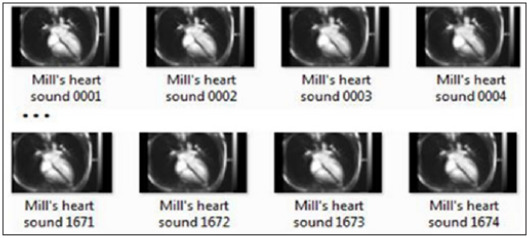
The information is obtained by extracting frames from CMR imaging video. There are different tools in order to extract frames from cardiac MRI Video (e.g. Free Video to JPG Converter, VLC or Virtual Dud) (Figure 1).
B. Step 2: Adjacent frames comparisons
The difference between two adjacent frames is used to estimate motion direction and magnitude. This process has been implemented within a tool called Image Diff. The Perforce image diff tool enables researchers to compare two adjacent frames. The following represents pixels difference value (Percent Changed; Pixels) and color difference value (Percent Change; Color) between adjacent Frames (Table 1).
C. Step 3: Presenting data in graphic form
In the final step the percentage of change (pixels and color) from one value to another, between frames are plotted as a graph (Figure 2 & 3).
Results
The results show that the proposed design approach works efficiently in the Cardio Kinesis System for clinical applications of cardiovascular assessment.
Conclusion
In conclusion, the proposed Cardio Kinesiograph System (CKinG) may become a robust and efficient tool for the clinical assessment of Cardiac and Vascular function. CKinGbased Computer-Aided Diagnosis (CAD) holds the promise of improving the diagnosis accuracy and reducing the cost.
Discussion
The developed system can be used as a prototype in the clinical sectors for the evaluation of cardiovascular diseases. However, a novel method for real time MRI of cardiac kinesis and simultaneously Kinesiographical Cardiac Analysis Based on Statistical Methods are proposed. The proposed methods may become efficient Medical Diagnostic Support tools (DSTs) for Heart Diseases.
Related Work
The publications below are based on Morbid Motion Monitor related topics:.
A. Dawoudi Mohammad Reza (2017) Morbid Motion Monitor. Current Treads in Biomedical Engineering & Biosciences. ISSN: 2572-1151.
B. Dawoudi MR (in press) (2017). Nursing and Technology foresight in Futures of a Complex World. European Journal of Futures Research.
To Know More About Current Trends in Biomedical Engineering & Biosciences Please Click on: https://juniperpublishers.com/ctbeb/index.php
To Know More About Open Access Journals Publishers Please Click on: Juniper Publishers
#Cardiovascular biomechanics#Statistical analysis methods#DNA modelization in biomedical image#Biomedical Engineering#biochemistry
0 notes
Text
Pacemakers Market show exponential growth by 2025
Crystal Market Research (CMR) render to you profound details in respect to leading participants, regions, application and type of the Pacemakers Market which is estimated to encounter substantial growth over the forecast period 2014 - 2025.
Pacemakers Market Opportunities:
The key opportunity for the players operating in pacemakers market lies in the development of various types of cost-effective pacemakers and increasing awareness towards cardiac diseases. Apart from that, emergence of various advanced features in artificial cardiac management devices will further broaden the opportunities of the key players to grow significantly over the forecast period. In addition, promotional activities related to product information and benefits in daily lives of individuals, will further increase demand of this market, creating a lucrative opportunities for growth, for the key players of this market.
Read Premium News from Openpr at @ https://www.openpr.com/news/1254231/Pacemakers-Market-offers-huge-growth-opportunities-for-the-future-Medtronic-plc-St-Jude-Medical-Biotronik-LivaNova-PLC-Vitatron-Holding-B-V-Boston-Scientific-Corporation.html .
Top Eminent Players:
The key players operating in the global pacemakers market emphasize on product development in order to introduce improved artificial cardiac management devices and capture a larger share of the market.
Some of the major players in this market are -
Medtronic plc
St. Jude Medical
Biotronik
LivaNova PLC
Vitatron Holding B.V
Boston Scientific Corporation
MEDICO S.p.A
Pacetronix
For more information, click on the below link @ https://www.crystalmarketresearch.com/report/pacemakers-market .
Pacemakers Market Segmentation:
By Product Type
External Pacemakers
Implantable Pacemakers-
Dual Chamber Pacemakers
Single Chamber Pacemakers
Biventricular Pacemakers
Regional Outlook:
North America (United States, Canada and Mexico)
Europe (Germany, France, UK, Russia, Italy and Rest of Europe)
Asia-Pacific(China, Japan, Korea, India, Southeast Asia and Rest of Asia-Pacific)
South America (Brazil, Argentina, Columbia and Rest of South America)
Middle East and Africa (Saudi Arabia, UAE, Egypt, Nigeria, South Africa and Rest of MEA)
Pacemakers Market Outlook and Trend Analysis:
A pacemaker is a small, low-voltage medical device implanted in the chest or abdomen to help manage irregular heartbeats. It monitors how slow or fast heart beats and the pattern in which the heart beats. When the heart beats too slowly, the pacing device provides electrical stimulation. This device helps to reduce symptoms of fatigue and dizziness due to slow heart rhythm. During pacemaker implantation, a thin insulated wire, lead, is placed through the veins and into the heart. The lead’s tip is connected to the heart tissue and the other end is attached to the pacing device. The lead provides electrical pulses from the pacing device to the heart and transmits information from the heart back to the device. Around 4 million people across the globe have pacing device implanted. Some pacemakers are designed to facilitate patients to safely undergo an MRI scan. These are known as MRI ready or MR-conditional pacemakers.
Click to Request a Sample @ https://www.crystalmarketresearch.com/report-sample/HC071131 .
Table of Contents:
1.Introduction
2.Executive Summary
3.Market Overview
4.Market Analysis by Regions
5.Pacemakers Market, By Product
6.Pacemakers Market, By Region
7.Company Profiles
8.Global Pacemakers Market Competition, by Manufacturer
9.Pacemakers Market Forecast (2018-2025)
10.Research Methodology
Ask Questions to Expertise at @ https://www.crystalmarketresearch.com/send-an-enquiry/HC071131 .
List of Tables:
Figure United States Pacemakers Revenue (Million USD) and Growth Rate (2013-2025)
Figure Canada Pacemakers Revenue (Million USD) and Growth Rate (2013-2025)
Figure Mexico Pacemakers Revenue (Million USD) and Growth Rate (2013-2025)
Figure Germany Pacemakers Revenue (Million USD) and Growth Rate (2013-2025)
Figure France Pacemakers Revenue (Million USD) and Growth Rate (2013-2025)
...
Key Growing Factors:
Increasing cases of Cardiovascular Diseases (CVDs), technological advancement in healthcare sectors and increasing reimbursement initiatives by the Government will primarily drive the growth of the pacemakers market. The increasing mortality rates due to inadequate treatment of cardiac diseases will encourage people to use artificial heart rate management devices owing to which the market is expected to have a steady growth in the upcoming years. For instance, as per CDC, in US, more than half of the Americans suffer from heart diseases every year including both men and women. This statistics indicate that artificial heart management device is the most commonly used device in this region. In addition, availability of adequate reimbursement facilities for implantation of pacemaker aids in reducing financial burden on patients thereby enhances the usage rates of these devices. Technological advancements in cardiac pacemaker are another positive aspect driving the market growth. Transitional tissue welding, dynamic peacemaking technology and microprocessor controlled devices are some of the advanced features that has been incorporated in the devices. However, high pricing of the devices often becomes unaffordable by the people despite of reimbursement facilities. The limitations of cardiac resynchronization therapy (CRT) pacemaker in pediatrics and risk of potential complications will impede the growth of the market. Thus, considering these drivers and restrains the pacemakers market is expected to exhibit steady growth over the forecast period.
Buy now Report @ https://www.crystalmarketresearch.com/checkout/HC071131 .
About Crystal Market Research:
Crystal Market Research is a U.S. based market research and business intelligence company. Crystal offers one stop solution for market research, business intelligence, and consulting services to help clients make more informed decisions. It provides both syndicated as well as customized research studies for its customers spread across the globe. The company offers market intelligence reports across a broad range of industries including healthcare, chemicals & materials, technology, automotive, and energy.
Contact Us:
Judy
304 South Jones Blvd, Suite 1896,
Las Vegas NV 89107,
United States
Toll Free: +1-888-213-4282
Email: [email protected]
0 notes
Text
Novel '6D' Cardiac MR May Simplify CMR, Avoid Pitfalls
Faster imaging time, less contrast agent, and no need for breath-holds or ECG triggering are among potential advantages of still-experimental 'six-dimensional' cardiac MRI.
0 notes
Text
Novel '6D' Cardiac MR May Simplify CMR, Avoid Pitfalls
Faster imaging time, less contrast agent, and no need for breath-holds or ECG triggering are among potential advantages of still-experimental 'six-dimensional' cardiac MRI.
0 notes
Text
Catheter Ablation Versus Medical Rate control in Atrial Fibrillation and Systolic Dysfunction (CAMERA-MRI)
Publication date: Available online 27 August 2017
Source:Journal of the American College of Cardiology
Author(s): Sandeep Prabhu, Andrew J. Taylor, Ben T. Costello, David M. Kaye, Alex AJ. McLellan, Aleksandr Voskoboinik, Hariharan Sugumar, Siobhan M. Lockwood, Michael B. Stokes, Bhupesh Pathik, Chrishan J. Nalliah, Geoff R. Wong, Sonia M. Azzopardi, Sarah Gutman, Geoffrey Lee, Jamie Layland, Justin A. Mariani, Liang-han Ling, Jonathan M. Kalman, Peter M. Kistler
BackgroundAtrial fibrillation (AF) and left ventricular systolic dysfunction (LVSD) frequently co-exist despite adequate rate-control. Existing randomized studies of AF and LVSD of varying etiologies have demonstrated modest benefits with a rhythm control strategy.ObjectiveTo determine whether catheter ablation (CA) for AF could improve LVSD compared to medical rate-control (MRC) where the etiology of the LVSD was unexplained, apart from the presence of AF.MethodsThis multi-center randomized clinical trial enrolled patients with persistent AF and idiopathic cardiomyopathy (LVEF ≤45%). After optimization of rate-control, patients underwent cardiac MR (CMR) to assess LVEF and late gadolinium enhancement (LGE), indicative of ventricular fibrosis, before randomization to either CA or ongoing MRC. CA included PVI and posterior wall isolation. AF burden post CA was assessed by implanted loop recorder, and adequacy of MRC by serial Holter-monitoring. The primary endpoint was ΔLVEF on repeat CMR at 6 months.Results301 patients were screened and 68 enrolled between November 2013 and October 2016 and randomized with 33 in each arm accounting for two dropouts. The average AF burden post CA was 1.6% ± 5.0% at six months. On intention to treat analysis, absolute LVEF improved by +18 ± 13% in the CA group compared to +4.4 ± 13% in MRC group, (p <0.0001) and normalized (LVEF ≥50%) in 58% vs 9%, p = 0.0002. In those undergoing CA, the absence of LGE predicted greater improvements in absolute LVEF (+10.7%, p = 0.0069) and normalization at 6 months (73% vs 29%, p = 0.0093).ConclusionAF is an underappreciated reversible cause of LVSD in this population despite adequate rate control. The restoration of sinus rhythm with CA results in significant improvements in ventricular function, particularly in the absence of ventricular fibrosis on CMR. This challenges the current treatment paradigm that rate control is the appropriate strategy in patients with AF and LVSD.
Teaser
Condensed AbstractThe value of restoring sinus rhythm in concurrent AF and systolic dysfunction is unclear with existing studies showing mixed results. This clinical trial randomised 66 patients with persistent AF and idiopathic cardiomyopathy to CA (n=33) or ongoing MRC (n = 33) with the primary endpoint of LVEF improvement at 6-months. Compared, to MRC, the CA group had significantly greater improvements in LVEF and associated reductions in ventricular and atrial size, NYHA class and BNP. Furthermore, MRI detected ventricular fibrosis predicted the extent of LV recovery. AF is an underappreciated reversible cause of systolic dysfunction in this population despite adequate rate control.
from # All Medicine by Alexandros G. Sfakianakis via alkiviadis.1961 on Inoreader http://ift.tt/2wKAHee
from OtoRhinoLaryngology - Alexandros G. Sfakianakis via Alexandros G.Sfakianakis on Inoreader http://ift.tt/2wTO5fe
0 notes
Text
A Case of Multiple Embolic Strokes Caused by a Congenital Left Ventricle Diverticulum Undetected on Echocardiography
Rovere G¹; Perazzolo A²; Moliterno E²; Giarletta L²; Brancasi A²; Marano R²*
Abstract
Congenital Left Ventricular Diverticulum (CLVD) is a rare cardiac malformation caused by a localized protrusion of the left ventricular wall and usually diagnosed with routine echocardiography. CLVD is often associated with other cardiac and noncardiac abnormalities, but it can also occur alone. When echocardiography is non diagnostic, other noninvasive techniques such as cardiac CT (CCT) and Cardiac MR (CMR) can help to rule in/out the diagnosis by providing additional information on myocardial structure, morphology, and kinetics of both ventricles and of the diverticulum itself. We report the case of a patient who arrived in the Emergency Department for an acute cerebrovascular ischemic event with embolic pattern, who underwent non invasive diagnostic tests in order to identify the etiology.
Abbreviation: CVLD: Congenital Left Ventricular Diverticulum; CCT: Cardiac CT; CMR: Cardiac Magnetic Resonance; CAD: Coronary Artery Disease; RV: Right Ventricle; LV: Left Ventricle; CLVC: Congenital Left Ventricle Cleft; LVA: Left Ventricle Aneurysm
#Cardiac malformation#Left ventricle wall abnormalities#Left ventricle diverticulum#Stroke#Echocardiography#Cardiac MR (CMR)
0 notes
Text
Nuclear Imaging Equipment Market Shows Expected Growth from 2018-2026
Nclear imaging allows physicians to diagnose and treat various diseases with the help of radiopharmaceuticals and gamma cameras. It offers valuable medical information to help diagnose a disease in its early stages. Nuclear imaging equipment provides promising prognosis, thereby allowing patients for early treatment to save additional therapy cost.
Market Dynamics
Increasing adoption of nuclear imaging equipment in healthcare settings is attributed to benefits of nuclear imaging equipment in improving selection therapy and monitoring patient response for a specific treatment. Furthermore, Medicare and private insurance companies cover the cost of various nuclear imaging procedures, thereby increasing the adoption rate of the equipment in emerging economies. Increasing prevalence of cardiovascular diseases also propels demand for nuclear imaging. Cardiovascular diseases require diagnostic tests to identify heart disease, severity of prior heart attacks, and the risk of future heart attacks. According to a report by the Centers for Disease Control and Prevention (CDC), 2015, around 800,000 people in the U.S. die due to stroke and other cardiovascular diseases.
The global Nuclear Imaging Equipment Market is expected to be driven by increasing prevalence of chronic diseases including cardiac diseases and cancer
Increasing prevalence of chronic diseases is expected to propel growth of the nuclear imaging equipment market. According to a report published by Center for Chronic Disease Prevention and Health Promotion 2012, around 117 million people were suffering from at least one chronic health condition and one in four adults had two or more chronic health conditions in the U.S. Moreover, according World Health Organization (WHO), chronic diseases are expected to account for around three-quarters of all deaths worldwide by 2020. WHO further stated that around 71% of these deaths would occur due to Ischaemic Heart Disease (IHD) and 75% due to stroke.
Furthermore, increasing funding for development of advanced nuclear imaging equipment is expected to propel the market growth. For instance, the National Institute of Biomedical Imaging and Bioengineering is focusing on offering funding to develop new radio tracers and technologies in order to enhance image quality of nuclear imaging equipment. Moreover, in November 2018, the Radiological Society of North America (RSNA) Research & Education (R&E) Foundation granted US$ 40,000 for its joint research grants with Canon Medical Systems.
Key Players
Key players in the global nuclear imaging equipment market are focused on new product launch to enhance their market share. For instance, in May 2018, the Food and Drug Administration (FDA) cleared— Biograph Vision— a positron emission tomography/computed tomography system developed by Siemens Healthineers. The system helps to reduce scan time to improve patient throughput and also reduces patient radiation exposure and tracer cost.
Major players operating in the global nuclear imaging equipment market include, Siemens Healthineers, Koninklijke Philips N.V.,GE Healthcare, Toshiba Medical Systems Corporation, Neusoft Medical Systems Co., Ltd., Mediso Medical Imaging Systems Co., Ltd., CMR Naviscan Corporation, Digirad Corporation, SurgiEye GmbH, and Positron Corporation
Ask For Sample Copy Of This Business Research Report : https://www.coherentmarketinsights.com/insight/request-sample/2366
Detailed Segmentation:
Global Nuclear Imaging Equipment Market, By Product Type:
Single-Photon Emission Computed Tomography (SPECT) Systems
Hybrid SPECT
Standalone SPECT
Hybrid Positron Emission Tomography (PET) Systems
Planar Scintigraphy
Global Nuclear Imaging Equipment Market, By Application:
Oncology
Cardiology
Neurology
Others
Global Nuclear Imaging Equipment Market, By End User:
Hospitals
Imaging Centers
Research Institutes
Others
Increasing technological innovations in nuclear imaging equipment and research grants are expected to bolster the market growth
North America is expected to hold a dominant position in the global nuclear imaging equipment market. This is owing to increasing adoption of new imaging equipment, which in turn is expected to improve cancer treatment with enhanced image quality. For instance, in October 2018, the Center for Quantitative Cancer Imaging at Huntsman Cancer Institute (HCI), University of Utah, installed— nanoScan PET/MRI 3T preclinical in vivo imaging system for screening rodent tumor models. Furthermore, increasing prevalence of cancer in North America is expected to propel demand for nuclear imaging equipment over the forecast period. A report by the National Cancer Institute projected around 1,735,350 new cases of cancer and 609,640 deaths due to the disease in the U.S in 2018.
About Coherent Market Insights:
Coherent Market Insights is a prominent market research and consulting firm offering action-ready syndicated research reports, custom market analysis, consulting services, and competitive analysis through various recommendations related to emerging market trends, technologies, and potential absolute dollar opportunity.
Contact Us:
Mr. Shah
Coherent Market Insights
1001 4th Ave,
#3200
Seattle, WA 98154
Tel: +1-206-701-6702
Email: [email protected]
#Nuclear Imaging Equipment Market#Nuclear Imaging Equipment Market size#Nuclear Imaging Equipment Market trends#Nuclear Imaging Equipment Market outlook
0 notes