
juniperpublishers-toxicology
Open Access Journal of Toxicology
54 posts
Don't wanna be here? Send us removal request.
Last Seen Blogs

dylan9b93rien
Senza titolo

bass-music
BASS MUSIC-and-everything-else-inbetween


yeol-daa
fairy tuan... i love you

duxetesopic
Untitled
Text
Juniper Publishers wishes Happy Easter to you and your family members

0 notes
Text
Fulminant hepatic failure induced by antipsychotic drugs (a case report)

Introduction
Fulminant hepatitis is a rare condition but has a very poor prognosis in the absence of liver transplantation. It is important to identify the cause as soon as possible to start the etiological treatment, which may be drug poisoning, viral hepatitis, or alcoholic ... [1-3]. N-acetylcysteine (NAC), a glutathione precursor, was first used as a treatment for paracetamol overdose in 1979. Since then, it has been firmly established as an effective and safe treatment for paracetamol induced IHC prevention. NAC has also been shown to be effective outside paracetamol intoxication. It has been evaluated as an option for acute IHC other than paracetamol in adults and children. In a randomized clinical trial comparing NAC with placebo in adults with paracetamol-free IHC, NAC was associated with a marked improvement in survival without liver transplantation [4]. NAC has also been evaluated for non-hepatic clinical conditions, these indications include its use in lung diseases (COPD and pulmonary fibrosis), in the prevention of contrast-induced nephrotoxicity and for the treatment of certain cardiac diseases [3,5]. In this article, we report the case of a neuroleptic overdose in a 16-year-old who has rapidly progressed to fulminant hepatitis. The oral administration of N-acetylcysteine has allowed a dramatic improvement [3].
Observation
A 16-year-old man consulted at the emergency reception service (j1) for asthenia, disabling diffuse myalgia and uncontrollable vomiting, in the context of cutaneous-mucous jaundice, which had appeared for 4 days. Her antecedents included herpetic keratitis since the age of 3 years and chronic epigastralgia for 4 years. In the emergency department, the clinical examination found a sleepy, sleepy patient, a conjunctival subitem, muscle pain with manual pressure. The temperature was 37.8°C, and blood glucose was 2.2mmol/l. The blood pressure was 85/38 mmHg, the heart rate was 104 beats per minute and 95% saturation in the air. The biochemical assessment was very disturbed, with: an inflammatory syndrome (C-reactive protein at 150 mg/L, fibrinogen at 6.6g/L), renal insufficiency (urea at 14.4mmol/L, creatinine at 159 , 12mmol/L), cytolysis (ASAT: 10454 IU/L, ALA at 4408 IU/L), cholestasis (conjugated bilirubinemia at 151mmol/L, gamma GT at 489 IU / L, PAL at 657 IU/L ) and a disturbance of its hemostasis (TP <15%, a TCA at 96.4 s and a very collapsed factor V). The hemogram showed the following: white blood cells at 9210mm-3, platelets at 159 000mm-3 and hemoglobin at 11.2g/dl.
On the diagnosis side
The patient was admitted to intensive care (1st day). Etiologically, an infectious hypothesis has been ruled out by the negativity of his liver serologies (anti-HVA Ab, anti-HBV Ab and HBs antigen and anti-HCV antibodies) as well as the serology of CMV, the abdominal ultrasound was without particularities, blood ceruloplasmin level was normal, anti-smooth muscle and antimitochondrial antibodies were achieved returning normal. The preferred toxic hypothesis was a neuroleptic overdose because the interview reported a prescription of 3 different neuroleptics, by his doctor for his chronic epigastralgia of psychogenic origin (Olanzapine, Mainspring and Metoclopramide). Supported: Symptomatic treatment consisted of stopping neuroleptics, infusion of fresh frozen plasma, vitamin K, laxatives and ciprofloxacin were initiated associated with administration of N-acetylcysteine with a dose of oral load of 140mg/kg followed by a dose of 70mg/kg/day maintenance for 48 hours.
Evolution
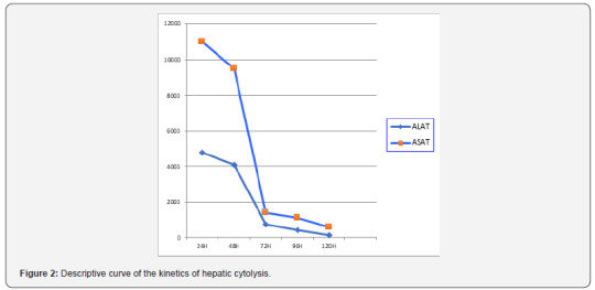
The evolution was quickly favorable. Hyper-bilirubinemia was divided by five in three days, the state of consciousness improved rapidly with appearance of an asterixis, the correction of the hemostasis disorder was more progressive with normalization towards the 4th day (Figure 1), a dramatic improvement in hepatic transaminases was observed as early as the second day (Figure 2). The patient left the intensive care unit to the gastric department on day 6. The symptomatic treatment was continued until day 10, the PBH was performed on day 11 without abnormality then he left the hospital on day 17.

Discussion
IHA is defined as a sudden failure of liver function in a patient with no history of liver disease. The cardinal signs of hepatic failure include coagulopathies and hepatic encephalopathy of any grade in the context of acute liver injury [4]. Currently, there is no scientifically proven beneficial treatment in the treatment of IHC, apart from liver transplantation Lee [5]. More than 1000 drugs have been listed as being responsible of hepatic side effects; 16% of these agents were neuropsychiatric drugs. Antidepressant drugs (tricyclic agents or SSRI), mood stabilizing agents and neuroleptic drugs have been implicated in biological or/and clinical hepatotoxicity. For these reasons, some psychotropic agents have been withdrawn of the pharmaceutical, On the contrary, in case of clinical hepatotoxicity, challenge or maintenance is absolutely inadvisable. Mechanism of the hepatic troubles: precise mechanisms of the hepatotoxicity remain unclear. Contrary to phenothiazine drugs, no information is available on the respective rule of the agents and their metabolites. Hypersensitivity syndrome or eosinophilia has been reported, suggesting a possible immuno-allergic mechanism. Presence of risk factors: risk factors have been retrieved, in some observations, like high daily dosage, high plasmatic concentration, age, alcoholism, obesity or antecedent of hepatic disorders like Gilbert syndrome. [6] Special care is advisable with these patients. As hepatotoxicity has been observed after surd Osage (or suicide attempt), a hepatic check-up has to be performed in these clinical situations [7]. Co-medication with hepatotoxic drugs may increase the risk as it has been suggested. Acetylcysteine is a precursor of glutathione. It is well known as an antidote for acetaminophen overdose due to its ability to increase glutathione levels, which inactivates the toxic metabolite of acetaminophen [8]. N-acetyl-pbenzoquinone mine. Glutathione is a major antioxidant that can serve as a scavenger for free radicals; therefore, acetylcysteine may increase glutathione stock during periods of oxidative stress, increase nitric oxide production, which causes vasodilation and therefore tissue oxygenation, and may also have an antiinflammatory effect. by inhibition of pro-inflammatory factors (TNF alfa and IL8) [9,10]. The majority of studies evaluated the use of NAC in acute IHC secondary to acetaminophen poisoning. There is little research on the use of NAC in IHC secondary to other causes. Hu [8]. evaluated the efficacy of NAC in patients with non-acetaminophen-overdosed IHC (safety and efficacy of NAC in patients with ALF not caused by acetaminophen overdose), in a meta-analysis, which consisted of analyzing four assays prospective clinical trials evaluating NAC versus placebo in the treatment of non-acetaminophen-induced IHC[3].
Conclusion
NAC is a beneficial treatment in the context of nonparacetamol induced IHC, it can prolong the survival of patients with or without liver transplantation and survival after transplantation, but it cannot improve overall survival. Therefore, due to the lack of available scientific evidence, current data is unable to conclusively determine the role of NAC in patients with IHC without paracetamol. Thus, they are unable to make recommendations for clinical practice.
1 note
·
View note
Text
Further Studies in Translatable Model Systems are Needed to Predict the Impacts of Human Microplastic Exposure

Abstract
Microplastics are a pervasive environmental contaminant that have been found in many media including water sources, soils, and foodstuff. Due to the worldwide presence and persistence of microplastic debris, human exposure is inevitable. Human exposure occurs predominantly through ingestion, although dermal and inhalation exposures are probable. Microplastic single exposure studies in aquatic species and fish have shown various toxic effects including those on reproduction and survival. In addition to potential intrinsic toxicity, microplastics often have chemicals adsorbed to their surfaces. Studies report that these chemicals can have innate toxicity that is modulated by the composition of microplastics. Both the impacts of microplastics alone and co-exposures with adsorbed chemicals exhibit size dependent effects. Analysis of the current literature has revealed published studies predominantly investigate the toxicity of microplastic exposure in fish and other aquatic species, with limited knowledge about the effects in mammals and cell lines. Toxicity has been shown to vary widely between taxonomic groups, suggesting inferring human health relevance will require model systems where human routes of exposure can be mimicked. Although it may be difficult to extrapolate the results from aquatic model systems to relevant human health impacts, they may suggest effects to investigate. In order to best estimate the short- and long-term impacts of human microplastic exposure, it is imperative that studies in model systems with increased similarity to human anatomy and cellular processes be done.
Origins of Microplastics
Since the introduction of plastic products in the early 1900s, plastic use and therefore waste has continued to increase. The mismanagement of plastic debris frequently results in plastic pieces, including microplastics, entering the environment [1]. Microplastics are a ubiquitous environmental problem and have been detected on all seven continents [2]. In addition to being found worldwide, microplastics have been detected in numerous forms of media including but not limited to oceans [3], lakes [4], drinking water [5,6], sediments [7], soils [8], sugar [9], and table salt [10]. The prevalence and persistence of microplastics in the environment is likely due to their physical properties.
Microplastics are small pieces of plastic less than 5 mm in size [11-13]. There are two main types: primary and secondary microplastics. Primary microplastics are those that are manufactured at the microplastic size. This includes those that are produced for use in consumer products, as well as those that are byproducts of other goods. Products that intentionally contain microplastics include personal care products [14,15], drug delivery systems [16] and air blasting media [17], while synthetic clothing [18] contains unintentional microplastics. Secondary microplastics are those that are produced through the environmental degradation of larger pieces of plastic debris through processes such as physical abrasion [19] and photodegradation [20]. Recently, researchers in Italy reported a potential new class of secondary microplastics. They found that some benthic crustaceans are able to break ingested plastics into smaller pieces, producing microplastics [21]. The small size of microplastics in addition to their properties, such as resistance to corrosion, that make plastics desirable product components contribute to their pervasiveness in the environment [22]. Microplastics can further be degraded to nanoplastics (<0.1 μm), but particles <1.0 μm are rarely collected in environmental studies, further pointing to a knowledge gap in understanding the health impacts.
Routes of Human Microplastics Exposure
Due to the widespread nature and persistence of microplastics, human exposure is inevitable. Humans are exposed through ingestion and possibly through dermal exposures and inhalation. Ingestion occurs via activities including consumption of aquatic species containing microplastics and drinking and cooking with microplastic contaminated water. Microplastics have been detected in aquatic species such as fish [23] and mussels [24]. Organisms may confuse microplastics with food, especially if their food sources are on the same size scale as the microplastics present [25], or accidentally ingest microplastics while feeding or drinking [26]. Additionally, microplastics have been observed to transfer up trophic levels in the environment [27,28], and this property is what allows humans to uptake microplastics from food. Dermal contact with microplastics is a less likely route of exposure, but it is possible that swimming or showering in water that contains microplastics could result in microplastic uptake [29]. This is more likely if an individual has a barrier defect in their epidermal layer, such as those that occur as a result of UV radiation exposure [30]. Lastly, microplastics have been detected in samples of both indoor and outdoor air [31], suggesting humans may be regularly inhaling microplastics. Given the likelihood of human exposure, it is vital that translatable studies be performed.
Impacts of Microplastics on Biological Systems
Microplastics have been studied in a variety of model systems, however the literature shows a bias towards fish and other aquatic species. These single exposure studies have demonstrated a wide variety of toxic effects. An inexhaustive list of these effects includes an increase in reactive oxygen species (ROS) production in copepods and rotifers [32,33], a disruption of lipid homeostasis due to a decrease in HDL levels in catfish [34], and a decrease in intestinal calcium concentrations in Caenorhabditis elegans [35]. Additionally, microplastics were observed to exhibit size dependent effects. Exposure to smaller microplastics in Tigriopus japonicus, a small aquatic crustacean, and the rotifer, Brachionus koreanus, resulted in greater reductions in survival [36] and lifespan [33], respectively, when compared to exposure to larger microplastics. Despite this variety in effects observed, these studies do not vary widely in model system used.
In addition to the plastic polymers that comprise the microplastic itself, many microplastic particles contain hydrophobic organic contaminants (HOCs) adsorbed to their surfaces. Some are chemical additives introduced during the manufacturing process to impart desired properties to the plastics being produced, such as plasticizers, flame retardants and pigments [37]. Additionally, once in the environment, microplastics tend to accumulate other contaminants such as metals [38], bacteria [39], and persistent organic pollutants (POPs) [40]. On their own these contaminants exhibit toxicity that may be modulated by co-exposure with microplastics. The results of microplastic-contaminant co-exposures reveals the toxic effects observed are distinct from exposure to the microplastics alone. A 2018 review that characterized results based on taxonomic group, plastic type, and contaminant present suggests that the toxicity of the contaminant is modulated by microplastic type present [41]. For example, when studies in Mollusca were analyzed the effect profiles of polycyclic aromatic hydrocarbons (PAHs) in the presence of polyethylene (PE), polystyrene (PS), or polyvinyl chloride (PVC) exhibited different toxic effects [41]. Reasons for this were not given but it is likely related to the solubility and permeability of the PAH in the bulk plastic. Similarly, to exposures to microplastics alone, co-exposures with contaminants show size dependent effects. A study that measured the estrogenic equivalency of populations of different sized microplastics containing endocrine disrupting contaminants, revealed a general trend where an increase in estrogenic activity was associated with a decrease in microplastic size [42]. Just like the microplastic single exposures, there is a clear bias in the literature towards studies in fish and aquatic species.
The general consensus thus far is that microplastics induce toxicity alone and in co-exposures. Because of the lack of mammal or cell exposure studies this conclusion cannot yet be applied to humans. A survey of the primary literature results of a Google scholar search of “microplastics toxicity” revealed 71% of the articles analyzed studied fish or other aquatic species. This same survey found only three and five percent of articles analyzed studied effects in cell lines and mammals, respectively. This is especially problematic as a different 2018 review that compiled the results of 43 studies of microplastic exposure found that effect sizes varied widely between taxonomic groups [43], suggesting it is extremely imperative a proper model system be used to estimate effects of human exposure. Additionally, the emerging routes of exposure of inhalation and dermal exposure cannot reliably be studied in aquatic species due to their lack of skin or lungs comparable to those of humans. Therefore, it is imperative that microplastic exposure studies be performed in a model system with relatively conserved biological processes, such as barrier function or cell signaling, and organ systems compared to humans. Without using these types of model organisms, it is a stretch to apply knowledge gained from studies when hypothesizing the short- and long-term effects of human microplastic exposure.
Existing studies using human cell models are sparse [44- 46] and they use model particles rather than testing materials recovered from the environment. Overall, the conditions tested in these studies showed little to no impacts on cell viability with microplastic exposure, with the exception of human dermal fibroblasts treated with 3 μm polystyrene particles at a very high dose of 1000 μg/ml microplastics [45]. Beyond viability the cellular effects observed varied between studies. ROS production was elevated in response to microplastic exposure in once study [46], while another found all but one exposure had no significant effect on ROS production [44]. Similarly, cytokine and histamine secretion varied widely between treatments [44,45]. These inconsistencies between human cell studies further motivate the need additional comprehensive studies with both commercial and environmental microplastic particle samples.
Conclusion
Microplastics are a ubiquitous environmental contaminant that inevitably leads to human exposure. Despite the high likelihood that humans are exposed, the majority of research on microplastic exposure has focused on the effects in aquatic species. Exposure to microplastics with and without the presence of organic contaminants has been shown to result in numerous toxic effects in these aquatic species. However, due to the routes of human exposure and the variability in toxicity observed between taxonomic groups, the amount of information that can be used to estimate the effects of exposure to microplastics in humans is extremely limited. In order to gain results that are more relevant to human health outcomes, proper model systems must be used. A couple systems that would be more appropriate are primary human cell lines and laboratory mice. The results observed in the literature thus far may not be applicable to human health but may provide endpoints to investigate in relevant systems. Further studies are needed in order to best understand the effects of microplastic exposure in humans.
0 notes
Text
Toxopathological Studies on Some Antimicrobial Drugs in Nile Tilapia (Oreochromis Niloticus) and Catfish (Clarias Gariepinus)

Introduction
Fish consider one of the healthiest food as it is low in fat and rich in protein and omega 3 Fayet-Moore [1] & Yipel [2]. The fish farming industry is rapidly expanding in Egypt, as well as in other countries, it has been associated with recurrent bacterial infectious diseases. Farmed Nile tilapia represents more than 58.45, while catfish production is about 3.08% of the total aquaculture harvest in Egypt Gafrd [3]. Antimicrobial medications are used extensively in aquacultures for prophylactic or therapeutic purposes during microbial infections which may result in environmental pollution, development of resistant bacteria and my induce toxicity to human and animals Aly [4] & Khalil [5]. The availability of adequate data on the pharmacokinetics of antimicrobial agents in farmed fish is very important in order to minimize the environmental impacts of the drugs used in aquaculture. Since the excess amount of drugs can do harm to people, the European Union (EU) and the U.S. Food and Drug Administration (FDA) prescribed a Maximum Residue Limits (MRLs) for these drugs. The EU MRLs of CPX and SDM in fish were established at 100μg/kg Rezk [6] and 6-8μg/kg for quinolens in the edible tissues of fish Victoria [7].
Quinolones are effective antibacterial drugs widely used in human and veterinary medicine because of their potential therapeutic efficacy Plakas [8], Guo [9], Victoria [7] & Koc [10]. Ciprofloxacin is one of the most potent quinolones used to treat infections with gram negative bacteria as Escherichia coli, Pseudomonas aeruginosa, Salmonella spp., Shigella spp. and Haemophilus spp., and is also effective against some grampositive bacteria such as Staphylococcus aureus Davis [11] & Van Bambeke [12]. Oxytetracycline (OTC) is an antibacterial agent of tetracycline family that is extensively used for treatment of certain bacterial diseases in aquaculture all over the world Ambili [13]. The withdrawal time for edible tissue is differing according to the water temperature and the type of aquatic system Jeffry [14]. Because of the wide spread and long-time use of OTC, many residue studies have been recorded Rigos [15-16] & Julie [17].
Sulfonamides are the oldest antimicrobial agents and still play an important role in aquaculture treatments. Sulfamethazine (SMZ) is the most used antimicrobial drug in Veterinary field. Sulfonamides residues have been repeatedly detected in the aquatic environment Kolpin [18] & Batt [19]. Moreover sulphamethoxazole residues have been reported in shrimp by Wang [20]. Sulfamethoxazole is an effective bacteriostatic against gram positive as well as gram negative bacteria; it affects bacteria by inhibiting folic acid synthesis Baran [21]. Antimicrobial drug residues may be transferred through food-chain to human and induce antibiotic resistance. To our knowledge, however, very few data are available about residues of ciprofloxacin, oxytetracycline and SDM in farmed Nile tilapia (O. niloticus) and catfish (C. gariepinus) reared under field conditions. However, this study aimed to investigate serum concentration peaks of ciprofloxacin, oxytetracycline and SDM post-treatment and their residues in liver, kidney and muscles together with serum biochemical estimation and histopathological examinations.
Materials and Methods
Animals and diet
Three hundred and sixty fish from each of Nile tilapia (O. niloticus), and catfish (C.gariepinus) (weight, about 50 and 75g for tilapia and catfish, respectively) were supplied from Central Lab for Aquaculture Research (CLAR), Egypt and used in this experiment that was performed in triplicates, following the Universal Directive on the protection of animals used for scientific purposes. Four different basal diets (control, CIP, oxytetracycline and sulfadimethoxine) were prepared in the form of pellets to use in the study. Basal diets were prepared by grinding the corn to granules using 0.5mm mesh (Thomes-Willey Laboratory Mill Model 4). Ingredients were mixed mechanically by horizontal mixture (Hobarts model D300T) at a low speed for 30 minutes. Oil (vegetable & cod liver) was added gradually to assure the homogeneity of the ingredients, the mixing speed increased for 5 minutes during the addition of water (600ml water) until the mixture began to clump. Pellets were then prepared using a pellet machine (CPM California pellet mill Co.) with 0.5cm diameter, and pellets were left to dry in air for 24 hrs (Table 1).
Fish with a history of no previous medication, were divided into 4 groups (each of three replicates, 30 fish each) and held in floating cages placed in fish farm ponds and group 1 fed a basal diet while groups 2-4 fed a medicated diet containing 1g CIP, 7.5g OTC and 25mg SDM/kg ration; respectively on a daily bases for five successive days. The temperature was recorded every 12h and adjusted to (26-30°C). The treatment was carried out once daily at 9 a.m. for 5 successive days at a rate of 1 .0% biomass using automatic feeders. Salinity, pH and total hardness were adjusted to, 3±1.1%, 8.21±0.21 and 38.9±1.9mg/L; respectively.
Sampling of the fish
The first sampling day was the 5th day of medication (0 day post treatment), and on the 1st, 3rd, 7th, 14th, and 21st days after the end of treatment with the antimicrobials. At each time of sampling, 15 fish from each group (5fish/replicate) were netted. Fish were anesthetized by immersion in water containing 0.1ppm MS-222 and blood samples were collected. Serum samples and muscle, liver and kidney specimens were collected from all groups. Muscle samples were taken from the dorso-lateral body area just posterior to the operculum. Each specimen was placed in a polyethylene bag and stored at -80°C until they were analyzed. CIP, OTC and SDM concentrations were estimated by ELISA.
Biochemical Studies
The activities of Asparate Aminotransferase (AST), Alanine Aminotransferase (ALT), Alkaline Phosphatase (AP), creatinine and urea, were estimated using commercial diagnostic Kits (Human Diagnostics, Germany). Methods were carried out according to the company directions.
Histopathological examinations
Tissues specimens from the muscles, liver and kidneys were collected at 5th day post-treatment and processed routinely according to Drury and Wallington (1980). Sections were stained with hematoxylin and eosin (H&E) and examined by light microscope.
Statistical analysis:
Statistical analysis was performed using the one way analysis of variance (ANOVA) followed by Duncan’s multiple range test to determine the differences among the six fish groups (mean at significance level of P<0.05). All analyses were run on the computer using SAS program Chris Hemedinger [2].
Results
Drug residues
Mean concentrations of the drugs (mean ± SE) vs. time in the serum, liver, kidney and musculature were recorded in (Table 1-3). The peak concentrations of the three drugs in serum were at 0 day. The lowest drug residues were seen in the muscles throughout the entire experiment.
Ciprofloxacine: Results obtained after oral dose of 1 g CIP/kg ration for 5 successive days were shown in (Table 2). The highest recorded concentrations of CIP in sera of Nile tilapia and Catfish were (1.91±0.38ug/ml) and (1.78±0.36ug/ml), respectively at 0 day. CIP concentrations were identified all over the experiment in kidneys with the highest concentrations (2.1±0.65ug/g) at 1st day in Nile tilapia and (1.80±0.64ug/g) at 0 day in kidneys in catfish. CIP neither detected in muscles of Nile tilapia nor of Catfish at 14th and 21st days post-treatment while, were not detect in livers of both kinds of fish at 21st days post-treatment.
Oxytetracycline: (Table 3) shows the serum, liver, kidney and muscle concentrations of OTC versus time in Tilapia and Catfish after oral administration of 75mg OTC/kg ration for 5 successive days. Peaks of OTC in serum were (2.15±0.41ug/ ml) and (2.02±0.31ug/ml) at 0 day in Nile tilapia and Catfish; respectively while, it was not detect in sera of both fish species after 14th and 21st days but detected only in one Catfish (0.03μg / ml) at 7th day post treatment. The highest tissue residues of OTC were (6.1±1.21ug/g) and (7.4±1.35ug/g) in liver of Nile tilapia and Catfish; respectively at 0 day of the treatment. In Nile tilapia and Catfish the OCT concentrations in kidneys were 0.08±0.04 and 0.05±0.02 (μg /g); respectively at 21st day post treatment. The lowest drug residues were in muscles throughout the entire experiment. OCT concentrations were detected in muscles of Nile tilapia and Catfish at (0.10±0.03ug/g) and (0.14±0.02ug/g); respectively after 21 days post treatment
Sulphadimethoxine: (Table 4) showed the mean concentrations of SDM in Nile tilapia and Catfish sera and tissues versus time profile after oral administration of 25mg SDM/kg ration for 5 successive days. The highest serum concentrations of SDM were (3.12±0.32ug/ml) and (2.98±0.46ug/ml) at 0 day in Nile tilapia and Catfish; respectively while it was detected in only one Tilapia fish (0.04μg /ml) at 7th day of treatment and not thereafter was detected. SDM was detected in kidneys of both Tilapia and catfish all over the experiment. SDM highest concentrations in kidney were at 0 day post-treatment (44.2±5.1ug/g) and (31.2±4.6ug/g) in Nile tilapia and Catfish; respectively. At 21st day of treatment; SDM was not detected in muscles and liver of Catfish but detected only in one Tilapia fish (0.11ug/g and 0.03ug/g in liver and muscles; respectively).
Biochemical results
(Figure 1,2) represented the biochemical results at 5th day of oral administration of CIP, OTC and SDM in both Nile tilapia and Catfish. ALT was significantly increased in both fish species after 5 days of oral administration of the three drugs compared with control. In Tilapia fish AST was significantly increased after administration of the three tested drugs while, in Catfish AST was significantly increased after administration of OTC and SDM in comparison with control. Creatinine was significantly increased in Nile tilapia with all three drugs but in Catfish it was significantly increased with OTC and SDM whereas not increased with CIP. Urea was significantly increased in Tilapia fish after administration of all drugs except OTC while, in Catfish urea was significantly increased in both OTC and SDM but not significantly changed in case of CIP compared with control.
Histopathological results
The oral administration of 1g CIP/kg ration for 5 successive days in Nile tilapia and Catfish at 5th days post-treatment, revealed minimal histopatholoigical alterations in comparison with the other treated groups. The musculature exhibited hyaline degeneration in few muscle bundles (Figure 3), the liver displayed nuclear pyknosis of some hepatocytes with mild parenchymal edema (Figure 4) while the kidneys showed proliferation of melanomachrophage cells and mild tubular nephrosis in the renal epithelium (Figure 5). The oral administration of 75mg OTC/kg ration, for 5 successive days in Tilapia and Catfish at 5th day posttreatment, revealed edema and focal hyaline degeneration in the musculature (Figure 6). Focal proliferation of melanomoacrophage cells was observed in the liver and kidney parenchyma. Wide spread vacuolar degeneration in the hepatocytes (Figure 7) and tubular nephroses in the renal tubular epithelium (Figure 8) were evident.
The oral administration of 25mg SDM/kg ration, for 5 successive days in both Nile tilapia and Catfish at 5th days posttreatment, revealed edema and hyaline degeneration as well as focal Zenker’s necrosis in the musculature with focal of mononuclear leukocytic infiltration (Figure 9). The liver exhibited wide spread vacuolar degeneration as well as coagulative necrosis in the hepatocytes with some mononuclear cells infiltration and melanomacrophages (Figure 10). The kidney showed tubular nephrosis mainly vacuolar degeneration with few cells exhibited coagulative necrosis, hyaline casts and few mononuclear cells infiltrations were evident (Figure 11).
Discussion:
Using of antimicrobial drugs in aquaculture production is one of the main sources of environmental pollution Pruden [23]; Rico & Van den Brink [24]. During the past years there was increase in the occurrence of antibiotic resistant bacteria and this is of critical implications on public health Gouvêa [25] & Rezk [6]. Quesada [26] & Guidi [27] mentioned that tetracycline, oxytetracycline (tetracyclines), enrofloxacin (quinolones), and sulfadimethoxine (sulfonamide) are most commonly used antibiotics in aquaculture worldwide and the presence of their residues in food could resulted in health hazards and toxic effects. Therefore, understanding the depletion of drugs from different tissues of fish is of extreme importance and the drug residues must be assessed in order to determine the time needed before the antimicrobials disappear from the tissues and to judge when the treated fish can be safely consumed. There are limited data about the occurrence of drugresidues in intensive culture of freshwater fishes in Egypt, hence the goal of this study was to estimate tissue distribution and residue depletion after oral administration of CIP, OTC and SDM in Nile tilapia (O. niloticus) and catfish (C. gariepinus).
The elimination and residues of antimicrobials depend upon dose, duration, fish species, and aquaculture conditions He [28]. Nile tilapia and catfish are kinds of tropical fish and the appropriate temperature for survival is ranging between 24– 32°C. The water temperature in this study was 26-30°C and the research was conducted on healthy fish in conditions those are quite close to actual aquaculture. In this study the withdrawal time of CIP from serum in both O. niloticus and C. gariepinus was almost 7days. Guo [9] concluded that CIP in eels eliminated from plasma for about 298h, after oral gavage of a single dose (10μg / kg). Wu [30] reported that, elimination half life of enrofloxacine and its metabolite ciprofloxacin were 15.61, 16.83, and 17.19h in muscle, liver, and plasma of Tilapia; respectively. Ciprofloxacin concentration was 0.3 and 0.1μg/g in liver and muscle of Chinese mitten-handed crab after single intramuscular injection of 5.0mg enrofloxacin/kg body weight Guanghong [31]. The maximum enrofloxacin concentrations in the muscle, liver and plasma of O. niloticus were 3.61μg/g, 5.96μg/g and 1.25μg/ml; respectively after oral dose of enrofloxacine (50mg/kg) for 7 days and the predicted withdrawal time was 22 days Weihai [32]. Withdrawal
time of CIP from muscle and liver under our experimental conditions was 14 days in both O. niloticus and C. gariepinus. Enrofloxacin metabolized into ciprofloxacin therefore, extended withdrawal time for enrofloxacin is recommended. Renal CIP concentrations in both O. niloticus and C. gariepinus were 0.12μg/g and 0.10μg/g; respectively at 21 days post-treatment. The main target organ for CIP metabolism is kidney Ole [33]. Our results showed that, serum OTC concentrations at 0 day posttreatment (5th day of medication) in Nile tilapia and catfish were 2.15 and 2.02μg/mL; respectively. Food and Drug Administration (FDA) regulations specify OTC treatment in finfish culture at 55 to 83mg/kg fish per day for 10 days with a 21-day withdrawal prior introducing for food. After 21 days, OTC concentrations must be below the tolerance of 2ppm (μg/g). The mean peak concentrations of OTC at 0 day post-treatment in fish muscle of O. niloticus and C. gariepinus were 0.94 and 0.99μg/g; respectively. Comparable to other studies carried out in farmed fish; Bjorklund & Bylund [34] found peak OTC concentrations of 0.6-1.5ug/g in farmed rainbow trout and salmon. Our study showed that, OCT concentration in muscle was 0.10μg/g and 0.14μg/g in O. niloticus and C. gariepinus at 21 day post-treatment. Rigos [16] recorded plasma and muscle concentrations of OCT were 0.9±0.2μg/ml and 3.0 ±1.1 μg/g in seabream 150 hours post single intravascular injection (40mg/kg) while, at 24h post-oral dosing (75mg/kg) muscle and liver concentrations of OCT were 0.008 and 6.2±1.8 (μg/g); respectively. Julie [17] observed that OCT concentrations in muscles of adult rainbow trout were below 2ug/g by 21 days after withdrawal of OTC medicated feed for 10 days. Bjorklund & Bylund [34] reported OCT concentration in muscle of rainbow trout (Salmo gairdneri ) to be below 1ug/g by 14 days after drug withdrawal. Josè [35] concluded OTC concentrations in sea bream muscle were lower than in all the other tissues and declined under 0.1ug/g 20 days after treatment ceased. Meanwhile, Rigos [17] concluded poor intestinal absorption of OCT and that oral administration was unsuccessful in sharp snout sea bream. Reda [36] found that, the OTC residues in O. niloticus muscles were 0.05ug/g after a withdrawal period of 15 days when supplemented in diet at 100mg/kg diet for 12 weeks, this level was lower than the MRLs of OTC (0.1ug/g) that established by commission regulations, EU [37]. The differences between these species are likely the result of physiological differences between species and/ or differences in experimental design. Hepatic accumulation of OCT in our work was observed in both O. niloticus & C.s gariepinus (0.51 and 0.98μg/g) 21 day post-treatment, respectively. Hepatic metabolism is the major route for OCT metabolism in different fish species. Rigos [17] and Bjorklund & Bylund [34] recorded OTC hepatic accumulation. Ole [38] recorded the highest average concentrations of SDM in plasma and muscles of Atlantic salmon (14.30μg/ml and 17.72μg/g, respectively) after oral administration in feed for 5 consecutive days as well as the withdrawal time was 288, 300 and 350 hrs in muscle, liver and kidney; respectively. The elimination half-life of SDM from blood of rainbow trout was 24.5 hours after a single oral administration (200mg/kg), at a water temperature of 15°C Kauzauki [39]. Our work showed that, the highest average concentration of SDM in liver, kidney and muscle were 8.95, 44.2 and 2.15μg/g; respectively in Nile tilapia at 0 day post-treatment. The corresponding values in catfish were 6.14, 31.2 and 2.02μg/g; respectively. SDM was not detectible at the 21th day post-treatment in muscle of C. gariepinus and detected only in one O. niloticus.
Conclusion
The antimicrobial drugs based on dose and type may negatively impact the liver and kidney functions with significant changes in enzymatic parameters and histopathological picture [48-55]. Also, the three tested medications had residues in the liver, kidney and muscles of Nile tilapia and catfish, the lowest drug residues were in muscles. CIP is considered as the safest one with the least residues. For the control of fish bacterial diseases, preventive measures should be applied and during urgent need, the selection of correct antimicrobial agent is very important through frequent antimicrobial sensitivity testing. An antimicrobial with minimal residue limit should be selected to protect animal and human health from potential hazards caused by contaminated fish. However, further studies are needed to estimate the toxicity of therapies in the aquatic creatures and environment.
Ethical approval
All the animals were maintained in accordance with the National and International Institutional Guidelines for the Care and Use of Animals for Scientific purposes.
Competing interest
The authors declare that they have no significant competing financial, professional or personal interests that might have influenced the performance or presentation of the work described in this manuscript.
0 notes
Text
Wish you a very happy new year

May Every day of the new year glow with good cheer & happiness for you &your family. Happy New Year!
0 notes
Text
Age Related Heavy Metal Accumulation in Sediment and Mangrove Roots in the Niger Delta Coastal Fringes, Nigeria

Introduction
Globally, marine and coastal ecosystems continue to be subjected to heavy metal pollution from municipal wastes, runoffs from agricultural and industrial sources [1-8] thereby, making coastal vegetations have a key function of trapping and storing pollutants [9-11]. The Niger Delta region remains a major concern in terms of heavy metal and hydrocarbon pollution due to increasing anthropogenic activities [12,13]. Accumulation of heavy metals in natural ecosystems is a threat to biodiversity and human health because of their persistence and toxicity [14-17] due to bioaccumulation and biomagnification effects [18,19]. The mangrove environment is very sensitive and vulnerable to pollution in view of its rich flora and fauna community. Mangroves are one of the most biologically important and productive ecosystems in the world [20]. MacFarlane reported that mangrove forests serve a key function of primary production in estuarine ecosystems and are an essential habitat for a wide variety of species such as birds, insects, mammals and reptiles [21,22].The proximity of mangroves to urban centers make them recipients of heavy metal contamination [23,24]. However, Mackey AP et al. [25] stated that mangroves are poor indicators of trace metals but found that large amounts of heavy metals are found in mangrove soils while few are found in plant tissues [26]. It is pertinent to state that even at low concentrations, heavy metals are poisonous due to bioaccumulation [27,28]. Mangrove forests are found in 118 countries around the globe with Nigeria’s Niger Delta area having the largest in Africa and fourth largest in the world in the order Indonesia>Brazil>Australia>Nigeria [29,30].
Biologically, six mangrove species make up these forests, three species in the family Rhizophoraceae (Rhizophora racemosa (red mangrove; tall), Rhizophora harrisonii (red mangrove; dwarf), Rhizophora mangle (red mangrove; dwarf)), and species in the family Avicenniaceae (white mangrove) and Combretaceae [31]. The mangrove forest of the Niger Delta is fast being depleted partly by Nypa palm invasion and wholly due to urbanization and industrialization. Accumulation of contaminants especially heavy metals occur in the roots but restricts its translocation to aerial portions of the plant hence less amount of heavy metals are found in the leaf compared to stem and root [26,32,33]. Heavy metals are not degradable but accumulate in plant tissues from soil which could cause long-term damage to plants particularly for mangrove soil with small grain size capable of accumulating such contaminants [34-36]. Most studies in the Niger Delta area had focused on the concentration of heavy metals in mangrove root and soil. The aim of this study was to assess the concentration of heavy metals (Pb, Cd and Ni) in the sediment and roots of two species of mangrove plants (Rhizophora, and Avicennia) in relation to the age of the plant root examined.
Materials and Methods
Site description
The study was conducted in mangrove forest areas on the coastal fringes of the Bonny estuary in the Niger Delta. The study areas included st.1: (Eagle island), St.2: (Bundu Ama) and St.3 (Borikiri) all in the southern Niger Delta region of Nigeria (Figure 1). The vegetation in the study area is mangrove with a mix of Avicennia, Rhizophora, Laguncularia and Nypa fruticans with Rhizophora as most dominant. The scanty mangrove trees in the area were irregularly disturbed with most at the fringes appearing under regenerative conditions of young age. Anthropogenic activities such as dredging, metal fabrication/maintenance works, oil servicing company activities, illegal oil bunkering activities and discharge of wastewater into nearby creeks characterized the study area.
Sample Collection
Mangrove Root Samples
Six (6) mangrove root samples were collected on a monthly basis for six months (December 2017 - May 2018). Samples for heavy metal determination were composited from triplicate samples to enhance wider coverage. Mangrove root (Rhizophora, Avicennia) samples were collected with sharp stainless-steel knife from three different sites studied. The plant roots were carefully collected from the part above the soil and divided into two parts. One part of each root sample was used to determine heavy metal concentration in roots while the corresponding part was used for age determination. The samples were properly labeled and immediately taken to the laboratory for analysis.
Sediment
Sediment samples were collected from the same stations as the root samples using soil/sediment sugar. Three spots around the root were sampled and composited. Samples were collected close to the root of the mangrove plant in order to correlate heavy metal content in both sediment and root samples. Sediment samples were put in properly labeled polythene bag and taken to the laboratory for analysis. All samples were preserved in mobile coolers while in transit.
Laboratory Analysis
Plant root samples were dried, grinded and digested with HCl/ HNO3 using the method of the American Society for Testing and Materials [37]. The concentration of heavy metals in plant root was determined with an Atomic Absorption Spectrophotometer (GB Avanta PM AAS, S/N A6600). Metal concentration was blank corrected and expressed as μgg-1 dry weight of sample for quality control.
Sediment
Samples were wrapped in properly labeled aluminum foils and put in ice coolers before taken to the laboratory for analysis. One (1g) of air-dried sediment was digested with Equia-Regia (mixture of HCI and HNO3 in the ratio of 3:1). The digested sediment samples were filtered with 20 ml of de-ionized water and the filtrates were stored in clean acid- washed and appropriately labeled 30 ml sample containers. Heavy metal analysis was done using Atomic Absorption Spectrophotometer.
Estimation of Age of Mangrove Root
A section of the mangrove root used for heavy metal analysis was air dried and surface polished to enable visualization of growth zones/ring bands. Triplicate samples were examined to obtain average age. Macroscopic and microscopic observations were made and ring-like formations (concentric circles) counted to estimate age of root. This is similar to methods used by other researchers to determine age of mangrove plants [38,39].
Data Analysis
Analysis of variance (ANOVA - General Linear Model) was used to test significant difference in metal concentrations across stations and also between the months of study. Tukey test was used for posting hoc analysis. Pearson correlation coefficient was used to determine relationship between metal concentration in root, sediment and age of plant root. The software Minitab 16 was used for the statistical analysis.
Results and Discussion
The concentration of heavy metals in sediments, roots of mangrove plants (Rhizophora-Rh and Avicennia- Av) and the age of plant roots examined is given in (Figure 2a-f). Table 1 gives a summary of the ANOVA output for metals in sediments, plant roots and age of roots. Table 2 has correlation of metal concentrations in sediments and plant roots while Table 3 presents that of metal concentrations in plant roots and the age of the plant root.
Heavy metals in Sediments
Both temporal and spatial variations were quite visible in terms of the variables examined. For purposes of clarity, trends and comparison, graphs were plotted to highlight spatial variations on monthly distinctions in line with discussions. First set of samples collected in December, only Ni was considerably observed in root and sediments while Pb and Cd were < 0.02 and <0.001 μgg-1 respectively across study stations. The concentration of Ni in sediments (<0.018 - 5.0 μgg-1) and those in root (<0.018 - 6.5 μgg-1) showed differences in the accumulation of the same heavy metal by different mangrove plants root within the same ecosystem. This implies that level of Ni observed in sediment did not transmit to a corresponding proportion in the root of the respective plants in the same month.
The sediments around the root of Rhizophora had elevated values of Ni compared to actual Ni concentration in the root. Interestingly, Avicennia roots bioaccumulated more of the heavy metal relative to the surrounding sediments. This is an implication for phytoremediation of such metal in polluted environments. This finding agrees with that of [40] who also recorded higher values (μgg-1) of Ni-33.12, Cd-0.33 and Pb-5.01 in the root of Avicennia compared to the respective values (27.42, 0.02 and 0.53) of the metals in sediments. The age of the plant roots also varied (2.3 - 4.2 years) as Avicennia root of higher age concentrated elevated amount of Ni unlike those of Rhizophora. The second set of samples in January also had metal levels in sediment generally higher than values in mangrove plant roots with St.1 and St.3 having higher values compared to St.2. But Ni (7.0 μgg-1) and Cd (0.6 μgg-1) were only observed in sediments at St.3 and St.1respectively while Pb in sediment (2.3 - 7.3 μgg-1) and root (1.2 - 7.3 μgg-1) were recorded in all stations examined. The levels of Pb obtained in this study were generally higher compared to those (0.34 mg/kg) for polluted soils and 0.001mg/kg for unpolluted mangrove soils in the Niger Delta [41] but Cd values in the sediment of this study were comparable to concentrations reported by [41]. Avicennia root also concentrated more of Pb relative to the surrounding sediment regardless of root age unlike Rhizophora suggesting a relationship with intake from the surrounding environment. However, Rhizophora with the highest age (4.3 years) at St.1 did not accumulate the highest metal but Avicennia at St.3 had the highest concentration of Pb (7.3 μg/g) at the mean age of 3.3 years. Age difference between periods of sampling was quite minimal. At the 3rd sampling in February, all three metals were recorded in all stations in the order Pb>Ni>Cd with St.3
Metal concentration in sediment was Pb (<0.02 - 7.6 μgg-1), Ni (<0.018 μgg-1), Cd (<0.001 μgg-1) while those in root were Pb (<0.02 - 8.7 μgg-1), Ni (<0.018 - 2.2 μgg-1) and Cd (<0.001 - 1.0 μgg-1). Plant roots accumulated more metals particularly Avicennia roots when compared to the root of Rhizophora and the surrounding sediments. Avicennia root with higher age generally accumulated more metals relative to Rhizophora roots with no clear pattern in relation to age of plant root. Increase in age with corresponding rise in metal concentration in the root was shown more in Avicennia plant compared to other mangrove plants examined. This result agrees with the findings of other researchers that Avicennia is a highly efficient plant for bioaccumulation of heavy metal contaminant [17,33,40,42]. In the month of March only Pb (1.2 μgg-1) was observed in sediment around the root of Avicennia at St.1 but the roots of the plants indicated higher levels of Pb, Ni and Cd compared to values in the surrounding sediment across the study sites. The oldest plant root (Av - 6.3yrs) however, did not accumulate the highest concentration of metal at specific locations. The preference for root was clearly demonstrated by Pb, Ni & Cd across study sites where such metals were detected in the root but not in the surrounding sediments. This pattern was also observed in April samples with values in sediment as follows Pb: <0.02 - 6.5 μgg-1, Ni: <0.018 - 4.2 μgg-1, Cd: <0.001μgg-1) and values in root (Pb: 1.3 - 6.2 μgg-1, Ni: 0.1 - 3.2 μgg-1, Cd: <0.001 - 0.4 μgg-1). The levels of Cd and Ni of this study corroborate the findings of Gbosidom VL, Obute GC and Tanee FBG who reported similar ranges within the Niger Delta mangrove (Rhizophora) but at variance in terms of Pb content [43]. However, researchers elswhere had reported Ni values (mg/kg) in sediments around Avicennia as 25.08, 54.12 and 1.9 - 7.7 [42,44,45]. The observed trend shows Avicennia roots indicating higher accumulation of metals compared to Rhizophora roots with St.3 having elevated levels of metals in plant roots and sediments. This is due to St.3 having more input of upland drainage and other anthropogenic activities compared to other sites.
The Pb content of Rhizophora at St.3 indicated a corresponding increase with the Pb content (6.2 μg/g) of the surrounding sediment but did not show a proportional increase with root age (2.7yrs). This implies that mangrove plant roots with lower age can also accumulate higher levels of heavy metal contaminants than older plant roots with respect to mangrove type. The heavy metals (Pb, Cd and Ni) content of both sediment and plant root in this study were lower compared to the findings of [46] who reported (sediment= Cd; 28.10, Pb:41.53, Ni:28.08 μgg-1 and root - Cd; 343.08, Pb:502.04, Ni:609 μgg-1) in a crude oil polluted area within the Niger Delta. The last set of samples in May examined also affirmed higher accumulation of heavy metals in Avicennia root than the surrounding sediments compared to values found in Rhizophora root. Metal concentrations in sediments and roots was in the order Pb>Ni>Cd with variations across sites studied, hence, Pb and Ni generally remained higher in concentration compared to Cd. In another study, Nazli MF et al. [47] reported higher concentrations of Pb (83.1±3.1) and Cd (0.8±0.5) in sediments above the values of this study but had Cd values (0.6) in mangrove plant roots was comparable to that of this study. The value of Pb (92.9) in the root of mangrove plant reported by is at variance with those of this study [47]. The age of the plant root also varied across sites but incremental concentrations of metals with increase in age of root was more apparent in Avicennia. However, one deviation occurred at St.2 where the root of Avicennia with the highest age (7.3yrs) did not accumulate the highest metal level, interplay of other environmental factors may be responsible for such difference.
The concentration of Pb in sediments surrounding the root of Rhizophora showed significant spatial and temporal variations (p<0.05). Post hoc analysis showed actual concentration to occur thus: location (St.3
The concentration of Pb in the root of Avicennia was also significantly different (p<0.05) between stations examined with post hoc analysis indicated as St.30.05) across study stations and months. Such differences suggested the discrete nature of the three stations in terms of their heavy metal content but in most cases with no clear seasonal pattern of significant variations. Such differences were mainly due to point and non-point sources of run-off and other anthropogenic activities including indiscriminate wastes dumps in the study area. The heavy metal concentrations obtained in this study were lower than critical concentrations recorded for soil/sediments and upper ranges in plants and also lower than values in European union guide for heavy metals in soil [48,49]. However, the metal values in this study were slightly within and higher in some cases than the standard values suggesting an area prone to heavy metal contamination particularly at St.1 & St.3 with drainage inputs and other anthropogenic activities [50]. S Rh- sediment around Rhizophora root, S Av Cd - sediment around Avicennia root, R Rh- Root of Rhizophora, R Av - Root of Avicennia, F-values outside parenthesis, P-values: in parenthesis, ns; not significant, ** : significant (p<0.01), *: significant (p<0.05).
Correlation (Heavy metals in sediment and root of plants)
Pearson correlation in Table 2 was used to determine the magnitude and direction of relationship between levels of heavy metals in sediment and those in plant roots since the variables in question are independent. Other significant correlations existed due to interaction of different heavy metals but fell outside our study interest. The level of Ni in sediments around Avicennia had significant positive correlation (p<0.01) with the level of the metal in the root of the plant. This implies an increase in Ni content of the surrounding sediment translated into an increase in the Ni content of the Avicennia plant root. The implication is that Avicennia plant accumulated more metals with corresponding increase in the environment, but this trend was different for the Rhizophora plant root. The higher concentration of metals in Avicennia sp root in this study also consents with the findings of who reported a value range of 0.6 - 5.5μg/g. in similar environments [51].
S Rh - Sediment around Rhizophora root, S Av - Sediment around Avicennia root
R Rh - Root of Rhizophora, R Av - Root of Avicennia
ns - not significant, ** - significant (p<0.01)
Correlation (heavy metals in plant roots and age of roots)
The strength and direction of linear relationship between the level of metal in the root of the plant and the age of the plant was also examined with Pearson correlation as given in (Table 3). The level of Cd in the root of Rhizophora had a significant (p<0.05) linear negative relationship with the age of the plant root while the concentration of Pb in the root of Avicennia had a strong positive correlation. This implies that as the age of the root of Rhizophora increased it absorbed less Cd and possibly other metals while increase in the age of the root of Avicennia correspondingly increased intake of Pb and likely other metals depending on metal interactions. The effective intake of heavy metals by the root of mangrove plants may be governed by the nature of the plant, availability of the metal in surrounding soil, metal interactions and other environmental factors.
Rh Age - Age of Rhizophora root, Av Age - Age of Avicennia root
R Rh - Root of Rhizophora, R Av - Root of Avicennia
* - significant (p<0.05)
Conclusion
Sediment and mangrove plant root samples were collected from three different stations in the southern region (Port Harcourt) of Nigeria for the analysis of heavy metals (Pb, Ni and Cd). The age of the plant roots were also examined with a view to establishing relationship between heavy metal levels in sediment, plant root and age of root. Results indicated spatial and temporal variations in the heavy metals accumulated in the roots and the sediment/soil surrounding plant roots examined. Heavy metal concentration both in sediment and plant root was generally in the order Pb>Ni>Cd with higher concentrations observed at St.3 followed by St.1 and St.2. due to the anthropogenic activities at the various stations. The study concluded that different plant roots contain different concentrations of heavy metals irrespective of the concentrations within their surrounding sediment. Avicennia concentrated more metals with increase in the metal content of the surrounding sediment but this relationship was not observed for Rhizophora root and sediment metal content. Though, there were variations in the age of the plants, there was no clear pattern that the plant root with the highest age also had highest accumulation of metal. Importantly, the root of Rhizophora showed that increase in age was met with reduced metal (Cd) concentration while Avicennia generally accumulated more heavy metal (Pb) with increase in root age. Observed discrepancies in heavy metal concentration with respect to study site was mainly human factor induced.
For more open access journal, please click on Juniper Publishers
For more Open Access journal of toxicology articles, please click on Open Access journal of toxicology articles
https://juniperpublishers.com/oajt/OAJT.MS.ID.555632.php
0 notes
Text
Toxopathological Studies on Some Antimicrobial Drugs in Nile Tilapia (Oreochromis Niloticus) and Catfish (Clarias Gariepinus)

Introduction
Fish consider one of the healthiest food as it is low in fat and rich in protein and omega 3 Fayet-Moore [1] & Yipel [2]. The fish farming industry is rapidly expanding in Egypt, as well as in other countries, it has been associated with recurrent bacterial infectious diseases. Farmed Nile tilapia represents more than 58.45, while catfish production is about 3.08% of the total aquaculture harvest in Egypt Gafrd [3]. Antimicrobial medications are used extensively in aquacultures for prophylactic or therapeutic purposes during microbial infections which may result in environmental pollution, development of resistant bacteria and my induce toxicity to human and animals Aly [4] & Khalil [5]. The availability of adequate data on the pharmacokinetics of antimicrobial agents in farmed fish is very important in order to minimize the environmental impacts of the drugs used in aquaculture. Since the excess amount of drugs can do harm to people, the European Union (EU) and the U.S. Food and Drug Administration (FDA) prescribed a Maximum Residue Limits (MRLs) for these drugs. The EU MRLs of CPX and SDM in fish were established at 100μg/kg Rezk [6] and 6-8μg/kg for quinolens in the edible tissues of fish Victoria [7].
Quinolones are effective antibacterial drugs widely used in human and veterinary medicine because of their potential therapeutic efficacy Plakas [8], Guo [9], Victoria [7] & Koc [10]. Ciprofloxacin is one of the most potent quinolones used to treat infections with gram negative bacteria as Escherichia coli, Pseudomonas aeruginosa, Salmonella spp., Shigella spp. and Haemophilus spp., and is also effective against some grampositive bacteria such as Staphylococcus aureus Davis [11] & Van Bambeke [12]. Oxytetracycline (OTC) is an antibacterial agent of tetracycline family that is extensively used for treatment of certain bacterial diseases in aquaculture all over the world Ambili [13]. The withdrawal time for edible tissue is differing according to the water temperature and the type of aquatic system Jeffry [14]. Because of the wide spread and long-time use of OTC, many residue studies have been recorded Rigos [15-16] & Julie [17].
Sulfonamides are the oldest antimicrobial agents and still play an important role in aquaculture treatments. Sulfamethazine (SMZ) is the most used antimicrobial drug in Veterinary field. Sulfonamides residues have been repeatedly detected in the aquatic environment Kolpin [18] & Batt [19]. Moreover sulphamethoxazole residues have been reported in shrimp by Wang [20]. Sulfamethoxazole is an effective bacteriostatic against gram positive as well as gram negative bacteria; it affects bacteria by inhibiting folic acid synthesis Baran [21]. Antimicrobial drug residues may be transferred through food-chain to human and induce antibiotic resistance. To our knowledge, however, very few data are available about residues of ciprofloxacin, oxytetracycline and SDM in farmed Nile tilapia (O. niloticus) and catfish (C. gariepinus) reared under field conditions. However, this study aimed to investigate serum concentration peaks of ciprofloxacin, oxytetracycline and SDM post-treatment and their residues in liver, kidney and muscles together with serum biochemical estimation and histopathological examinations.
Materials and Methods
Animals and diet
Three hundred and sixty fish from each of Nile tilapia (O. niloticus), and catfish (C.gariepinus) (weight, about 50 and 75g for tilapia and catfish, respectively) were supplied from Central Lab for Aquaculture Research (CLAR), Egypt and used in this experiment that was performed in triplicates, following the Universal Directive on the protection of animals used for scientific purposes. Four different basal diets (control, CIP, oxytetracycline and sulfadimethoxine) were prepared in the form of pellets to use in the study. Basal diets were prepared by grinding the corn to granules using 0.5mm mesh (Thomes-Willey Laboratory Mill Model 4). Ingredients were mixed mechanically by horizontal mixture (Hobarts model D300T) at a low speed for 30 minutes. Oil (vegetable & cod liver) was added gradually to assure the homogeneity of the ingredients, the mixing speed increased for 5 minutes during the addition of water (600ml water) until the mixture began to clump. Pellets were then prepared using a pellet machine (CPM California pellet mill Co.) with 0.5cm diameter, and pellets were left to dry in air for 24 hrs (Table 1).
Fish with a history of no previous medication, were divided into 4 groups (each of three replicates, 30 fish each) and held in floating cages placed in fish farm ponds and group 1 fed a basal diet while groups 2-4 fed a medicated diet containing 1g CIP, 7.5g OTC and 25mg SDM/kg ration; respectively on a daily bases for five successive days. The temperature was recorded every 12h and adjusted to (26-30°C). The treatment was carried out once daily at 9 a.m. for 5 successive days at a rate of 1 .0% biomass using automatic feeders. Salinity, pH and total hardness were adjusted to, 3±1.1%, 8.21±0.21 and 38.9±1.9mg/L; respectively.
Sampling of the fish
The first sampling day was the 5th day of medication (0 day post treatment), and on the 1st, 3rd, 7th, 14th, and 21st days after the end of treatment with the antimicrobials. At each time of sampling, 15 fish from each group (5fish/replicate) were netted. Fish were anesthetized by immersion in water containing 0.1ppm MS-222 and blood samples were collected. Serum samples and muscle, liver and kidney specimens were collected from all groups. Muscle samples were taken from the dorso-lateral body area just posterior to the operculum. Each specimen was placed in a polyethylene bag and stored at -80°C until they were analyzed. CIP, OTC and SDM concentrations were estimated by ELISA.
Biochemical Studies
The activities of Asparate Aminotransferase (AST), Alanine Aminotransferase (ALT), Alkaline Phosphatase (AP), creatinine and urea, were estimated using commercial diagnostic Kits (Human Diagnostics, Germany). Methods were carried out according to the company directions.
Histopathological examinations
Tissues specimens from the muscles, liver and kidneys were collected at 5th day post-treatment and processed routinely according to Drury and Wallington (1980). Sections were stained with hematoxylin and eosin (H&E) and examined by light microscope.
Statistical analysis:
Statistical analysis was performed using the one way analysis of variance (ANOVA) followed by Duncan’s multiple range test to determine the differences among the six fish groups (mean at significance level of P<0.05). All analyses were run on the computer using SAS program Chris Hemedinger [2].
Results
Drug residues
Mean concentrations of the drugs (mean ± SE) vs. time in the serum, liver, kidney and musculature were recorded in (Table 1-3). The peak concentrations of the three drugs in serum were at 0 day. The lowest drug residues were seen in the muscles throughout the entire experiment.
Ciprofloxacine: Results obtained after oral dose of 1 g CIP/kg ration for 5 successive days were shown in (Table 2). The highest recorded concentrations of CIP in sera of Nile tilapia and Catfish were (1.91±0.38ug/ml) and (1.78±0.36ug/ml), respectively at 0 day. CIP concentrations were identified all over the experiment in kidneys with the highest concentrations (2.1±0.65ug/g) at 1st day in Nile tilapia and (1.80±0.64ug/g) at 0 day in kidneys in catfish. CIP neither detected in muscles of Nile tilapia nor of Catfish at 14th and 21st days post-treatment while, were not detect in livers of both kinds of fish at 21st days post-treatment.
Oxytetracycline: (Table 3) shows the serum, liver, kidney and muscle concentrations of OTC versus time in Tilapia and Catfish after oral administration of 75mg OTC/kg ration for 5 successive days. Peaks of OTC in serum were (2.15±0.41ug/ ml) and (2.02±0.31ug/ml) at 0 day in Nile tilapia and Catfish; respectively while, it was not detect in sera of both fish species after 14th and 21st days but detected only in one Catfish (0.03μg / ml) at 7th day post treatment. The highest tissue residues of OTC were (6.1±1.21ug/g) and (7.4±1.35ug/g) in liver of Nile tilapia and Catfish; respectively at 0 day of the treatment. In Nile tilapia and Catfish the OCT concentrations in kidneys were 0.08±0.04 and 0.05±0.02 (μg /g); respectively at 21st day post treatment. The lowest drug residues were in muscles throughout the entire experiment. OCT concentrations were detected in muscles of Nile tilapia and Catfish at (0.10±0.03ug/g) and (0.14±0.02ug/g); respectively after 21 days post treatment
Sulphadimethoxine: (Table 4) showed the mean concentrations of SDM in Nile tilapia and Catfish sera and tissues versus time profile after oral administration of 25mg SDM/kg ration for 5 successive days. The highest serum concentrations of SDM were (3.12±0.32ug/ml) and (2.98±0.46ug/ml) at 0 day in Nile tilapia and Catfish; respectively while it was detected in only one Tilapia fish (0.04μg /ml) at 7th day of treatment and not thereafter was detected. SDM was detected in kidneys of both Tilapia and catfish all over the experiment. SDM highest concentrations in kidney were at 0 day post-treatment (44.2±5.1ug/g) and (31.2±4.6ug/g) in Nile tilapia and Catfish; respectively. At 21st day of treatment; SDM was not detected in muscles and liver of Catfish but detected only in one Tilapia fish (0.11ug/g and 0.03ug/g in liver and muscles; respectively).
Biochemical results
(Figure 1,2) represented the biochemical results at 5th day of oral administration of CIP, OTC and SDM in both Nile tilapia and Catfish. ALT was significantly increased in both fish species after 5 days of oral administration of the three drugs compared with control. In Tilapia fish AST was significantly increased after administration of the three tested drugs while, in Catfish AST was significantly increased after administration of OTC and SDM in comparison with control. Creatinine was significantly increased in Nile tilapia with all three drugs but in Catfish it was significantly increased with OTC and SDM whereas not increased with CIP. Urea was significantly increased in Tilapia fish after administration of all drugs except OTC while, in Catfish urea was significantly increased in both OTC and SDM but not significantly changed in case of CIP compared with control.
Histopathological results
The oral administration of 1g CIP/kg ration for 5 successive days in Nile tilapia and Catfish at 5th days post-treatment, revealed minimal histopatholoigical alterations in comparison with the other treated groups. The musculature exhibited hyaline degeneration in few muscle bundles (Figure 3), the liver displayed nuclear pyknosis of some hepatocytes with mild parenchymal edema (Figure 4) while the kidneys showed proliferation of melanomachrophage cells and mild tubular nephrosis in the renal epithelium (Figure 5). The oral administration of 75mg OTC/kg ration, for 5 successive days in Tilapia and Catfish at 5th day posttreatment, revealed edema and focal hyaline degeneration in the musculature (Figure 6). Focal proliferation of melanomoacrophage cells was observed in the liver and kidney parenchyma. Wide spread vacuolar degeneration in the hepatocytes (Figure 7) and tubular nephroses in the renal tubular epithelium (Figure 8) were evident.
The oral administration of 25mg SDM/kg ration, for 5 successive days in both Nile tilapia and Catfish at 5th days posttreatment, revealed edema and hyaline degeneration as well as focal Zenker’s necrosis in the musculature with focal of mononuclear leukocytic infiltration (Figure 9). The liver exhibited wide spread vacuolar degeneration as well as coagulative necrosis in the hepatocytes with some mononuclear cells infiltration and melanomacrophages (Figure 10). The kidney showed tubular nephrosis mainly vacuolar degeneration with few cells exhibited coagulative necrosis, hyaline casts and few mononuclear cells infiltrations were evident (Figure 11).
Discussion:
Using of antimicrobial drugs in aquaculture production is one of the main sources of environmental pollution Pruden [23]; Rico & Van den Brink [24]. During the past years there was increase in the occurrence of antibiotic resistant bacteria and this is of critical implications on public health Gouvêa [25] & Rezk [6]. Quesada [26] & Guidi [27] mentioned that tetracycline, oxytetracycline (tetracyclines), enrofloxacin (quinolones), and sulfadimethoxine (sulfonamide) are most commonly used antibiotics in aquaculture worldwide and the presence of their residues in food could resulted in health hazards and toxic effects. Therefore, understanding the depletion of drugs from different tissues of fish is of extreme importance and the drug residues must be assessed in order to determine the time needed before the antimicrobials disappear from the tissues and to judge when the treated fish can be safely consumed. There are limited data about the occurrence of drugresidues in intensive culture of freshwater fishes in Egypt, hence the goal of this study was to estimate tissue distribution and residue depletion after oral administration of CIP, OTC and SDM in Nile tilapia (O. niloticus) and catfish (C. gariepinus).
The elimination and residues of antimicrobials depend upon dose, duration, fish species, and aquaculture conditions He [28]. Nile tilapia and catfish are kinds of tropical fish and the appropriate temperature for survival is ranging between 24– 32°C. The water temperature in this study was 26-30°C and the research was conducted on healthy fish in conditions those are quite close to actual aquaculture. In this study the withdrawal time of CIP from serum in both O. niloticus and C. gariepinus was almost 7days. Guo [9] concluded that CIP in eels eliminated from plasma for about 298h, after oral gavage of a single dose (10μg / kg). Wu [30] reported that, elimination half life of enrofloxacine and its metabolite ciprofloxacin were 15.61, 16.83, and 17.19h in muscle, liver, and plasma of Tilapia; respectively. Ciprofloxacin concentration was 0.3 and 0.1μg/g in liver and muscle of Chinese mitten-handed crab after single intramuscular injection of 5.0mg enrofloxacin/kg body weight Guanghong [31]. The maximum enrofloxacin concentrations in the muscle, liver and plasma of O. niloticus were 3.61μg/g, 5.96μg/g and 1.25μg/ml; respectively after oral dose of enrofloxacine (50mg/kg) for 7 days and the predicted withdrawal time was 22 days Weihai [32]. Withdrawal
time of CIP from muscle and liver under our experimental conditions was 14 days in both O. niloticus and C. gariepinus. Enrofloxacin metabolized into ciprofloxacin therefore, extended withdrawal time for enrofloxacin is recommended. Renal CIP concentrations in both O. niloticus and C. gariepinus were 0.12μg/g and 0.10μg/g; respectively at 21 days post-treatment. The main target organ for CIP metabolism is kidney Ole [33]. Our results showed that, serum OTC concentrations at 0 day posttreatment (5th day of medication) in Nile tilapia and catfish were 2.15 and 2.02μg/mL; respectively. Food and Drug Administration (FDA) regulations specify OTC treatment in finfish culture at 55 to 83mg/kg fish per day for 10 days with a 21-day withdrawal prior introducing for food. After 21 days, OTC concentrations must be below the tolerance of 2ppm (μg/g). The mean peak concentrations of OTC at 0 day post-treatment in fish muscle of O. niloticus and C. gariepinus were 0.94 and 0.99μg/g; respectively. Comparable to other studies carried out in farmed fish; Bjorklund & Bylund [34] found peak OTC concentrations of 0.6-1.5ug/g in farmed rainbow trout and salmon. Our study showed that, OCT concentration in muscle was 0.10μg/g and 0.14μg/g in O. niloticus and C. gariepinus at 21 day post-treatment. Rigos [16] recorded plasma and muscle concentrations of OCT were 0.9±0.2μg/ml and 3.0 ±1.1 μg/g in seabream 150 hours post single intravascular injection (40mg/kg) while, at 24h post-oral dosing (75mg/kg) muscle and liver concentrations of OCT were 0.008 and 6.2±1.8 (μg/g); respectively. Julie [17] observed that OCT concentrations in muscles of adult rainbow trout were below 2ug/g by 21 days after withdrawal of OTC medicated feed for 10 days. Bjorklund & Bylund [34] reported OCT concentration in muscle of rainbow trout (Salmo gairdneri ) to be below 1ug/g by 14 days after drug withdrawal. Josè [35] concluded OTC concentrations in sea bream muscle were lower than in all the other tissues and declined under 0.1ug/g 20 days after treatment ceased. Meanwhile, Rigos [17] concluded poor intestinal absorption of OCT and that oral administration was unsuccessful in sharp snout sea bream. Reda [36] found that, the OTC residues in O. niloticus muscles were 0.05ug/g after a withdrawal period of 15 days when supplemented in diet at 100mg/kg diet for 12 weeks, this level was lower than the MRLs of OTC (0.1ug/g) that established by commission regulations, EU [37]. The differences between these species are likely the result of physiological differences between species and/ or differences in experimental design. Hepatic accumulation of OCT in our work was observed in both O. niloticus & C.s gariepinus (0.51 and 0.98μg/g) 21 day post-treatment, respectively. Hepatic metabolism is the major route for OCT metabolism in different fish species. Rigos [17] and Bjorklund & Bylund [34] recorded OTC hepatic accumulation. Ole [38] recorded the highest average concentrations of SDM in plasma and muscles of Atlantic salmon (14.30μg/ml and 17.72μg/g, respectively) after oral administration in feed for 5 consecutive days as well as the withdrawal time was 288, 300 and 350 hrs in muscle, liver and kidney; respectively. The elimination half-life of SDM from blood of rainbow trout was 24.5 hours after a single oral administration (200mg/kg), at a water temperature of 15°C Kauzauki [39]. Our work showed that, the highest average concentration of SDM in liver, kidney and muscle were 8.95, 44.2 and 2.15μg/g; respectively in Nile tilapia at 0 day post-treatment. The corresponding values in catfish were 6.14, 31.2 and 2.02μg/g; respectively. SDM was not detectible at the 21th day post-treatment in muscle of C. gariepinus and detected only in one O. niloticus.
Conclusion
The antimicrobial drugs based on dose and type may negatively impact the liver and kidney functions with significant changes in enzymatic parameters and histopathological picture [48-55]. Also, the three tested medications had residues in the liver, kidney and muscles of Nile tilapia and catfish, the lowest drug residues were in muscles. CIP is considered as the safest one with the least residues. For the control of fish bacterial diseases, preventive measures should be applied and during urgent need, the selection of correct antimicrobial agent is very important through frequent antimicrobial sensitivity testing. An antimicrobial with minimal residue limit should be selected to protect animal and human health from potential hazards caused by contaminated fish. However, further studies are needed to estimate the toxicity of therapies in the aquatic creatures and environment.
Ethical approval
All the animals were maintained in accordance with the National and International Institutional Guidelines for the Care and Use of Animals for Scientific purposes.
For more open access journal, please click on Juniper Publishers
For more Open Access journal of toxicology articles, please click on Open Access journal of toxicology articles
https://juniperpublishers.com/oajt/OAJT.MS.ID.555635.php
0 notes
Text
Wishing you Merry Christmas

May Christmas bring joy to your heart and happiness to your home. Wishing you a magical and blissful holiday.
0 notes
Text
Transdermal Methimazole for Feline Hyperthyroidism

Introduction
Over the past 20 years, the prevalence of feline hyperthyroidism has increased astoundingly [1-3]. It has become the most common endocrine disorder in cats, and the risk worsens with each year of increasing age, being most common in middle to older-aged felines [1]. The disease is primarily characterized by an excessive production and release of the thyroid hormones thyroxine (T4) and triiodothyronine (T3) most commonly due to a functional, benign adenomatous hyperplasia of the thyroid gland. At present, there is not a feline specific thyroid stimulating hormone (TSH) assay test available, therefore unlike human hyperthyroid diagnosis, veterinarians do not commonly depend on a low TSH value for primary hyperthyroid diagnosis. Hyperthyroidism diagnosis in cats is generally based on a high free T4 level and the presence of clinical signs and symptoms. Some of the clinical complications of hyperthyroidism that may be present include emotional lability, hyperactivity, palpitations, tachycardia, and a plethora of other manifestations of the disease (Table 1). Although the exact etiology is unknown, many nutritional and environmental causes are suspected, including canned cat food products containing iodine, soybean, phthalates, polyphenols, and polychlorinated biphenyls [2,4,5].
Regardless of the etiologic origin, medical management of prolonged thyroid hormone elevation is very important. Untreated hyperthyroidism can have many consequences on the cat. Many cats initially present with a change in personality or behavior, often being more easily agitated and mean, as well as with unexplained weight loss, changes in eating habits, accelerated heart rates, and a goiter. Hyperthyroidism, if left untreated, can also have life threatening adverse effects, such as causing hypertension, cardiac tachyarrhythmia, atrial fibrillation, and even death [6,7]. These result from elevated thyroid hormone levels and cause up-regulation of various gene expressions involved in the body’s metabolism, thermogenesis for heat regulation, nerve function, and muscle and bone function [7]. They also function to increase activation of the sympathetic nervous system, which elevates the heart rate, the heart’s force of contraction, and increases cardiac output overall [8,9]. Clearly, both the symptoms of the disease, as well as the enhancement of these biochemical pathways, can pose serious health risks to the feline patient. The longer a cat goes without treatment, the worse their complications become [6,10,11].
Like the management of hyperthyroidism in humans, there are several different treatment options available for cats. The top three recommended therapies include surgical thyroidectomy, radioiodine therapy, and medication treatment. Thyroidectomy and radioiodine treatment can be permanent solutions to the disease. However, limitations such as expense and permanent hypothyroidism prevent these from being practical options for most feline patients [7,11] (Table 2). Medication therapy is often the most practical and accessible way to manage hyperthyroid cats. Methimazole (Tapazole, Felimazole) is the most common and favored agent in the United States [12]. Other alternatives include carbimazole (a prodrug of methimazole marketed only in the UK), iodine-containing agents, iodine dietary restricted food, and homeopathic regimens [6,12-14]. Dietary iodine restriction is another option, however, there is limited supporting data to determine a true benefit.
Although several treatment options are available for hyperthyroid cats, each therapy option has considerable drawbacks to both the client and the feline patient. Oral methimazole has historically been the most accessible and affordable choice. However, gastrointestinal side effects and an unfavorable twice-a-day oral administration schedule often limit its ultimate therapeutic outcomes in the cat. Both negative attributes are avoided with use of the transdermal methimazole gel compound. Due to the limited amount of data available on transdermal methimazole, this review aims to evaluate whether the pluronic lecithin organogel (PLO) compound of methimazole is effective in treating hyperthyroid cats. In addition, it also serves to provide insight on the recommendations for its use.
Methods
A PubMed search was conducted to identify articles in which the safety or efficacy of transdermal methimazole for treatment of feline hyperthyroidism was assessed. Key MeSH search terms included feline hyperthyroidism with a subheading for treatment. In addition, feline hyperthyroidism plus one of the following search terms were searched: treatment, drug-related side effects and adverse drug reactions. A free-text search was also conducted to identify articles not included in the MeSH term search. Metaanalyses, randomized controlled clinical trials, and case reports were included in the review if the primary focus of the article related to the use of oral or transdermal methimazole for feline hyperthyroidism. Studies were excluded if published in languages other than English. In addition, studies highlighting mechanisms of action, studies of pharmacodynamics or pharmacokinetic effects were excluded.
Results
Clinical data on the topic of feline hyperthyroidism treatment is limited. A PubMed search revealed 14 articles with transdermal methimazole and feline hyperthyroidism as a subheading. Of the articles used in this review, there were six that directly assessed the use and efficacy of transdermal methimazole in the treatment of feline hyperthyroidism. Of those six, five were small clinical studies and one was a case report/series.
Evaluation of oral methimazole
Oral methimazole has remained the mainstay of feline hyperthyroidism treatment since the early 1980’s. It reversibly suppresses thyroid hormone levels by inhibiting thyroid peroxidase. It does not inactivate circulating T4 and T3, resulting in a 2 to 4-week delay before serum T4 concentrations begin to normalize [8]. While it accumulates in the thyroid gland, it does not block the release of preformed hormone, nor does it help reduce goiters [8,15]. Oral methimazole has variable bioavailability ranging from 27 to 100% so its efficacy varies from patient to patient [6]. The recommended dose for maximum efficacy is 2.5mg administered twice daily.
In a randomized, unblinded, clinical trial by Trepanier et al. [11], forty methimazole naive cats with newly diagnosed hyperthyroidism were studied to compare the efficacy of one daily dosing of oral methimazole to twice daily dosing. Owners completed a questionnaire of their cat’s baseline behavior status and reported any changes that occurred during the study. The overall efficacy of once daily methimazole was found to be less effective than twice daily dosing. Serum T4 concentrations were considerably higher in cats receiving once daily dosing, and only 54% (13/24) were found to be euthyroid at two weeks, compared to 87% (13/15) euthyroid in the twice daily group [16]. Both treatment groups showed considerable clinical improvement of many complications caused by hyperthyroidism. However, among the initial 40 cats studied, one cat in the once daily dosing group was removed prior to the 2-week point due to considerable gastrointestinal (GI) upset. Of the remaining 38 feline patients, 17 (44%) developed some type of adverse event throughout the four-week duration. Throughout the remainder of the study, 23% (9 cats) reported similar GI upset. Among the 24 cats treated once daily, 42% (10/24) required discontinuation of therapy, in order to resolve oral methimazole induced adverse events. Facial excoriation was reported in six patients, five reported from the once daily dosed group alone. Five of the six total facial excoriation cases reported were from the once daily dosed group. Manifestations of blood dyscrasias and hepatopathy were not significantly reported in either group [16].
Not only were adverse events such as GI upset and facial excoriations, found to be less prevalent in cats dosed twice a day, but also these cats were also more likely to obtain the goal euthyroid state. Cats also show rebound increases in serum T4 concentrations and a return to hyperthyroid state within 24 to 48 hours of methimazole discontinuation [3,16,17]. This likely correlates with the need for twice daily dosing in cats, and further research should be performed to help determine methimazole’s true intrathyroidal residence time in cats. Oral methimazole is not a cure for feline hyperthyroidism, and treatment must be continued indefinitely. With the intolerable GI upset from the oral tablets and the difficulty many owners face administering the medication twice daily to uncooperative cats, the alternative transdermal route of administration poses significant benefits [16].
Transdermal methimazole formulation
Despite the limited clinical studies on transdermal methimazole, some clinicians have achieved a good therapeutic benefit to using this dosage form in cats. Pluronic lecithin organogel is a microemulsion-based gel containing lecithin, isopropyl palmitate, and pluronic acid to effectively deliver both hydrophilic and lipophilic drugs topically across the stratum corneum and may aid in the administration of methimazole [18- 22]. PLO is composed of both an oil phase (lecithin phase) and an aqueous phase (pluronic phase). It includes isopropyl palmitate acts as a solvent and permeation enhancer while lecithin also serves as a permeation enhancer by increasing the fluidity of the stratum corneum, and slightly disorganizing the skin structure to permit substance permeation [23-25]. PLO reversibly turns into a thick gel at body temperature, leading to an increase in dehydration of the aqueous solution, forming a shell-like structure of aggregated micelles [7,24-28]. Methimazole is an ideal drug for transdermal delivery due to its low molecular weight, high lipid solubility, water solubility, low daily dose, and is non-irritating and non-sensitizing to the skin [20,24].
Efficacy of the PLO methimazole
In a small retrospective study examining dispensing records for 16 hyperthyroid cats undergoing transdermal methimazole treatment, the transdermal formulation was effective at reducing serum T4 concentrations in 15 of the 16 cats studied. One cat showed an increase in serum T4 level, but there is no mention or clarification of appropriate application or other possible contributing factors. The only adverse event reported was a single case of increased blood urea nitrogen level, thought to be the unmasking of prior renal disease. This study also demonstrates variability in dosing and administration frequency of the topical, ranging between 5 mg once a day to a twice daily dose of 7.5mg every morning and 5 mg every night. This wide variation between each feline patient, limits our ability to recommend a standard dose or administration frequency, but does indicate the need for patient-specific doses and frequencies in order to effectively reach the euthyroid goal [29].
In a randomized clinical trial conducted by Sartor et al, 47 newly diagnosed hyperthyroid cats were used to investigate whether PLO formulated transdermal methimazole was safe and efficacious in controlling feline hyperthyroidism. At two weeks of treatment, more cats in the oral methimazole group had serum T4 concentrations within the reference range (14 of 16 [88%], p=0.035). By week four, there was no difference between the oral and transdermal methimazole. The PLO transdermal methimazole group took longer to reduce serum T4 concentrations to the acceptable reference range, however, it was as effective as oral administration in producing euthyroidism by the fourth week of treatment [30]. Fewer GI adverse events were reported with the transdermal formulation (1/27 vs 4/17 in the oral group). The reduction of GI upset deems consideration as it is often the cause of discontinuation of oral methimazole [30,31].
Lecuyer et al evaluated the efficacy of transdermal methimazole in 13 newly diagnosed hyperthyroid cats. The feline patients received 5mg methimazole concentrated in PLO, applied to the inner ear twice daily. In addition to reaching the euthyroid state, all 10 cats that completed the study also showed improved clinical signs related to hyperthyroidism consistent with other previously reported studies [16,32-33]. No GI adverse events were reported, and investigators concluded that PLO transdermal methimazole is a safe and effective alternative to oral methimazole [6].
Duration of t4 suppression
A study by Boretti et al. [33] evaluated the duration of serum T4 suppression among newly diagnosed hyperthyroid cats treated with once daily transdermal methimazole versus twice daily dosing. Twenty cats were treated with the PLO-based methimazole formulation dosed either 2.5mg every 12 hours (10 cats, group 1) or 5mg every 24 hours (10 cats, group 2). Serum T4 concentrations were measured one and three weeks after initiation of therapy, immediately before and every two hours after gel application for up to 10 hours. Cats were limited to a maximum of five blood samplings in one day [33]. A sustained suppression of T4 concentration for at least 24 hours was seen following gel application and there was no significant difference in change in serum T4 concentration immediately before or any time after gel administration in either group. As also discussed in Lecuyer’s study [6], further research is needed concerning the duration of intra thyroid methimazole accumulation [6,33,34]. Among the twice daily dosing group, reductions were required in three cats, and a dose increase was required in one patient. Of the once daily dosing group, two cats required a decrease in dose, and one cat required an increased dose, after three weeks of treatment as a result of sustained hyperthyroid levels [33]. Investigators concluded that once daily application of the PLO methimazole compound can effectively reduce serum T4 concentrations in most hyperthyroid cats. Once a day dosing is most convenient for the owner, and thus promotes better compliance [33]. The compounding of this preparation allows for changes in dose or frequency and allows for the individualization of therapy.
PLO vs. novel lipophilic base
In a 12-week prospective study by Hill et al, a novel lipophilic formulation of methimazole was investigated. The study included 45 cats newly diagnosed with untreated, naturally occurring hyperthyroidism [12]. The study used a novel lipophilic formulation prepared with methimazole, “carrier compounds” (propylene glycol, polyethylene glycol 4000, dimethyl formamide, and cyclodextrin), and several penetration enhancers, chosen from fatty acids, terpenes, pyrrolidones, a short chain alcohol, glycol ethers, acetins, and triglycerides. The formulation was determined to be stable for 12 months after preparation, by the International Cooperation on Harmonization of Technical Requirements for Registration of Veterinary Products. Cats were treated with a starting dose of either oral carbimazole (5mg twice a day) or the novel transdermal methimazole formulation (10mg, or 0.1mL applied to the inner ear once a day). Both the once daily novel transdermal methimazole and twice daily oral carbimazole were effective in the treatment of feline hyperthyroidism in cats with compliant owners. All owners were satisfied with the improved clinical symptoms.
The novel lipophilic transdermal formulation had several advantages over the oral carbimazole, as the transdermal medication was tolerated better, and caused no gastrointestinal side effects in the cats. Owners reported that administering tablets to their cats was a challenge, and 35% admitted to missing doses or cats spitting out the medication [12]. Unlike the rare occurrences of pruritus reported with the PLO formulation of methimazole, no adverse events of pruritus or erythema of the inner ear were reported [6,12]. The study suggests that since methimazole is a lipophilic drug, a lipophilic vehicle might more suitable than the PLO base.
Although this study clearly highlights the effectiveness of once a day use of this novel lipophilic formulation, it would have been more appropriate to study it in comparison with the PLO methimazole formulated topical. The novel lipophilic formulation appears to be less irritating to the skin among cats than the PLO. However, this has not been shown clinically significant in any study, and thus does not provide enough evidence to recommend one transdermal formulation over the other [6,12,33]. Further evaluation and study are needed to compare the costs, efficacy, stability, accessibility, and adverse event rates between the PLO and novel lipophilic formulations of methimazole.
Discussion
Transdermal drug delivery is an appealing route of administration for veterinary medicine, especially for clients with uncooperative pets. PLO used for methimazole is recognized as a viable transdermal delivery tool because of its enhanced drug transport capabilities. It can effectively deliver both hydrophilic and lipophilic drugs. Transdermal methimazole circumvents the liver’s first pass metabolism, potentially allowing a lower drug dose for an equal effect while also avoiding the intolerable GI upset often caused by oral drugs leading to discontinuation. Following chronic daily application of PLO formulated methimazole to the inner ear of cats with hyperthyroidism, successful resolution of clinical signs and lower T4 levels have been noted [6,18,30,31,33].
Although ultimately effective, delayed onset of action was noted and transdermal methimazole takes longer to achieve therapeutic serum T4 concentrations compared to oral methimazole activity. Oral administration may be more suitable in cats with very severe hyperthyroidism, requiring rapid reduction of thyroid hormone levels. Repeated dosing with the PLO formulation can lead to exfoliation of the inner ear, mild inflammation, and may cause a depot of drug in the skin [30,35]. As the PLO works to compromise the skin barrier over time, more drug is absorbed. Therefore, maximum effectiveness is not seen immediately, but most feline patients will reach a euthyroid level by week 4 of treatment. Transdermal methimazole can be deemed noninferior to the widely approved oral formulation.
Oral methimazole has only been proven effective if dosed twice a day in cats [16]. Once daily dosing of transdermal methimazole was successful, however, the need for twice daily dosing was recognized early in treatment. Once daily dosing presents an obvious advantage as it is most convenient for the owner and aids in promoting good compliance. Near perfect compliance is imperative when treating hyperthyroidism, because serum T4 concentrations can return to their hyperthyroid level within 48 hours after the last dose. Another unique advantage of the transdermal formulation is that it can be compounded into any dosage concentration needed.
In the past, transdermal methimazole was recommended only for short-term use in cases of oral methimazole induced GI upset or an uncooperative cat. Oral methimazole was indirectly favored due to the cost, variable stability, and unknown pharmacokinetic information of the transdermal form. However, more recent studies have suggested extended effectiveness with long-term use of the transdermal methimazole. Also, upon diagnosis of hyperthyroidism, most cats are near the end of the life and shortterm treatment is usually enough in resolving the hyperthyroid illness until the cat expires due to other unrelated diseases. Although the transdermal formulation is more expensive, it is still a more reasonable cost compared to the expense of thyroidectomy and radioactive therapy. Cat owners reported missing oral doses or cats spitting tablets, thus the transdermal gel may be worth the extra cost in order to manage the disease. Clients at large reported satisfaction with the compounded medicine, with only a few reports of precipitation of the gel [6].
Conclusion
Transdermal use of PLO compounded methimazole is an effective therapy for lowering serum T4 concentrations in cats. It is safe, posing fewer adverse effects than the oral formulation. It can be effectively used to treat feline hyperthyroidism through individualized dosing and frequency of administration. Owners should rotate ears each application and remove any residue with a damp cotton ball prior to the next application. Cats tolerate it very well, and it is favored by owners for its convenience and resolved GI upset events. Frequent monitoring of the cat’s liver function tests, BUN, creatinine, CBC, platelet count, and serum T4 concentration is recommended. Very little data exists regarding its pharmacokinetic properties and formulation stability, and the significance of the information available is limited by the small sample sizes studied.
For more open access journal, please click on Juniper Publishers
For more Open Access journal of toxicology articles, please click on Open Access journal of toxicology articles
https://juniperpublishers.com/oajt/OAJT.MS.ID.555630.php
0 notes
Text
Anti-Contractile Mechanism of Resveratrol in Non-Vascular Smooth Muscle Under α1-Adrenoceptor Stimulation involves IP3-Receptor, Protein Kinase-C and NADPH Oxidase

Introduction
Reactive oxygen species (ROS) are products of most metabolic reactions [1]. When their production exceeds the physiological antioxidant protection that cells can withstand, harmful and damaging effects occur, such as lipid peroxidation and DNA oxidation. This imbalance is responsible for several diseases such as cancer, cardiovascular diseases like atherosclerosis and hypertension, and several complications of Diabetes Mellitus [2]. The production of ROS, however, is not always harmful, as can be demonstrated by phagocytic cell actions against invading microorganisms [3], and by the physiological effects of nitric oxide (NO) as an important neurotransmitter and vasodilator [4,5].
Important mechanisms induce an influx of ions from the extracellular environment to the intracellular medium, directly or indirectly, through the production of ROS, which results in increased Ca2+ concentration in the cell cytoplasm [6,7].
Ca2+ mobilization is not only related to diseases and worrisome lesions, but also to muscle contraction, which will be covered in this article. Cytosolic calcium concentration ([Ca2+]c) increases triggering of the Ca2+-calmodulin complexation, which is essential for the muscle contraction due to activation via phosphorylation of myosin light chain (MLC) [8]. Smooth muscle contractions are substantially dependent on adrenergic stimulation, through α1-receptor.
Norepinephrine (NE), non-selectively, and Phenylephrine (PE), selectively, stimulate α1-adrenoceptors coupled to Gq protein, to produce second messengers. Such receptors are located primarily in vascular and non-vascular smooth muscle but have also been found in cardiomyocytes [9].
Through Gq protein activation, the phospholipase C (PLC) triggers the production of second messengers: inositol 1,4,5-triphosphate (IP3) and diacylglycerol (DAG) [10,11]. The IP3 interacts with receptors (RIP3) in the sarcoplasmic reticulum, the principal, but not the unique reservoir of intracellular calcium [12], releasing Ca2+ into the cytoplasm. DAG, in turn, activates protein kinase C (PKC), which leads to the opening of Ca2+ channels on the plasmatic membrane. Furthermore, DAG activates the enzyme NADPH oxidase located in the membrane to produce ROS [13].
The NADPH oxidase system is commonly recognized as the main source of ROS production in the vessel wall. Decreasing mRNA expression of NADPH oxidase [14] has been successfully studied by using atorvastatin, the synthetic 3-hydroxy-3-methylglutaryl coenzyme A (HMG-CoA) reductase inhibitor. Besides its well-known lipid-lowering effects, atorvastatin presents pleiotropic effects including anti-atherogenic anti-inflammatory actions, inhibition of the in vitro oxidation of LDL, and reduction in various oxidative stress markers [15,16].
Studies aiming to associate α1-adrenoceptors and ROS production are greatly benefited by preparations presenting sympathetic innervation and/or high populations of such receptors. Anococcygeus smooth muscle is a smooth muscle widely used as an essential tool for studying the mechanisms involving sympathetic activation. Its adrenergic innervation corresponds to 60% compared to other innervations and α1 is the primary receptor [17-19]. Furthermore, another advantage of the anococcygeus smooth muscle is its easy isolation and the presence of a long lineal structure with a thin layer of muscle cells, allowing for prompt diffusion of drugs and ions [11,20]. As retractor penis muscles, the anococcygeus is part of the erectile machinery in male rodents [21]. Concerning the potential clinical importance of anococcygeus smooth muscle and its relation to male genital apparatus, thus, it is a useful pharmacological and physiological tool to study issues such as benign prostate hypertrophy, etc [22]. Currently, α1- antagonists are commonly used to relax the smooth muscle in the prostate for treating benign prostate hypertrophy related to lower urinary tract symptoms [23,24].
Antioxidant substances are being investigated with the intent to minimize or even solve pathological effects triggered by ROS. Among these is Resveratrol (RESV), an agent that has been attracting researchers’ attention since the 80’s after the “French paradox”, which linked red wine consumption to the reduction of cardiovascular problems [25,26]. RESV inactivates free radicals [27-29] and decrease the activity of the contractile machinery of vascular [30,31] and non-vascular smooth muscle [32,33] by regulating the phosphorylation of MLC stimulated by PE. Nevertheless, many of these data were obtained from studies in vascular smooth muscle. Moreover, there are few studies of non-vascular smooth muscle in the literature and the correlation between RESV effects and α1-adrenoceptors stimulation.
It is believed that RESV may be able to prevent the action of ROS related to its activation by adrenergic stimulation. In this sense, RESV could lead to a reduction in contractile response in non-vascular smooth muscle. Thus, this study aimed to investigate the mechanism of RESV on the contractile reactivity in isolated rat anococcygeus muscle after α1-adrenergic stimulation.
Materials and Methods
Drugs
The following drugs were used: phenylephrine (PE), trans-resveratrol (RESV), prazosin, 2-aminoethoxydiphenyl borate (2-APB) [20] and 2-[1-(3-Dimethylaminopropyl)-1H-indol-3-yl]-3-(1H-indol- 3-yl)-maleimide (GF109203X) (Sigma-Aldrich, Inc., St. Louis, MO, USA), atorvastatin (ATV; Fagron, São Paulo-SP, Brazil), NaCl, KCl, KH2PO4, CaCl2, MgSO4, NaHCO3 and C6H12O6 (Lab Synth®, Diadema- SP, Brazil), isoflurane (AstraZeneca®, Cotia-SP, Brazil). Phenylephrine was dissolved in distilled water; resveratrol was dissolved in 70% v/v ethanol, and 2-APB was dissolved in methanol with further dilution in distilled water before use. Working concentrations of ethanol and methanol in the bath were <0.01% (v/v). Previous experiments showed that the solvents used had no effects on preparations at the applied concentrations.
Tissue preparation and isometric force measurement
This study was approved by the Ethics Committee of Animal Experiments of UNAERP-CEP/UNAERP number 019/2012. Male Wistar rats (200g) were anesthetized with isoflurane and killed by decapitation. Anococcygeus is a paired smooth muscle arising from the vertebral column to the ventral side of the colon. It comes from tendinous origins on the posterior sacral vertebrae and runs caudad around both sides of the rectum to unite on its ventral aspect.
As both anococcygeus smooth muscles were isolated [16] and dissected from each rat, the right muscles were used to test the drugs while the left ones were used in control experiments in which the respective drug was dissolved [34]. After isolating the pair of muscles, they were separated, and each one was carefully freed of connective tissue, tied at both ends by cotton thread ligatures and placed in 5mL organ baths, which were oxygenated (95% O2 and 5% CO2) and warmed (37 °C). The baths of Krebs’ physiological salt solution (PSS) had the following composition (in mmol/L): 118.0 NaCl, 4.7 KCl, 1.2 KH2PO4, 2.5 CaCl2, 1.2 MgSO4, 25.0 NaHCO3 and 2.0 C6H12O6 (pH 7.4). Muscle strips were connected to a force transducer (Scientific Instruments®, West Palm Beach-FL, EUA) set to a resting tension of 0.5g and allowed to equilibrate for 1 hour before the protocols. During the resting periods, the bath solution was replaced every 15 minutes. After 1 hour, muscle preparations were stimulated twice with 60mmol/L KCl-PSS (equimolar), to generate reproducible contractions and then washed out back to the resting tension.
Effect of RESV on the contractile response stimulated with pe
This set of experiment was designed to discover the minimal concentration of RESV would interfere with the potency or efficacy of the concentration-response curves of PE.
Muscle preparations were stimulated with increasing and cumulative concentrations of PE (1nmol/L-100μmol/L) before and after 20 minutes incubation with different concentrations of RESV (10pmol/L to 1mmol/L). Each concentration of RESV was tested in different preparations.
Effect of RESV on the contractile response stimulated with PE in the presence of PZ, 2-APB, GF109203x or ATV
Concentration-response curves were performed in anococcygeus muscles with increasing and cumulative concentrations of PE (1nmol/L-100μmol/L) before and after 20 minutes incubation with RESV 100μmol/L in the presence or absence of Prazosin (Pz, 10nmol/L-α1-adrenoceptor antagonist), 2-APB (100μmol/L-IP3 receptor antagonist), GF109203X (5μmol/L - PKC inhibitor) or Atorvastatin (ATV, 100μmol/L - NADPH oxidase antagonist).
This set of protocols was intended to investigate the mechanism involved in the cascade under α1-adrenoceptor activation, in which RESV could interfere.
Data presentation and statistical analysis
Data are expressed as mean ± SEM. Significant differences between two groups (p<0.05 or p<0.001) were determined by Student two-tailed t-test for paired data or by One-way ANOVA followed by Newman-Keuls post hoc. The cumulative concentration- response curves to PE allowed us to analyze the pharmacological parameters are the maximal effect (Emax, also referred to as efficacy) and potency (pD2 = -log EC50). Contraction values are presented as normalized data (percentage, %) of KCl contraction.
Results
RESV reduced the efficacy and the potency of PE
To determine whether ROS are involved in the activation of α1-adrenoceptor upon stimuli with PE, different concentrations of RESV (10pmol/L to 1mmol/L) were tested. As shown in Figure 1, incubation with RESV 100μmol/L reduced maximal contraction induced with PE (101.78 ± 1.13% vs. 69.39 ± 5.31%, n=6; p<0.001), and RESV 1mmol/L reduced efficacy (100.31 ± 0.77% vs 8.92 ± 4.95%, n=6; p<0,001) and potency (6.47 ± 0.05% vs 5.58 ± 0.27%, n=6; p<0.05) of contraction induced with PE. The results suggest that ROS sensitive to RESV are possibly derived from activation of α1-adrenoceptors with PE.
The concentrations of RESV that altered the PE-induced contractions were 100μmol/L and 1mmol/L. At lower concentrations, neither efficacy nor potency of the concentration-response curves of PE was changed (data not shown). The next experiments were done using 100μmol/L of RESV.
RESV in a combination of prazosin further decreased PE-induced contraction
It was examined whether ROS are directly involved in the activation of α1-adrenoceptor by using Pz, an α1-receptor antagonist. As shown in Figure 2, Pz 10nmol/L did not reduce efficacy but reduced the potency (6.27±0.09 vs. 5.35±0.09, n=6-7; p<0.001) of PE.
On the other hand, pre-treatment with Pz 10 nmol/L and RESV 100 μmol/L significantly reduced efficacy (100.06±0.68% vs. 55.78±4.37%, n=6-7; p<0.001) and the potency (6.13±0.06 vs. 5.44±0.05, n=6-7; p<0.001) of cumulative concentration-response curves to PE.
RESV in combination with 2-APB further decreased PE-induced contraction
To verify if ROS, sensitive to RESV, play a role in the intracellular contractile mechanisms after activation of α1-adrenoceptor we tested the contribution of IP3 receptors (RIP3) in this response using 2-APB, a RIP3 antagonist on the cumulative concentration- response curves to PE. Figure 3 shows that 2-APB 100μmol/L reduced both the efficacy (101.85±0.73% vs. 16.57±4.83%, n=6-7; p<0.001) and the potency (6.83 ± 0.27 vs. 5.76 ± 0.11, n=6-7; p<0.001) of PE. Furthermore, RESV 100μmol/L promoted an additional reduction to the inhibitory effect caused by 2-APB 100μmol/L. Therefore, the efficacy was practically abolished (99.3 ± 0.57% vs. 5.37±1.42%, n=6-7; p<0.001).
RESV in combination with GF109203X further decreased PE-induced contraction
The participation of ROS sensitive to RESV, under α1- adrenoceptor stimulation, and PKC activation was tested via GF109203X, a non-selective PKC inhibitor on the cumulative concentration-response curves to PE. As depicted in Figure 4, GF109203X 5μmol/L reduced the efficacy (100.21 ± 0.84% vs. 85.64 ± 4.02%, n=6-7; p<0.001) and the potency (6.46 ± 0.10 vs. 5.69 ± 0.08, n=6-7; p<0.001) of PE. The combination of RESV with GF109203X promoted a further reduction of the efficacy (100.21 ± 0.84% vs. 40.55 ± 7.36%, n=6-7; p<0.001). However, PE potency was not changed.
RESV in combination with ATV further decreased PE-induced contraction
The contribution of NADPH oxidase as a source of ROS, sensitive to RESV, was also investigated using ATV as a non-selective NADPH oxidase inhibitor. As seen in Figure 5, ATV 100 μmol/L did not reduce the efficacy but decreased the potency (6.37 ± 0.09 vs. 5.78 ± 0.05, n=6-7; p<0.001) of the cumulative concentration-response curves to PE. However, the addition of RESV 100μmol/L in a combination of ATV promoted a further decrease of the efficacy (99.96 ± 0.93% vs. 90.54 ± 4.29%, n=6-7; p<0.001) but did not change the potency of the contractions stimulated by PE, which were previously reduced by incubation with ATV alone.
Discussion
The present study is the first to demonstrate the anti-contractile effect of RESV, a compound with antioxidant activity, on the contractile machinery triggered by α1-adrenoceptors stimulation on anococcygeus smooth muscle. Different studies demonstrated ROS release promoted vasoconstriction [30,35]. Furthermore, it has been reported that activation of α1-adrenoceptor induces ROS releasing in vascular smooth muscles and some non-vascular smooth muscles [30]. Our data shows that RESV reduces both the potency and the efficacy of the cumulative concentration-response curves to PE in smooth muscle. Considering the already accepted antioxidant property of RESV [31], this finding confirms the hypothesis that contraction mediated by α1-adrenoceptor activation is positively associated with ROS activity in non-vascular smooth muscle.
It is known that superoxide increases the release of Ca2+ from intracellular stores and promotes an increase in its inflow to the intracellular environment [36]. Complementarily it is also known that ROS mediate α1-adrenoceptor-stimulated hypertrophy of vascular smooth muscle and cardiomyocytes, a long-term effect of catecholamines [9,37,38]. The contribution of ROS to the acute vasoconstrictor effect of α1-adrenoceptors was already characterized [30] for vascular smooth muscle.
Based on this observation, we tested the mechanism of RESV after the α1-adrenoceptor stimulus. Experimental protocols were designed to evaluate PE contractions in the presence of RESV, and the intracellular mechanisms activated after this stimulus, related to Ca2+ mobilization via IP3, DAG, the participation of PKC, and NADPH oxidase activation.
First, to evaluate the direct α1-adrenoceptors involvement, prazosin (Pz), a selective α1-adrenoceptor antagonist, was used alone or in combination with RESV so testing the contribution of ROS to PE-induced contractions. As a result, the concentration-response curves induced by PE in the presence of Pz presented a reduced potency, but not efficacy. Pz resulted in rightward shifts of the concentration-response curves to PE, with no depression of the maximum response. This finding is supported by literature [39]. Our original contribution is that, in the presence of RESV in a combination of Pz, PE stimulated contraction through α1- adrenoceptor on the anococcygeus smooth muscle isolated from rats was further reduced, suggesting the contribution of ROS, sensitive to RESV, on PE-induced contraction.
It is well known the activation of α1-adrenoceptor promotes an increase in [Ca2+]c leading to smooth muscle contraction and ROS play an essential role in this process [40]. The contribution of RIP3 on PE contraction was evaluated using the RIP3 antagonist, 2-APB [41]. To test the participation of ROS in the contraction via Ca2+ mobilization from internal stores, 2-APB was used alone and in combination with RESV. Our results identified a dynamic contribution of RIP3 on PE-induced contraction since the contraction was strongly reduced when the RIP3 was antagonized. Moreover, the presence of RESV in combination with 2-APB further decreased this contractile response, suggesting PE-induced contraction through ROS-dependent Ca2+ mobilization from sarcoplasmic reticulum on rat anococcygeus smooth muscle.
Slater et al. [42] demonstrated that RESV causes inhibition of protein kinase C (PKC) in endothelial cells. Considering that, besides Ca2+ from internal stores, the contraction induced by α1- adrenoceptor is partially dependent of increasing cytosolic Ca2+ concentration due to its influx in anococcygeus smooth muscle [12]; we have investigated the involvement of PKC on RESV effects. Similarly, to the IP3 receptor inhibition, PKC is also important to PE-induced contraction. Interestingly, RESV enhances the reduction of the PE-induced contraction in the presence of GF109203X, a non-selective PKC inhibitor, suggesting PE contraction triggers the production of ROS, and this is important to the activation of PKC.
The NADPH oxidases are the most important enzymes, whose role is to generate superoxide/ROS [43]. Since we observed ROS participated in the PE contractile response, the possible source of this ROS was investigated using atorvastatin (ATV) as a non-selective NADPH oxidase inhibitor [14,44,45]. According to Orallo et al. [46], the vasorelaxant activity of RESV enhancing nitric oxide signaling in the endothelium has been attributed to the inhibition of the vascular NADH/NADPH oxidase activity, leading to a reduction in basal superoxide production, and, consequently, decreased inactivation of nitric oxide. Our data shows that PE-induced contraction seemed to be partially dependent on NADPH oxidase activity in rat anococcygeus smooth muscle.
In our hands, in the presence of RESV and ATV, PE contraction was further reduced, pointing to a ROS-dependent activation of NADPH oxidase that contributes to PE-induced contraction in a non-vascular smooth muscle.
The main limitation of the present study is the lack of molecular and biochemistry studies. However, by pharmacological tools, it was possible to reach our aims.
Go to
Conclusion
The results support the view that ROS play a crucial role in the PE-induced contraction of rat anococcygeus smooth muscle. In addition, this contraction was dependent of α1-adrenoceptors, RIP3, PKC, and NADPH oxidase activation. Furthermore, the wellknown antioxidant RESV exerts an anti-contractile effect on PE contraction by mechanisms involving ROS-dependent RIP3, PKC and NADPH oxidase activation.
Figure 6 depicts, as a graphical conclusion, the proposed mechanism of RESV acting on the classical intracellular pathway triggered by alpha1 adrenoreceptor stimulation associated with H2O2 production by NADPH oxidase.
Acknowledgment
The authors thank the Brazilian National Research Council (Conselho Nacional de Pesquisa: CNPq) and University of Ribeirão Preto (UNAERP). The authors also thank Carla R. K. Antonietto for technical assistance.
0 notes
Text
An Improved Method to Conjugation of KLH-Diazinon

Abstract
Monoclonal antibodies are now widely used for hapten detection by immunoassay kits. The first and critical step in development of an immunoassay kit is selection and synthesis of a suitable hapten-carrier. Diazinon as a non-immunogenic hapten should be attached in an appropriate way to a carrier molecule. The suitable design of active hapten is an important stage and challengeable problem for monoclonal antibody production. This study indicates that our procedure for making an active hapten is time saving, easier and less expensive in comparison with previous study findings and, furthermore, resulting in production of a high affinity anti-diazinon monoclonal antibody
Keywords: Hapten; Diazinon; Carrier; Conjugation
Introduction
The immunoassay techniques are recently used in the assay and detection of low molecular weight poisons, like the residue of organophosphate pesticides in agricultural and food products. The first and critical step in development of an immunoassay kit is selection and synthesis of a suitable hapten[1].Traditionally, gas chromatography, high performance liquid chromatography and mass spectroscopy are validated techniques for the determination of small molecules, however, these methods are not cost effective and user-friendly[2].The immunoassay techniques such as enzyme-linked immunosorbent assay (ELISA) have been developed as a simple, rapid, and cost-effective alternative to above mentioned techniques[3]. Diazinon is one of the organophosphate pesticides with wide application in agriculture that are toxic to humans[4].
Monoclonal antibodies are now widely used for hapten detection by immunoassay kits. The application of immunization is essential as an adjunct to monoclonal antibody production to do so, diazinon as a hapten, should first be attached correctly to a carrier molecule[5]. The suitable design of hapten is an important stage for immunization in animal models to monoclonal antibody production against small molecules[6]. A precise attachment of the hapten to a carrier is very challenging and requires high skilled researchers. The chemical cross-linkers strongly influence a hapten structure and limit availability of its antigenic active sites leading to a loss of effective interaction between hapten and antibodies in immunoassay techniques[6]. Small organic molecules such as diazinon are non-immunogenic. Therefore, design and synthesis of suitable carrier conjugate is a high fundamental parameter for its immunoassays. Unfortunately, in most cases the complexity of hapten synthetic process, leads to the masking of its functional groups and so lowers titer and affinity of monoclonal antibodies against hapten structure. Another problem is selection of a suitable spacer between hapten and carrier. Conformational effects of the spacer arm on hapten most probably cause invalidity of some immunoassays. Furthermore, type and length of spacer arms and most noteworthy, attachment site is very critical and affect the affinity of the resulting antibodies[1].
Few cases have been reported regarding the correct attachments of a diazinon to carrier molecules. Unfortunately, the developed assays had low sensitivity and specificity for diazinon detection. Furthermore, one of the strong problems is selection of spacer with suitable length (n = number of atoms) between hapten and carrier protein to prevent possible changes in electrostatic effects of hapten-protein conjugates and, hence, perturbation in the three-dimensional conformation of the hapten[6]. In previous studies for diazinon hapten synthesis, spacer arms such as 3-aminopropanol (n=3) or 6-aminocaproic acid (n=6) have been used[7].
Commonly, two strategies are discussed for the design of diazinon hapten synthesis. One approach is linker attachment directly to the its aromatic ring[7]. In this strategy, the attachment of the hydrocarbon spacer arm to the oxygen atom of the diazinon aromatic ring has been used[8]. This approach is an easy synthesis pathway; however, it is suitable for production of polyclonal antibodies against diazinon[7]. The other approach is the connection of a spacer to the aromatic ring through thiophosphate moiety that is suitable for maximizing exposure of aromatic ring[9]. At first, this method was introduced by Heldman et al and was developed later by McAdam et al.& Tae Lee [5,9,10]. Although, this strategy has difficult synthesis pathway, but it can be used for monoclonal antibody production against diazinon[8]. We encountered some difficulties in synthetizing the reactive hapten diazinon by McAdam [9,11] and Tae Lee [5] methods. So, we improved their method in order to optimize reaction yields. Herein, we described an effective synthesis of diazinon-linker (6-aminocaproic acid) conjugate, attached to the diazinon thiophosphate group (Figure 1). Finally, we have prepared conjugated diazinon (hapten)-KLH which could be useful as an immunogen.
Materials and Methods
Buffers and solutions
The following buffers were employed: PBS, 10mM phosphate buffer, 137mM NaCl, 2.7mM KCl, pH 7.4; carbonate-bicarbonate buffer, pH 9.6; 1 and 2NHydrochloric acid; chloroformpetroleum- methanolether(30:70:1); chloroform- ethyl acetate (30:70).
Chemicals and apparatus
Sodium hydride (NaH), ethyl dichlorothio phosphate, 6- aminocaproic acid, N-hydroxy succinimide (NHS), dichloro methane, 4-dimethyl aminopyri dine, N, N-dicy clohexyl carbodiimide (DCC), Keyhole limpet hemocyanin (KLH), were purchased from Sigma Aldrich (USA). Technical diazinon was gifted by Farad Company, Iran.
All the other reagents, including KOH, anhydrous dimethylformamide (DMF), Toluene, MeOH, EtOH, silica gel/ hexane/ethyl acetic acid (200:65:7), Sephadex G-25, MgSO4, Fatty acid free bovine serum albumin, were of the highest quality available from commercial sources. Dialysis membrane (cut off 12000 Da) was obtained from Himedia Laboratories (India).
The Chemical reactions were monitored and purified by thinlayer chromatography (TLC) plates (Silica Gel 60 F 254, Merck Germany) eluting by chloroform-petroleum ether, methanol (30:70:1) solution and detected with a UV-transilluminator at wavelength of 365nm. H NMR spectra were run on Bruker spectrometer (300MHz) using TMS as an internal standard. UV spectra were taken on a CE9500 Cecil Spectrophotometer (England).
Hapten synthesis
Preparation of pyrimidone [2]: To prepare Pyrimidone intermediate [2], Technical diazinon 1 (23g) dissolved in 4ml of EtOH70%, containing 50mg KOH, mixed and refluxed for 6 hours in 100°C under stirring.The reaction mixture was neutralized with 2N HCl. Afterwards, pyrimidone [2] was extracted from the reaction mixture by addition of an equal volume of, chloroform in petroleum ether (1:9) (×2). The extract was vacuum-dried for 1h at 40-45 °C to give 6.4280g (yield of 55.6 %) of pyrimidone 2. The 1H-NMR spectrum was then recorded. 1H NMR (CDCl3): δ 6.74 (1H, s, H-py), 2.93 (1H, m, J = 6.9, CH), 2. 30(3H, s, py-CH3), 1.33(6H, d, C H3).
Preparation of O-Ethyl O- [6-methyl-2-(1-methylethyl)- pyrimidinyl] phosphorochloridothioate [3]: Next, Pyrimidone [2] (150mg, 0.98mmol) dissolved in 1 mL anhydrous DMF. Sodium hydride (NaH, 60mg, 2.5mmol) was dissolved into anhydrous toluene (5mL) and added spontaneously to pyrimidone [2] solution. The mixtures were stirred for 15min at RT. Salt sodium of 2 was prepared and ethyl dichlorothiophosphate was added (140μL) and stirred (rpm= 100) at RT for 3 days. Then, the product was isolated by a TLC method with chloroform-ethyl acetate-methanol (30:70:1) to give oily 164.64mg, 0.56mmol (yield of 57%) of 3. The 1H-NMR spectrum was then recorded.1H NMR (CDCl3): δ 6.73 (1H, s, H- py), 4. 47 (2H, q, CH2O), 3.15 (1H, m, CH), 2.53(3H, s, py-CH3), 1.37 (3H, t, CH3), 1.32(6H, d, CH3).
Preparation of O-Ethyl O-[6-methyl-2-(1-methylethyl)- pyrimidinyl] N-(4-carboxypentyl) phosphorami dothioate[4]: A stirred solution of 100mg (0.34 mmol) of 3 in 0.3mL of MeOH cooled in an ice-water bath was added dropwise to a solution of 50mg (0.9 mmol) of KOH. 53mg (0.41mmol) of 6-aminocaproic acid in 1 mL MeOH was added and stirred for 15 min at RT. After stirring, to transfer the product [4] from aqueous phase to organic phase, the mixture was filtered and acidified with adding 1N HCl (2ml) extracted with 2ml of chloroform The organic extract was dried over MgSO4, and concentrated in a rotary evaporator at 40-45°C to about 500μl and then purified by chromatography on silica-gel column eluting with hexane-ethyl acetate-acetic acid (200:65:7). A yellow oil (95mg,0.24mmol, 71.7% yield)and stored at -20°C until used. The 1H-NMR spectrum was then recorded.1H NMR (CDCl3): δ 11.5(1H, bs, COOH), 6.31 (1H, s, py), 4.07 (2H, m, CH2O), 3.72 (3H, m, NHCH2), 3.14(1H, m, CH) , 2.54(3H, s, py-CH3), 2.25 (2H,m, CH2CO2), 1.55 – 1.20(9H, m, CH2, CH3).
Preparation of Hapten active ester [5]: For immunization purposes, the carboxylic acid group of the above prepared Hapten [4] was covalently attached to amino groups of carrier protein (KLH or OVA), using NHS and DCC as an activating ester reagent. Briefly, NHS, N-hydroxysuccinimide (11mg, 0.0095mmol) dissolved in 1.5mL dichloromethane and Hapten (40mg, 0.10mmol), 4-dimethylaminopyridine (1.1mg, 0.0087mmol), and DCC, N, N-dicyclohexylcarbodiimide (20mg, 0.095mmol) were added to it. The reaction mixture was incubated for 4h at RT with stirring. Then filtered to remove the dicyclohexylurea, and the solvent was evaporated under reduced pressure at 40- 50°C to give crude yellow oily active ester [5]. This crude product was used in the next step without any further purification.
Preparation of Hapten KLH conjugate [6]: Crude activated hapten [5] was dissolved in anhydrous DMF (1ml). A solution of carrier protein (KLH) in 800μL, 0.1M Borate buffer (pH 9), was added dropwise to 5 solution. Immediately, 2μL of 2N NaOH was added. The mixture was stirred overnight at 4°Cand then centrifuged for 5min at 10000rpm to remove any precipitate. Finally, KLH-hapten conjugate 6 were separated from uncoupled haptens by dialysis.The reaction solution was dialyzed using dialysis membrane (cut-off 12 KDa, Himedia, India) against PBS (10mM phosphate buffer, 137mM NaCl, 2.7 mM KCl, PH 7.4) at 4°C with 3 changes per day for 2 days. The maximum absorption of native carrier was at 280nm and it was found to be 414nm in conjugate.
Results and Conclusion
This study is an effort to modify the synthetic process in preparation of diazinon hapten (Figure 1). Mc Adam and then Yang Tae Lee have previously reported method for preparation of diazinon hapten. In their method diazinon [1] is converted to pyrimidone intermediate [2] which is later is converted to chlorinated intermediate [3]. The latter is subsequently linked to 6-aminocaproic acid to produce [4]. Acid derivative is activated by NHS to give active ester [5] of hapten.Purification of each product was performed by recrystallization and / or column chromatography. And finally, active ester is conjugated to suitable carrier protein such as KLH to give [6]. We repeated the method exactly as described previously. The major problem we encountered during our work was insufficient product yield for in each step and so we could not get enough active ester as our finished product. So, we have used an improved and modified approach in developing diazinon hapten conjugates. At the first stage in preparation of 2 we omitted crystallization step and after evaporation of organic phase we used the oily residue 2 without any further purification. In the preparation of 3 and 4 we did not change the procedure. In the next step in preparation of active ester 5 we deleted the final purification process of 5 by column chromatography and used crude 5 directly in the next step of carrier protein conjugate 6 preparation. This approach was significantly successful in an increased production. After conjugation of 5 to carrier protein, the purification process by dialysis bag was performed to delete any small molecule impurity, including unreacted active ester 5 and acid derivative 4 from reaction mixture. The overall yield of our process was significantly increased, and we isolated carrier protein conjugate 6 in enough quantity.
Due to deleting final time-consuming crystallization and column chromatography procedure the overall reaction time in our study was relatively shorter than that of McAdam and others. The setting of column chromatography usually takes a lot of time and it is a relatively expensive technique. The yield reported by Lee for column chromatography of active ester 5 was relatively low (28%). As it was mentioned above, we repeated the method exactly as described previously and had low yields. Therefore, we attempted to replace it by purification of carrier protein conjugate 6 through dialysis. Of course, this did not affect our results and we got the final product 6 with a less expensive and easier way.
This study involved an improvement in immunoassay method by using monoclonal antibody against haptenic structure. The preparation of specific antibodies has a lot of complexity using an immunizing hapten. The synthesis of the immunizing hapten has been the most important key in monoclonal antibody production. Developing immunoassay technology is often expensive, timeconsuming and challengeable for researchers. Nevertheless, we well know that we are not allowed to do any major modifications in the hapten synthesis procedure. Some attempts have been reported regarding production of monoclonal antibody against diazinon but none of them were successful.
The structure of carrier conjugate was confirmed through comparing UV maximum absorption of native KLH and conjugate and a shifting from 280 to 414nm was observed. Although, there are many factors that influence on making a monoclonal antibody, one of the most important of these factors is correct hapten synthesis. The sensitivity of our monoclonal antibody detected was found to be 10-100pg/ml (data patented 139650140003001715, Iran).This was significantly lower than that reported by Lee et al. [5](4.0ng/ml).
In conclusion, we can say that our reported procedure for hapten synthesis was time saving, easy and less expensive, resulting in production of diazinon monoclonal antibody with high affinity.
For more open access journal, please click on Juniper Publishers
For more Open Access journal of toxicology articles, please click on Open Access journal of toxicology articles
https://juniperpublishers.com/oajt/OAJT.MS.ID.555621.php
#Juniper Publishers#open access journals#Open Access Journal of Toxicology#journal of toxicology#toxicology reports
0 notes
Text
Maternal Cigarette Smoking and Maternofetal Thyroid Dysfunctions

Introduction
Optimum levels in the gestational thyroid hormones (THs; 3,5,3'-Triiodothyronine (T3) and Thyroxin (T4)) are important for a regular pregnancy outcome [1-78]. Modulation of maternal thyroid function throughout pregnancy depends on several factors such as TH binding proteins, placental deiodinases, placental human chorionic gonadotropin, and dietary iodine [79,80]. Cigarette smoke comprises >4800 compounds, including at least 200 endocrine disruptors or toxicants and 80 recognized carcinogens [81,82]. As well, cigarette tobacco smoke is classified as human carcinogen according to the International Agency for Research on Cancer [82-84]. On the other hand, there are associations between the cigarette tobacco smoke and thyroid disorders. Cigarette smoking can increase the risk of multinodular goiter and Graves' orbitopathy in the iodine- deficient region [85-94]. In addition, exposure to cigarette tobacco decreases the levels of serum Thyroid-Stimulating Hormone (TSH) [67,68,72,85,89] and increases or decreases the levels of serum T4 and T3 [67,69,70,87,90,92,94-96]. In fact, the reduction in the levels of T3 and T4 was observed in both active and passive smokers [82]. On the other hand, the action of cigarette smoking on Thyroperoxidase Antibodies (TPO-Abs), Thyroglobulin Antibodies (Tg-Abs), and TSH Receptor Antibodies (TSHR-Abs) is debated. Some investigations have shown that cigarette smoking causes a reduction in the concentration of TPO-Abs and Tg-Abs [68,95]. Effraimidis et al. [75] reported that smoking cessation can increase the concentration of TPO-Abs and Tg-Abs. However, Pedersen et al. [81] stated a negative relation between the cigarette smoking and the presence of Tg-Abs in a moderate and mild iodine-deficient population. Moreover, Cho et al. [72] failed to found a link between the cigarette smoking and the concentration of TPO-Abs. Alternatively, Quadbeck et al. [93] postulated that the cigarette smoking increases the risk of the presence of TSHR-Abs.
During the third trimester, Shields et al. [80] reported that exposure to cigarette smoking decreases the concentration of maternal TSH and increases the concentration of maternal free T3. Also, smoking before gestation or during the first trimester increases the concentration of free T3 and decreases the concentration of free T4, Tg-Abs, and TPO-Abs [88]. These disorders can increase the risk of hypothyroidism in the future. Smoking during the 2nd trimester increases the concentration of free T3, but the concentration of free T4 remains unchanged [88,94]. The conflicting results are possibly due to differences in sample size, study design, and definitions of cigarette smoke exposure [86,94]. There are numerous probable mechanisms by which smoking disrupts THs levels [80].
a) Nicotine may induce the secretion of THs either directly or through sympathetic activation [73].
b) Thiocyanate (a toxin in the cigarette smoke) may deplete the intrathyroidal iodine to elevate thyroid nodularity and autonomous THs secretion [85].
c) Smoking may alter the activity of deiodinases and then THs levels [80]. Thus, it is also worth remarking that smoking during or before pregnancy may perturb the maternofetal thyroid .
This maternal disruption may increase the risk of mortality, morbidity, and several developmental disorders. Further studies are required to investigate whether the variations in the maternofetal thyroid function related to maternal active and passive smoke causes any adverse gestational consequences.
Go to
Conflict of Interest
The author declares that no competing financial interests exist.
For more Open Access Journals in Juniper Publishers please click on: https://juniperpublishers.com
For more articles in Open Access Journal of Toxicology please click on: https://juniperpublishers.com/oajt/index.php
For more Open Access Journals in Juniper Publishers please click on: https://www.zoominfo.com/c/juniper-publishers-inc
0 notes
Text
The Aetiology of 'Aerotoxic Syndrome'- A Toxico- Pathological Viewpoint-Juniper Publishers
Background
The term 'aerotoxic syndrome' was coined in 2000 [1] to describe a collection of predominantly neurological and respiratory signs and symptoms found in some commercial aircrew, which includes pilots and cabin crew. From the 1950's, aircraft were redesigned to provide pressurized cabin air directly from the compressor stage of the engine, known as bleed air. This allowed for the fuel consuming turbo compressors used prior to that date to be dispensed with. However, it also led to the exposure of aircrew and passengers to fugitive emissions from aircraft engines. This bleed air remains, to date, supplied to the cabin unfiltered.
Gas turbine engines require the use of vapors-phase lubricants, including additives, which can withstand the extremely high operating temperatures and pressures found in normal use. The most widely used anti-wear additive is tri- cresylphosphate (TCP), an organophosphate compound which is neuro toxic. TCP used in gas turbine lubricating oils is not pure but comes as a technical grade complex mixture of isomers and other associated triaryl phosphate (TAP) compounds. The complexity of this mixture is further enhanced by an ester base stock, amine antioxidants, proprietary ingredients and the addition of pyrolosis products, as the oil ages through use.
There are two main exposure scenarios:
1. acute higher dose 'fume events’ where there is generally only a detectable odour and, on occasion, a visible haze. These are associated with acute irritation of mucosa, primarily of the respiratory system, eye irritation, breathing problems and nausea. Acute neurological /neuro behavioral side effects have also been reported, ranging from mild headache, cognitive problems, dizziness to incapacitation.
2. chronic repeated low-dose exposure of aircrew on a day to day basis to a complex mixture of fugitive jet engine emissions. The current design of the majority of commercial airliners guarantees this continual background exposure because all oil seals 'weep' small amounts of lubricant in normal operation [2]. The exposure times can amount cumulatively to thousands of hours per individual. Frequent flyer passengers would also accumulate large numbers of hours of exposure, though not to the same extent as professional aircrew. The symptoms reported are predominantly neurological and respiratory and tend to be diffuse in nature. They include inability to concentrate, memory problems, breathing problems, headache, fatigue, numbness, tingling, confusion, dizziness, cardiovascular effects and performance decrement. These symptoms setting out a clear pattern of acute and longer-term effects, in many cases supported by medical findings and diagnoses, have been reviewed recently by Michaelis, Burdon and Howard [3].
Currently:
1. On the one hand there are a number of aircrew who have been acutely impaired in flight incidents, with others becoming chronically ill, many of whom have had to retire as a result. This is acknowledged by many aircrew organizations and some doctors and scientists.
2. On the other hand, arguments are put forward that the levels of contaminants in aircraft cabin air are too low to be able to cause the illnesses complained of, which are largely dismissed as being psychological/psychiatric in nature, common in the general population or, alternatively, as hyperventilation. This opposed position is acknowledged by a number of airline operators, aircraft manufacturers, some scientists and regulatory and airline industry bodies. It is that conundrum which this paper addresses.
Aetiological Considerations
There is an enormous literature on the acute toxicity of organo-phosphate pesticides and nerve gas agents through acetyl-cholinesterase inhibition, which will already be familiar to the readership. This aspect was reviewed by the UK Committee on Toxicity in the context of cabin air quality [4]. They concluded, considering only the one isomer tri-orthocresyl phosphate (TOCP), that it would not cause organo-phosphate delayed neuropathy (OPIDN), which is acknowledged to be a sequel of high dose exposure involving acetyl-cholinesterase inhibition. However, OPIDN is definitely not the clinical picture observed in Aerotoxic Syndrome [3].
The effect of repeated low dose OP exposure has been reviewed by Terry [5]. He describes a number of mechanisms by which OPs can cause harm at exposure levels below those required to cause lowering of acetylcholine esterase. These include covalent binding of OPs to tyrosine and lysine residues, which suggests that numerous proteins can be modified by OPs. In addition, the mechanisms of oxidative stress and neuro inflammation and the known OP targets of motor proteins, neuronal cytoskeleton, axonal transport, neurotrophins and mitochondria. This type of exposure has been associated with prolonged impairments in attention, memory, and other domains of cognition, as well as some chronic illnesses where these symptoms are manifested, precisely the spectrum of symptoms reported for air crew by Michaelis et al. [3]. A more recent paper by Terry's group [6], detected antero grade axonal transport deficits associated with the oxon metabolite of chlorpyrifos at 0.1nM in vitro, in cultured embryonic rat neurons, a very low concentration.
The clinical picture is further complicated by at least three factors:
1. the complexity of the mixture to which air crew are being exposed. Some work has been done on the enhancement of OP toxicity in mixtures [7] and it is clear that the traditional 'one chemical at a time' toxicology will not suffice.
2. The wide variability between individuals to metabolize and detoxify OP compounds. A good example of this is provided by studies on British farm workers who developed 'dippers flu' as a consequence of handling OP sheep dips. A paper by Cherry et al showed that R allele at position 192 on PON1 was associated with a higher probability of illness from dippers flu’s [8].
3. Low dose repeated exposure to OPs has been demonstrated in vitro to increase the vulnerability of neurons to a subsequent high dose event [9]. Thus the prospect of an 'acute-on-chronic' mechanism must be considered, where those cumulatively pre-exposed for hundreds or thousands of hours would be more vulnerable to harm from a subsequent high dose fume event.
Discussion
There is no dispute about the fact that fugitive jet engine emissions are found in aircraft cabin bleed air. The recent detailed study investigating the pattern of effects, findings and diagnoses, was supported by maintenance investigations confirming oil fume leakage in 87% of the identified incident cases with suspected oil contamination in another 7% [3]. The difficulty in maintenance investigations identifying the oil leakage sources has been acknowledged within the aviation industry, along with recognition [10] of permanent low-level oil leakage with additional discontinuous fume events sourced to engine oil leakage. However, there is debate about the significance of this. When there is a differential susceptibility of various exposure groups to harm then we have to seek an explanation, this is one of the tenets of occupational medicine. What is observed is an increased vulnerability of aircrew, when compared with passengers, to the set of signs and symptoms collectively known as aero toxic syndrome. This is seen after reported acute fume events as well as in the absence of high dose events. An Airbus A380 diverted into Vancouver due to 'toxic gas type fumes', with 25 crew taken to hospital [11]. Fumes on a Boeing 767 with a confirmed oil leak led to crews being hospitalized with 5 of the 6 crew, including the 2 pilots, no longer able to fly [12]. Other reports commonly refer to chronic lower-dose exposures [13].
Following the logic of Sir Bradford Hill in considering causation: Temporality-Aero-toxic syndrome was never reported prior to the introduction of engine bleed air pressurization systems, though it was detected soon afterwards [14]; it is biologically plausible that the mixture of chemicals in bleed air, many of which are known neuro-toxins, could lead to the symptoms described; animal experimental data supports the diagnosis [5]; there is epidemiological evidence [3] to support the causation argument. Specificity -The fact that the symptomatology is rather non-specific is seized upon to explain clinical findings as being of a psychological/psychiatric nature, rather than an organic illness. However, consistency of the pattern of symptoms exists, supporting an organic aetiology [3].
On the balance of probabilities, a causal link between repeated exposure to a low dose mixture of fugitive turbine engine oil emissions, based on current scientific knowledge, seems more likely than not and, in our opinion therefore, to be responsible for aero-toxic syndrome.

0 notes
Text
Nanobiotechnology in Understanding Cancer Biology-Juniper Publishers
Open Access Journal of Toxicology

Abstract
The biosynthesis of nano particles with a great deal of effort by using a 'Green technology' that gives an innocuous, inexpensive and environmental friendly approach has been widely used. The technology also leads to fabricate wonder materials for biomedical applications. The in vitro green approaches for the reduction of metal ions furnishes a flexible method to obtain nano particles with control over their size and shape that can be attributed to the flexibility of changing the medium pH and reaction temperature. This review provides an outlook on a range of devices and tools that can make a system for detection of a therapeutic agent and to determine its action on an intended target, facilitating the research in diagnosis and prevention of cancer. The validation of nano particles with these exciting approaches may serve a strong foundation for modified chemotherapies in the next phase of clinical trials which would lead to profound changes in oncological practices by facilitating the realization of personalized medicines through demonstration of safety as well as efficacy in human clinical trials. Keywords: Green technology; Wonder materials; Personalized medicines; Cancer; Modified chemotherapy.
Keywords: Green technology; Wonder materials; Personalized medicines; Cancer; Modified chemotherapy
Introduction
Since the first preparation of the nano-particles that was carried out by Michael Faraday as early as in 1857, nano has become a flavor in the world of science. Nanoparticles, because of their exciting phenomenon of small size and variable shapes as spherical, wiry, tubular or sheet like has gained tremendous importance in the areas of medical diagnostics, drug delivery, chemical industry, textile industry and electronics. The utilization of this technology for the preparation of nano based products in area of research and development is growing at a great pace and is still expected to grow further in the coming years. The revolutionary impact of nanoscience in today's world is associated with the unforeseen hazards of these particles related to its method of synthesis.
The intersection of nanotechnology and biotechnology has led to a fairly new area of technology; Nano biotechnology. This new area of research has been used in the development of nanomedicine that covers health care related areas of nanoscience and technology and serves structured nanodevices to analyze the specific biological system.
Top-down and bottom-up approaches
The synthesis of nanomaterials and effective fabrication of nanostructures follows two basic approaches; the top down approach involves successive cutting of larger parts to get nano sized particles of smaller and smaller dimensions. Bottom up approach follows building of material from atoms or molecules or by clusters. However, the disadvantage associated with the top down approach is the structural damage leading to imperfection of surface structure and patterns. Bottom up approaches provides a better chance to form nano structure with fewer defects although; the process frequently in Nanotechnology is not a newer concept.
Nano synthesis: a green remedy
A remarkable area of nanoresearch is often concern with the global environment. A great deal of effort has been put on that provides a better platform for the biosynthesis of nano particles by using plants [1] that are more innocuous, inexpensive, and environmentally friendly as they do not leave hazardous residues to pollute the atmosphere [2-6]. Although, the chemical method of synthesis requires less time for the fabrication of large quantity of nano particles, but are considered toxic and often lead to products that are non-eco-friendly [7]. In recent years, the in vitro green approaches for the reduction of metal ions provides a flexible, method to obtain nano particles with control over their size and shape that can be attributed to the flexibility of changing the medium pH and reaction temperature [8]. Variety of different plant species in combination with acid and salts of metals can be used to reduce ions of gold, copper, silver, platinum, iron and many others [9].
Current appearance in cancer diagnosis and drug delivery
Facilitating the research in diagnosis and prevention of diseases, Nanotechnology offers a range of devices and tools that can make a system for detection of a therapeutic agent and to determine its action on an intended target. In recent years, nanotechnology has become a boon in cancer research by helping the oncologist to spot the cancer in early stages by detecting biomarkers that are undetectable through conventional detection techniques. Nanotechnology researchers have provided nano medicine based approaches that have been considered safe and effective treatment of cancer. Of the advances driven by National Cancer Institute (NCI), the discrimination of a healthy and cancerous cell by the use of photo luminescent nano particles will enable the clinician to identify the precancerous lesions thereby providing an early signal to reverse the premalignant changes and also allowing a time release of an anticancer drug sequentially at a desired location (www.cancer.gov). Tumors targeting objective has also influenced the role of Gold Nano particles (AuNP's) by their conjugation to Polyethylene Glycol (PEG) and unique biomarker binded antibodies on tumor cells. The fabrication of AuNP's with PEG prevented the unwanted aggregation and lengthened the retention time in blood by preferential accumulation of the particles in the tumor [10]. In another study, researchers at Cornell University have figured out the attachment criteria of gold nano particles by merging with iron oxide into colorectal cancer cell seeking the role of antibodies that can deliver the gold to the cancerous cell which can be heated by passing infrared laser because of the efficient property of the tiny particles of gold alloy which in turn will kill the cancerous cells [11].
Nano particle based drug delivery have also gain considerable potential for effective drug delivery in cancer therapy. The major challenge in the treatment of the disease is to get the drug at a specific place that is needed thereby avoiding side effects to other non-targeted organs. The limitations associated with the chemotherapeutics used against such dreaded disease are their non-restricted cytotoxicity in context to increasing dosage concentration. The nano particle formulation resulted in enabling the strategy of targeted drug delivery and these includes benefits of their small size which allow an easy penetration into the cell membrane, binding and stabilization of protein and lysosomal escape after endocytosis [12] thereby leading to the development of faster and safer medicines. Recently, the emergence of numerous proteinic and other drugs for targeting various cellular process have created a demand for the development of intelligent drug delivery system [13]. To meet the requirements for intelligent release of therapeutic agents to perform various function of detection, isolation and treatment of diseased conditions, a smart delivery system such as stimuli responsive nano materials will be a promising approach [14].
Carbon nano tube with its hollow structure is one of the wonder nano material that have motivated the researchers to explore their potential in the application of drug delivery to transport drug molecules, proteins and nucleotides. The size and shape of these materials allow them to enter living cells by adhering covalently or non-covalently to the surface without causing cell damage [15]. The potential application of carbon nano tubes in biotechnology is of much interest for exhibiting its advantages in biosensors [16], biomedical devices [17] and drug delivery systems [18]. However, the fictionalizations of CNTs is needed to reduce the chances of cytotoxicity and improving their biocompatible properties. The surface properties of the CNTs greatly influence their internalization behavior into the cell that is aided by the hydrophilicity of the tube. Also, the shorter length nano tubes are more effectively transported across the cell than the bundled CNTs [19].
Engineering of polymeric nanostructures for drug delivery inputs the use of a highly branched polymer known as Dendrimers that resemble the architecture of a tree. These multi branched macromolecules have attracted the researchers for various application in many fields due to its low polydispersity and high functionality. Dendrimers have offered escalating attention in scientific research particularly in the area of biomedical and pharmaceutics as a potential drug vehicle. A well-defined globular structure of these materials ensures a reproductive pharmacokinetics besides causing an increased cellular uptake of the drugs conjugated to them [20].
Mesoporous silica nanoparticles have reported exponential increase in research and are one of the hottest areas in the field of nanomedicine and nano biotechnology for its functional application as biocompatible nanocarriers. With a mesoporous structure, MSNs have been explored to treat various kind of disease parameters including tissue engineering [21] diabetes [22] inflammation as well as cancer [23]. The unique tailor able structure of mesoporous silica nano particle with their high surface area to large pore volume endow them to encapsulate variety of therapeutic agent to emphasize the targeted delivery into desired location [24]. Currently, delivery of variety of molecules of pharmaceutical interest has been appeared by employing mesoporous materials [25]. Mesoporous Silica Nanoparticle of size 50 to 300nm is considered facile for endocytosis without cytoxicity. Materials including MCM-41, SBA-15, SBA-1, SBA-3, HMS and MSU are groups of mesoporous biocompatibility and release kinetics of various drugs [13] materials that have been functionalized for improving the (Figure 1).
Nanotechnology in toxicity outlook; a concern/ lacunae
Although the use of wide variety of nanostructures continued to alter the current scenario of cancer disease and diagnostics as a carrier system due to its biocompatibility and ability to reduce systemic toxicity, a crucial investigation regarding the toxicological effect of nanoparticles and the route of particle administration as a potential source of toxicity has to be emphasized which may arise due to its size, shape, dosage, charge as well as surface chemistry. The effect of these Nano materials results from its interaction particularly with the proteins that may lead to clumping of the protein molecules and linking up of various medical conditions. The large sized particles, once inside will move to circulation and may accumulate in organs including liver, spleen heart and brain. Also, direct cell to cell transfer of these particles is very unlikely as the pores between the cells are even smaller than their size.
The absorption and opsonisation of nanomaterials or nanoparticles by serum protein may alter the effective size of the particles resulting in the change of an in vivo hydrodynamic diameter which is often lager than the size of in vitro Nanoparticles. There may be different trends of bio toxicity of nanomaterials in different ranges. Therefore, with the explosive increase in the research of this robust technology, it is necessary to have a concern outlook to fulfill the biomedical demand by well controlled fabrication of nano materials prior to be implemented in clinical practices.
Nanotechnology; validation in clinics.
The tremendous effort of the scientist towards protective utilization of nano particle based medicines or Nano medicines in fighting against cancer are showing promising outcomes. Concerning the issues associated with the drug circulation time and a localized therapy to the site of the disease, the utilization of Nano based therapeutics have a clear benefits than the unmodified drugs.
The progress route of Nano therapeutics has already been demonstrated in the clinic. Doxorubicin contained in a hollow nanoparticle used to treat ovarian cancer was the first Nano based cancer drug approved by Food and Drug Administration. Likewise, the evidence of nanoparticle delivered clinical RNA interference (RNAi) published in Nature [26], first demonstrated by Calando Pharmaceuticals was approved by FDA in various stages of trials.
The reduction of lung and toxillar lesion with a nanoparticle based therapeutic whereby the particles were combined with prostrate specific membrane antigen (PSMA) was reported by BIND Biosciences [27]. The outcome of the trial was greater efficacy compared to a lone drug at substantially lower doses. Furthermore, an albumin functionalized paclitaxel formulation of Celgene's Abraxane has got recognition for its necessary effect in the treatment of lung and pancreatic cancer along with breast cancer therapy by FDA (The-Scientist.com).
Drs. Ciaus Radu, Owen Witte and Micheal Phelps have designed a series of positron emission tomography (PET) at the Nano system Biology Cancer Center. The system was used for assigning chemotherapy to the patients such as gemcitabine, cytarabine, fludarabine and others to treat metastatic breast cancer, ovarian, lung as well as leukaemia and lymphomas. A bio distribution study was also conducted in eight healthy volunteers. A nanoparticle magnetic resonance imaging contrast agent found on the surface of newly developing blood vessels associated with early detection of tumor was developed by Dr. Gregory Lanza and his team at Siteman Center of Cancer Nanotechnology Excellence, Washington University. Phase I clinical trial was performed for assessing the utility of the agent in early detection of tumor.
A Nano sphere diagnostic company founded by Dr. Chad Mirkin at Nanomaterial for cancer diagnostic and therapeutic center has received approval by FDA for detecting cancer biomarkers by using Nano sensor. A clinical study using human tissue sample was performed to monitor low level of Prostate Specific Antigen (PSA) successfully
Nanomaterial using silica, metal, polymers as well as carbon based particles have been demonstrated on preclinical front which shows satisfactory results. Recently, a report on multi drug delivery action and efficacy of nanoparticles to mediate resistance in relapsing cancer and improving triple negative breast cancer was by a team of researchers (The-Scientist.com). Other approaches including layer by layer siRNA delivery for breast cancer, sequential administration of Nanoparticles for pancreatic cancer treatment and tumor penetrating peptides against ovarian cancer are very recent.
Thus, the validation of nanoparticles with these exciting approaches may serve a strong foundation for modified chemotherapies in the next phase of clinical trials which would lead to profound changes in oncological practices by facilitating the realization of personalized medicines through demonstration of safety as well as efficacy in human clinical trials.
Future prospects
Dealing with the most significant issue of cancer cells of Multi Drug Resistance (MDR) the heightened technology has shown inimitable benefits owing to a targeted delivery with its small sized vectors. The clinical prospects of nano materials are tremendously affecting the treatment of malignant cells which are more likely to possess the scene of multi drug resistance. The use of dendrimers as a promising material in nanooncology has been proved as an ideal candidate for delivering drugs to the tumor region, Besides this, dendrimers have been investigated for its use in killing bacterial cells as well as an agent for gene transfer and trans-membrane transport [12]. The case of synthesis of carbon nanotubes are considered as one of the strongest nano materials for considering the pathobiology of the disease under treatment. The efficient possibility of the nano tubes to target the cell receptors and blocking the cellular pathway of the disease by enabling the drug through the cell membrane is however a preferable system to kill the tumor. The promise of a successful cancer treatment using gold nano particles have led to bio affinity of gold nano particle probes for molecular and cellular imaging for early screening of the cancerous cells [28].
Mesoporous silica nano particles also meet the demand of cancer therapy by reducing the toxicity issues of many chemotherapeutic drugs. Due to the highly dynamic and heterogenous nature of the cancer, they can readily adapt to the stress imposed onto them. MSN-based nanocomposites target different phenotypes of a tumour thus holding a promising way to develop a co-operative therapy. FDA has recently approves a kind of ultrasound multimodal silica nanoparticles(Cornell dots) against advanced melanoma for even more specific diagnosis [24]. Besides that, the green method of synthesizing nanoparticles generated using plant phytochemicals can be also used in the discovery of new biomarkers and thus forming the basis of new drugs to fight cancer with refining diagnosis [29].
Conclusion
Nanotechnology covers a lot of domain today and will cover a lot more in near future. The creation of nanodevices with their changing form and multiple purposes as in cancer research will help in understanding the behavior of physiological markers of a disease and responsiveness of a drug [30-33]. Thus, exploiting the materials at atom and molecular level for the promising production of new materials controlling their shape and size at nano scale level has become a matter of potential concern. Also, it is necessary to envision that green method of synthesis of the base product of these devices has number of substantial benefits in context to several parameters including non-toxicity and cost effectiveness. However, the assessment of nano materials into human body while treating several disparities, the release of particulate materials into the disease environment as well as the extent to which they enter the intended sites of penetration will determine the ultimate risk of exposure particularly for those that cannot be metabolize by our body. Therefore, it is worth considering before formulating them into such scenarios.
Acknowledgement
The authors acknowledge the support from the entire biotechnology department, Assam down town University,Panikhaiti, Guwahati, Assam, India.
For more Open Access Journals in Juniper Publishers please click on: https://juniperpublishers.com
For more articles in Open Access Journal of Toxicology please click on:
https://juniperpublishers.com/oajt/index.php
For more Open Access Journals please click on: https://juniperpublishers.com
0 notes
Text
Novel Cell-Based Techniques in Management of Osteoarthritis

Juniper Publishers-Open Access Journal of Arthritis & Bone Research
Authored by Mihovil Plečko
Abstract
Articular cartilage is a hyaline cartilage 2-4 mm thick. It has a unique role to resist physical loads and to lower the friction between opposing articular surfaces, providing the possibility of movement in joints. Cartilage lesions can be a result of trauma or degeneration. Regeneration of the cartilage is a process starting from perichondrium, a layer of extra-articular connective tissue that surrounds hyaline cartilage and contains undifferentiated cells with the capacity to differentiate into chondrocytes. In physiological conditions, chondrocyte’s synthetic activity is in balance with proteolytic enzymes that moderate processes of degeneration of the ECM. When this homeostasis is compromised, disease called osteoarthritis (OA) starts to arise. OA is currently the leading cause of physical disability in the modern world. Pathogenesis of OA is still being broadly investigated. Current conventional biological treatment options are often unsatisfying and patients have to undergo total joint replacement operations. For these reasons, novel techniques are being developed. One of the most promising group of this techniques is one based on cells being implanted into joints, which includes autologous chondrocyte implantation and matrix-associated chondrocyte implantation, bone marrow mesenchymal stem cells and autologous microfragmented fat tissue with adipose tissue-derived mesenchymal stem cells. This mini-review shows current data available from preclinical and clinical trials considering this three-novel cell-based techniques in management of OA.
Keywords: Articular Cartilage; Osteoarthritis; Autologous Chondrocyte Implantation; Bone Marrow Mesenchymal Stem Cells; Autologous Microfragmented Fat; Adipose Tissue Derived Mesenchymal Stem Cells.
Abbrevations: OA : OsteoArthritis; ECM: Extra-Cellular Matrix; PG: Proteoglycans; GAG glycosAminoGlycans; ACI: Autologous Chondrocyte Implantation; MACI: Matrix Assisted Autologous Chondrocyte Implantation; CaReS: Cartilage Regeneration System; MSCs: Mesenchymal Stem Cells; BMSCs: Bone Marrow Mesenchymal Stem Cells; AdMSCs: Adipose Tissue-Derived Mesenchymal Stem Cells; SVF: Stromal Vascular Fraction.
Introduction
Articular cartilage is a hyaline cartilage 2-4 mm thick. It has a unique role to resist physical loads and to lower the friction between opposing articular surfaces, providing in that manner the possibility of movement in joints. Cartilage consists of chondrocytes located in lacunas, surrounded by extra-cellular matrix (ECM). This type of tissue has no blood vessels, lymph vessels or nerves, hence obtaining nutrients by diffusion from synovial fluid inside of the joint capsule and through capillaries in the surrounding connective tissue (perichondrium) [1]. Chondrocytes, which take up to 5% of total cartilage tissue, produce and maintain ECM which is formed from collagen, proteoglycans, hyaluronic acid, water, calcium salts and other glycoproteins [2] Proteoglycans (PGs) are molecules formed by covalent bonding of centrally positioned protein called aggrecan and glycosaminoglycans (GAGs) (except hyaluronic acid), long unbranched polysaccharides made of repeating disaccharide units. Proteoglycans bond with chain of hyaluronic acid forming structures called proteoglycan aggregates [3].Due to their characteristics they can bind a large number of cations, mostly Na+, through ionic bonds, thus making them extremely hydrated. Firmness of the cartilage depends on this bondage of water to GAGs and on electrostatic bonds between collagen fibers and GAGs. Cartilage lesions can be a result of trauma or degeneration. They can be described as full-thickness or partial, focal or generalized. Regeneration of the cartilage is a process starting from perichondrium, a layer of extra-articular connective tissue that surrounds hyaline cartilage and contains undifferentiated cells with the capacity to differentiate into chondrocytes [4]. In physiological conditions, chondrocyte’s synthetic activity is in balance with proteolytic enzymes that moderate processes of degeneration of the ECM. When this homeostasis is compromised, disease called osteoarthritis (OA) starts to arise [5]. OA is currently the leading cause of physical disability in the modern world. It is a heterogeneous condition with many risk factors (ie. obesity, overuse, previous trauma) that cause or promote progression of the disease [6].Osteoarthritis is divided into four grades by the International Cartilage Repair Society [7]. Pathogenesis of OA is still being broadly investigated. Studies indicate the importance of macro/ microtrauma, destruction of ECM, inflammatory cytokines, TGF-β1, subchondral bone, bone marrow lesions etc. [8-13] Still, current conventional biological treatment options are often unsatisfying and patients have to undergo total joint replacement operations. For this reasons, novel techniques are being developed. One of the most promising group of this techniques is one based on cells being implanted into joints.Chondrocyte ImplantationAutologous chondrocyte implantation (ACI) is hyaline-like cartilage restorative cell therapy, used to treat medium to large full-thickness cartilage lesions in the knee [14]. Initially, it was performed as a two-stage procedure. Firstly, a small biopsy of autologous articular cartilage is taken from a minimal weightbearing area or from the damaged tissue of cartilage defect itself. Release of chondrocytes by enzymatic digestion in laboratory follows. After being cultured, chondrocytes are returned to the surgeon for a second surgical procedure in which they are implanted into the defect, under periosteum, which is harvested from the proximal tibia and sutured to the surrounding cartilage of the defect, thus creating a sealed space. This technique is referred as first generation of ACI. ACI is indicated for younger patients (15 to 50 years of age), with moderate symptoms and well-contained full-thickness chondral lesions measuring between 2 and 10 cm2 with an intact bone bed [14].Advantage of this technique is that it uses autologous cells which do not cause tissue rejection due to immune response. Disadvantages are that it is a two-stage procedure, thus lasting for several weeks, that it requires an open incision and fullthickness cartilage around the defect. Furthermore, periosteum is often hard to suture, leading to significant possibility of cell leakage, and is prone to hypertrophy, calcification and delamination [15,16]. Nevertheless, ACI demonstrates good long term outcomes with over 70% of success [17]. The next generation eliminated the necessity of harvesting periosteal flaps, introducing collagen membrane to cover and seal chondrocytes in the defects. Collagen membrane is sutured to cartilage surrounding the defect and covers and seals the defect. Implantation of chondrocytes follows. As well as in first generation, suturing of collagen membrane is extensive and cell leakage is a possible complication of the surgery. This led to development of the third generation of ACI, called matrix assisted autologous chondrocyte implantation (MACI).It introduced matrices or 3D scaffolds that are precultured with chondrocytes, following implantation to the affected cartilage lesion site. The biocompatible scaffolds secure the delivery of chondrocytes to the location of the lesion. Materials used as matrix are collagen hydrogels or membranes, copolymer of polyglycolic/polylactic acid, polydioxanone and hyaluronic acid, where chondrocytes are placed into the matrix and then fixed to the chondral defect with fibrin glue [18]. Benefit of MACI is the ability to perform surgery without having to suture the periosteum/collagen membrane to the surrounding cartilage, thus avoiding all the complications associated with it. A representative of MACI is a 3D hydrogel called Cartilage Regeneration System (CaReS), based on colagen type I prepared from rat tail tendons.Autologous chondrocytes are derived from a cartilage biopsy specimen and are embedded into matrix without any additional processing. This implants are manufactured custom made in height and size to fit precisely into the chondral defect. Recent study [15] showed that CaReS is clinically effective and leads to significant functional improvement and reduction of pain. All in all, patients undergoing ACI/MACI treatment have favorable mid to long-term results.Bone Marrow Mesenchymal Stem CellsRegenerative medicine has a role to support and stimulate natural mechanisms of reparation within the body in order to help them heal defects that they could not repair on their own. Various cell types have been studied to find those with the potential to enhance regeneration processes in the body. One of those cell types are adult mesenchymal stem cells (MSCs) [19]. MSCs are undifferentiated cells with the capacity for selfrenewal and capability of proliferation and differentiation into various cell lineages. They were first isolated from the bone marrow [20]. Bone marrow mesenchymal stem cells (BMSCs) have the potential to differentiate into a variety of cells, including chondrocytes [21]. Because of their specific characteristics, both autologous and allogenic BMSCs are being used in different conditions. Preclinical studies showed the potential of BMSCs to promote regeneration of cartilage tissue in goats when injected into joint which had prior surgical induction of OA [22].Furthermore, BMSCs embedded into hyaluronan-based scaffold had positive outcome when used in rabbit OA models [23]. A clinical study was conducted to compare outcomes of the first ACI generation therapy and BMSCs therapy, concluding that BMSCs are as effective as ACI therapy. Advantages of using BMSCs over ACI are that it requires one less surgery, it is cheaper and donor-site morbidity is lower [24]. One clinical trial showed thatm patients with OA who underwent intra-articular administration of allogenic BMSCs have significantly lower level of pain than the placebo group [25]. Another study was conducted on 30 patients with chronic knee pain which was unresponsive to conservative treatments. All patients had radiological evidence of OA. They were randomized and divided into two groups. The test group was treated with allogenic BMSCs by intra-articular injection, while the control group received intra-articular injections of hyaluronic acid. Results showed significant improvement in pain and function levels over a period of 1 year as well as a significant decrease in poor cartilage areas with cartilage quality improvements measured by MRI T2 relaxation [26]. Mechanisms how BMSCs induce or promote cartilage regeneration and/or patient’s quality of life still have to be clarified, but currently available data is encouraging.Autologous Microfragmented Fat Tissue With Adipose Tissue-Derived Mesenchymal Stem CellsMesenchymal stem cells (MSCs) have the ability to differentiate into a variety of cell lineages as well as the ability to secrete many bioactive molecules. Paracrine secretion of cytokines, chemokines, growth factors etc. leads to trophic, immunomodulatory and anti-microbial effects [27]. Because of this features MSCs are often referred to as “mini-drugstores” or “medicinal signaling cells” [28]. As importance of MSCs started to rise in recent years, researchers often focused on searching for a source of MSCs that would be easier to harvest and would have sufficient quantity of this cells. Studies showed that MSCs reside in perivascular niches, therefore most of the tissues contain a certain amount of MSCs [29]. Moreover, perivascular cells named pericytes possess qualities similar to MSCs and could be precursors of MSCs [30]. As well as this two cell types, perivascular niches also contain endothelial progenitor cells, which were just recently presented to have regenerative potential in several conditions related to musculoskeletal system pathology [31]. Because of its sufficient level of vascularization and its abundance, human adipose tissue was introduced as a new source of MSCs [32]. Adipose tissue-derived mesenchymal stem cells (AdMSCs) are considered to be ideal for application in regenerative therapy. Fat tissue that contains AdMSCs can be easily harvested by minimally invasive techniques (ie. lipoaspiration) with a low percentage of complications and without leaving any true deficit on the harvesting site. Lipoaspirate contains derivate called stromal vascular fraction (SVF). SVF contains adipocytes, preadipocytes, MSCs, pericytes, endothelial progenitor cells, mastocytes, macrophages etc [33]. Lipoaspirate can be processed enzymatically or mechanically, releasing these cells and giving the ability to implant them directly to joints or to undergo prolonged ex vivo expansion. Enzymatic processing and/or ex vivo expansion lead to decrease in multipotency of this cells, as well as this type of procedures have complex regulatory issues [34]. Different approach is mechanical processing of the lipoaspirate which gives a product named autologous microfragmented fat tissue with AdMSCs. It contains preserved adipose structural niches of optimal size and allows transplantation of patients own AdMSCs by injection into joints, following clinical point of care principles, thus avoiding additional manipulation of this cells [34].
Conclusion
Recent studies show that the use of autologous microfragmented adipose tissue with AdMSCs in patients with knee OA significantly reduces level of pain, improves cartilage GAG content and slows down expected GAG decrease over time, suggesting that this therapy is slowing down progression of OA [35,36]. This procedure is simple, minimally invasive and quick with low percentage of complications. It is also important to underline that no malignant behavior of AdMSCs was reported in clinical studies so far [37]. Because AdMSCs are easily obtained and have a significant regenerative potential, this procedure could play an important role in future OA management.
For more Open Access Journals in Juniper Publishers please click on: https://juniperpublishers.com/
For more articles in Open Access Journal of Arthritis & Bone Research please click on: https://juniperpublishers.com/ntab/
To know more about Peer Review Journal of Arthritis & Bone Research
To read more…Fulltext please click on: https://juniperpublishers.com/ntab/NTAB.MS.ID.555598.php
#Juniper Publishers#juniper publishers publons#juniper publishers journals#juniper publishers in california usa
32 notes
·
View notes
Text
High Temperature Transformation of Tar-Asphaltene Components of Bituminous Sand Bitumen
Material Science
Juniper Publishers-Open Access Journal of Material Science
Authored by Yerzhan Imanbayev
Abstract
Transformations of high-molecular-weight compounds of bituminous sands natural bitumen under the heat treatment are studied. Cracking and extraction of Kazakhstan’s bituminous sand bitumen were carried out. Thermal processing of natural bitumen leads to a general change in the chemical composition of components and an increase in the output of certain factions. Molecular structures of the tar and asphaltene components of natural bitumen are built from the data of elemental analysis, 1HNMR spectroscopy and molecular weight. The content of oil, tar and asphaltenes were determined and the elemental composition of tar-asphaltene compounds was evaluated. The high molecular compounds were presented as giant molecules containing small aromatic islands with some linked by aliphatic chains.Keywords:Natural bitumen; Asphaltene tar; Molecular structure; Fourier transform infrared spectroscopy (FTIR)
Introduction
One of the areas of increasing the resource base of hydrocarbon feedstock is the development of unconventional sources of hydrocarbons, that including natural bitumen and heavy oil, which close to bitumen on the physical-chemical properties. Over the next few decades, their role in the energy balance will increase significantly as a result of changes in the reserves of conventional petroleum to the downside and necessity for their reproduction [1]. According to the US and Canada’s Geological Survey in North America hydrocarbon feedstock reserves of contained in the untraditional sources, larger than traditional reserves of oil and gas in all of the Middle East, more than 1 trillion barrels in Canada and in the United States as well. Their development while these vast reserves are a matter of great difficulty [2]. Replenish the hydrocarbon resource base through unconventional hydrocarbon material is necessary search and commercialization of cost-effective technologies for their development. Bituminous sand natural bitumen in their composition and physical-chemical properties differ significantly from conventional oils high content oftar-asphaltene compounds, petroleum acids, sulfur compounds and metals as well as high density and viscosity [3]. Production methods of natural bitumen are mainly related to two issues:Adverse collector properties– low thermal conductivity, low permeability, lack of reservoir energy;The organic part of bitumen layers is an inactive state.The main reserve for the effective solution of this problem is study the chemical composition of heavy or high-molecular-weight of petroleum component in the current refining technology 25-30% of the incoming crude oil processing and has been called “heavy oil residues”. If we consider that more than half of these “residues” are so-called non-carbon components of oil or tar-asphaltene substances, it becomes clear what kind of great scientific importance and practical relevance is the problem of studying the composition, structure, properties, chemical reactions and the main areas of chemical processing and technical use of petroleum tars and asphaltenes [4]. Currently, it accumulated a considerable amount of empirical data on the molecular structure of asphaltenes, obtained with the use of chemical and physical methods. The most reliable information on the structure of asphaltenes as a whole give physical methods that allow to work out the average idea of their structure and is of great practical value. However, without the involvement of chemical methods in the case where it is necessary, instrumentalmethods cannot give a full picture of the object being studied. Research of asphaltenes chemical transformations are shown that asphaltenes components may serve as a perspective cheap natural source for practically important products for many areas of the economy. Among obtained products has radiationion exchange materials having high dynamic and kinetic characteristics, agents non-sulfur vulcanization of unsaturated rubbers, fillers, curatives and others.Asphaltene components are in the colloidal state in highviscosity petroleum, heavy oil residues and natural bitumen. In the presence of an excess of low-molecular-weight alkenes or other non-solvents with respect, which the lyophobic tars and aromatic hydrocarbons in liquid medium is forming dispersion partially or completely dissolved then asphaltenes coagulated [5].
Experimental Part Materials
The object of the study was collected as a sample of the bituminous sand Beke field (Kazakhstan), extract of sand and extracted asphaltenes from organic part, as well as products of their transformations during thermal processing. A distinctive feature of this field is the output of bituminous sands to the ground surface. In such reopen reservoirs and areas of active water exchange was a loss of light fractions and residual oil components were affected by various supergene factors, which led to the weighting of their composition and the formation of inactive extra heavy oil and solid bitumen [6,7]. Bitumen deposits occur in them in small sized lens and inter-layers. Extraction of natural bitumen from bituminous sands was carried out in Soxhlet apparatus, chloroform used as solvent. The amount of extracted natural bitumen from bituminous sands of Beke field is 12 wt. %. The sulfur content in the bitumen is 1.5 wt. %. Natural bitumen is characterized by low fraction content from boiling point to 200 and 350 °C.Experimental procedureThe experimental scheme of cracking process and analysis of the obtained products is given in (Figure 1).The cracking of bitumen and study of the cracking products were described in detail in the article [6]. The molecular weight of tars and asphaltenes of natural bitumen and their cracking products measured by cryoscopic in naphthalene created in the Institute of Petroleum Chemistry on “Krion” instrument. Fourier transform infrared spectra of the tar-asphaltene components were filmed by Spectrum-65 Fourier IR spectrometer, which are used for the measurement of various organic and inorganic substances, with KBr cuvettes and KBr tablets in the range from 4000 to 400 cm-1.Elemental characteristics of samples were determined by elemental analyzer Vario MICRO cube (Germany). Oil samples are fed automatically into a combustion zone. Combustion gases are passing a catalytic post combustion zone and then a reduction zone, operating temperature was 1200°С. The formed analyte gases N2, H2O, CO2 and SO2 carried by helium (He) gas are sequentially separated by a temperature programmable desorption column (TPD) and quantitatively determined on a thermo-conductivity detector (TCD). An attached computer calculates the element concentrations from detector signal and sample weight.
Results and Discussion
Component composition of the products
Thermal upgrading of natural bitumen reduces the viscosity and tar-asphaltene components of heavy oil. Cracking process of natural bitumen leads to formation gas and liquid oil products, solid residues as coke (Table 1). Heat treatment results in increasing of oil content and the amount of bitumen highmolecular- weight components are decreased. High temperature leads to increasing formation of coke and gas as well as the destruction of tar-asphaltene components by formation light petroleum products. The content of the tars in the composition of liquid cracking products are decreased 19.14 wt. % and the content of gas and coke after process were 1.4% and 30.9 wt. %, respectively
.Elemental characteristics of tar and asphaltenes
Asphaltenes play an important role when the extraction and processing of heavy oil and natural bitumen. The asphaltenes are highly complex ring structures, the carbon skeleton containing naphthenic, aromatic and aliphatic hetero atomic radical cycles. Large fragments of molecules linked by bridges containing methylene groups and hetero atoms, sometimes by metals (V, Ni, Fe). The most typical substituent’s cycles with a small amount of alkyl carbon atoms and functional groups, for example, oxygencontaining groups: carbonyl, carboxyl, hydroxyl and ether, and the sulfoxide group.Heat treatment of natural bitumen Beke field reduces the molecular weights high macromolecular components by 30 % compared to initial data. It means high temperature processing directly asphaltene under similar experimental conditions, it leads to the same results: there are significant changes in the structure and properties of asphaltenes that change their phase composition. Asphaltenes loses the main feature and solubility in aromatic solvents, then change to the insoluble substances such as carbenes and cokes. According to elemental analysis (Table 2) in asphaltenes structure is reduced oxygen, sulfur content and increased content of carbon.The elemental composition of tar-asphaltene components of cracking products shows a decrease hetero element content in liquid products. At high temperatures within the molecular cyclization, recombination with benzyl and heterocyclic radicals, dehydrogenation, condensation – all these processes lead to an increase in the degree of condensation and aromaticity of system. Chain transfer processes and recombination can occur between two or more asphaltene plates, which go to the formation of a cross linked insoluble product – coke. C/Hratio varies from 7.69 to 11.69 in the range 300-500 °C, it is assumed that the transition to coke is carried out at a higher additional cross-linking.Infrared spectroscopy analysis of high macromolecular componentsThe chemical composition of tar and asphaltene of natural bitumen was studied by Fourier transform infrared spectroscopy (FTIR). FTIR spectrum of tars (Figure 2) peaks absorbance bands 754, 817, 877.8, 1600.7 cm-1 provides information condensed aromatic hydrocarbon tar structure. FTIR spectrum of the figure observed absorption band 721.2 cm-1, characteristic of the alkyl substituent groups (CH3, CH2) with long branched structures. It should be noted (Figure 2a) a rather high intensity of the band in 1376.8 cm-1, which belongs to the compounds of methylbenzene. Absorption band at 3355.5cm-1 shows the acidic hydroxyl group, absorption spectra bands 1600.7 and 1703 cm-1 give information stretching vibrations C-O-C and C-OH in conjunction carbonyl groups. After heating intensity peaks of these oxygen compounds of tar components were declined (Figure 2b), otherwise reduction of peaks area outcome of transformation reaction cyclic alkenes and removal of oxygen by formation water, carbon dioxide and molecular oxygen. At spectrum 1462.3 cm-1 absorption bands belong to the stretching vibrations of methylene groups and characterize the degree of branching of paraffin compounds.Based on the obtained FTIR spectrums (Figure 3) can be concluded that having broad absorption bands in asphaltenes 3000-3700 cm-1 are characteristic for polycyclic aromatic hydrocarbons and aliphatic chains. The cracking results of bitumen (Figure 3b) clearly indicated that polycyclic aromatic hydrocarbon units were produced from asphaltene macromolecular structures and some bigger polycyclic aromatic hydrocarbon compounds have been transferred to aliphatic component to one containing shorter chains. These hydrocarbons are at the ends of free functional groups (carboxyl, carbonyl, and hydroxyl) which forming hydrogen bonds. The peaks of the initial asphaltenes (Figure 3a) absorption bands at 3694.29, 3619.09 cm-1 is due to stretching vibrations OH groups that actively involved in the formation of intermolecular hydrogen bonds. At the FTIR spectrum absorption bands with minimum at 2850 and 3055.9 cm-1 which characteristic for alkyl substituent (CH3, CH2). Stretching and deformation vibrations CH2 and CH3 groups presented at absorption bands 1455.76 and 1376.11 cm-1. As shown presence intensity bands of stretching vibrations C-O-C and in the region 1000-1200 cm-1 is combined with intense carbonyl groups absorption bands in the 1600.26 and 1696.64 cm-1, which C-OH groups are missing after heat treatment of bitumen. These absorption bands are included aromatic compounds and due to the presence of hydrocarbons C=O groups. The absorption band in the spectrum 1032.5 cm-1 has given information about functional group S=O, and it is expressed by organic form RSO3H, RSO3 groups. Low intensity peaks present oxygenates (1100-1300 cm-1) is not clearly allocated at this spectrum, indicating that oxygen content is low,while the aromatic structure (753.5, 799, 874.14 cm-1) captured most clearly in asphaltenes spectrum. Fluctuations stretching benzene ring presented at 874.14 cm-1 band.Usually, thermal reactions result in the formation of highmolar- mass aromatic components in solution in the liquid phase. Reactions that contribute to this process are the cracking of side chains from aromatic groups, dimerization reactions, dehydrogenation of naphthenes (to form aromatics) and ring closure reactions (to form larger ring groups). Loss of side chains always accompanies thermal cracking. The formation of oligomers is enhanced by the presence of olefins, which themselves are products of cracking. When aromatic components reach a critical concentration, phase separation occurs to give a denser and more aromatic liquid phase. Aromaticity is the dominant factor that controls this phase separation. This observation suggests that increasing the molar mass is a significant factor in the formation of coke from bitumen.Molecular structure of tar and asphaltenesMonte Carlo method was used to construct the molecular structure of macromolecular compounds. This method is used in cases where it is impossible to obtain accurate information about the structure of the chemical compound, or when there are many possible embodiments of structures of a class of compounds, such as asphaltenes. Constructing molecular structure program is written in Fortran Version 6.6. The developed program for the construction of molecular structure used as input parameters analytical data and calculated using the structural-group analysis, which describes the structural fragments of average molecular structure tars and asphaltenes. In the constructing program expanded polycyclic structures library, which used in the construction of high molecular weight components of bitumen by comparison with similar published scientific literature procedures and has been included distribution of hetero atoms into functional groups of molecules. The input parameters are the analytical data obtained from elemental analysis, 1HNMR spectroscopy, molecular weight of tars and asphaltenes (Figures 4 & 5).1HNMR spectroscopy (Table 3) was employed to characterize aromaticity of hydrocarbons and to measure the inside changing of molecules. From the results of 1H NMR the aromatic carbon content was the key structural information. The feed asphaltenes and tars contained about 14.28 and 4.82 wt. % aromatic carbon, respectively, which was consistent with high content of coke. The easily reacted side chains attached to aromatics and sulfides would be cracked and evolved as gases which reduced hydrogen content (Table 2). In contrast, the aromatic compounds were intact and preserved in the course of thermal cracking, except for the significant yield of coke. The net concentration of aromatics in liquid product kept going higher with removal of aliphatic as fragments in the gas fraction. Aromatics in liquid product can be created either from naphthenes or side chains in aromatic bitumen. The olefins from cracked side chains can build aromatics by free-radical additions followed by rearrangements [8-11].The calculations data determined the most stable conformation of tars and asphaltenes molecules. It was found that the stability of the molecules affect structural characteristics such as the number of structural blocks, their size and spatial arrangement of atoms with respect to each other. Consisting of more structural blocks of smaller size molecules are more thermodynamically stable in compared with mono block structure. Molecules which constituting the burst structure are the thermodynamically more stable than a non-layered structures.
For more Open Access Journals in Juniper Publishers please click on: https://juniperpublishers.com/
For more articles in Open Access Journal of Material Science please click on: https://juniperpublishers.com/jojms/
To know more about Peer Review Journal of Material Science
To read more…Fulltext please click on: https://juniperpublishers.com/jojms/JOJMS.MS.ID.555552.php
#Juniper Publishers#juniper publishers journals#juniper publishers publons#juniper publishers in california usa#Juniper publishers usa address
42 notes
·
View notes
Text
Rapid Progression of Parkinsonian Symptoms in Lewy-Body Dementia after Administration of Antipsychotics: Case Report- Juniper publisher
For more Open Access Journals in Juniper Publishers please click on: https://juniperpublishers.com For more articles in JOJ Horticulture & Arboriculture (JOJHA) please click on:
https://juniperpublishers.com/jojha/index.php For more Open Access Journals please click on: https://juniperpublishers.comJUNIPER PUBLISHERS-
Open Access Journal of Toxicology (OAJT)
Author: Ali M K
Abstract
This case is of a 60 year old female patient who lives in an apartment and has no prior psychiatric history. Recently the patient had been getting more and more agitated, paranoid, increasingly withdrawn and did not maintain her ADLs. Patient mentioned being tired, not being able to sleep as people living upstairs play loud music. Upon investigation, there was no loud music being played by the patient's neighbors at night or at any time. Patient also complained of the people living upstairs did not like her and disturbs her which in fact was not true. Patient lives alone in the apartment and used to be active and all household chores were performed by the patient herself but recently patient has been really lazy and keeps lying on the couch and does not perform her chores. Patient has also started threatening people including her daughter. Patient worked in the city before and has been married twice but both resulting in separations and divorce. As per daughter, patient is easily forgetful, repeats things again and again. Patient denies using any drugs or illicit substances and has no significant medical problem. However according to the daughter patient has a past history of IV heroin abuse (20 years ago) and alcohol abuse.
Introduction
The research is being conducted to find out that if treating lewy body dementia with anti-psychotics rapidly increases Parkinsonian symptoms. According previous researches, anti psychotics have shown to cause drug induced Parkinsonism. This research is based upon whether or not there is any relationship in between the two.
Case Presentation
This is a case of a 60y/o Female, domiciled in an apartment, unemployed, no prior psychiatric history, brought in by EMS activated by daughter because patient was increasingly withdrawn, getting more and more agitated , paranoid and not maintains her ADL. On questioning, patient mentions that she feels tired, did not sleep for many days and does not want to give enough information but reported that, "lady upstairs in my apartment is against me, does not like me, playing loud music in the middle of the night and disturbed me, and I can't sleep."
As per daughter, patient was living alone in an apartment, doing everything on her own, but since the past few months patient is gradually becoming more and more isolated and withdrawn, not taking care of herself. As per daughter, her mother was always active, cooked on her own; always cleaned the apartment, good hygiene, always did her nail , but since the past few months patient is not cooking, the apartment is a mess, not clean at all, they found no food in the refrigerator, and losing weight. As per other family members, the patient is also increasingly paranoid, irritable and thinks that lady upstairs is against her and disturbs her by playing loud music. Patient's daughter spoke with the lady upstairs on her mom’s persistence, who told the patient's daughter that she is not playing any loud music and in fact goes to work and sleeps early Patient additionally has been threatening people including her daughter lately. As per daughter; patient has become more forgetful, with perseveration. According to her daughter, patient had past history of IV heroin abuse (20 years ago) and alcohol abuse,patient denied recent substance abuse and on testing urine toxicology was negative. No recent history of trauma.
The patient was admitted to treat the symptoms of paranoid delusions. Primary pathology at the time of admission most likely was Major depressive disorder with psychotic features with possible dementia as patient reported anhedonia, poor concentration, reduced sleep, and poor attention along with her paranoid delusions. She also has recent stressors including recent divorce, unemployment, and poor family support.
On testing performed CT scan showed frontal lobe atrophy, ventricular dilatation, and micro-vascular ischemia. Labs: TSH, B12, CMP, LFTS were all normal and within limits. Patient's strengths were her, previous employment history, no family psychiatry history, no past psychiatric history and higher education. Risks included her poor insight into her disease, unemployment, heroin/alcohol abuse history, delusions, and paranoid behavior.
Patient worked in the city for many years. Patient has married twice, last marriage seven years ago and they were separated in April, 2012 and after few months eventually divorced. Since then patient has been single. No significant medical problem.
Patient was started on low dose typical antipsychotic, Haldol 0.5mg per-oral q12h, with gradually increasing the dose to reduce psychosis. As the patient was started on the medication, her delusions significantly improved, on the contrary, patient developed severe parkinsonian symptoms such as akinesia, bradykinesia and impaired swallowing as the dose of Haldol was increased up till 2mg BID. This episode significantly increased the suspicion of Lewy Body Dementia. This suspicion with CT scan changes made the suspicion more concrete. On CT scan there was severe dilatation of the ventricles and sulci, particularly in frontal lobes, consistent with atrophy. Additionally there were mild peri-ventricular white matter hypo-densities with preservation of the distinction between grey and white matter. After witnessing the changes seen on CT scan, neurology was consulted, who performed the MOCA, on which the patient scored 16/30. They recommended discontinuation of Haldol and initiation of Seroquel 25mg QHS. After initiation of Seroquel, the patient improved considerably, as his parkinsonian symptoms resolved, and his swallowing greatly improved. Over the course of stay in the hospital, patient's Seroquel was increased up till 125mg QHS. Patient was discharged from the hospital on this dose, clinically stable.
Discussion
Lewy body dementia is a type of progressive brain disorder in which lewy bodies (protein alpha synuclein) build up in areas of the brain that regulate behavior, cognition and movement. This condition impairs functions such as memory retention, thinking, executive functions and the ability to understand visual information [1]. Patients may have fluctuations in attention or alertness, problems with movement including tremors, stiffness, difficulty walking, hallucinations and alterations in sleep and behavior. It is also said to be probably the second most common cause of degenerative dementia in older people, only Alzheimer’s disease is more common [2].
Parkinsonism is a general term used to describe neurological or brain disorders that cause symptoms similar to those seen in Parkinson's disease [3]. These symptoms include an ongoing loss of motor control system which causes resting tremors, stiffness, postural instability and slow movement [4,5]. It also causes a wide range of non motor symptoms which include depression, loss of olfactory function, and cognitive changes. The symptoms mentioned are also similar to those observed in patients suffering from lewy body dementia therefore it is difficult to distinguish lewy body dementia from Parkinson’s disease [6].
Anti psychotics are drugs used to treat symptoms of psychosis such as delusions, hallucinations, paranoia or confused thoughts. It is used in diseases such as schizophrenia, severe depression and severe anxiety [7]. These drugs are also useful at stabilizing episodes of mania in patients suffering from bipolar disorder. The main action of antipsychotic drugs is that they act on dopamine receptors reducing the levels of excessive dopamine. They may also affect levels of other neurotransmitters namely acetylcholine, nor-adrenaline and serotonin [8]. Older antipsychotics are called typical or first generation antipsychotics. First generation antipsychotics are now rarely used and are only used when second generation antipsychotics are not effective. Atypical antipsychotics are less likely to produce drug induced disorders such as Parkinsonian symptoms, restlessness, rigidity, tardive dyskinesia, tremors and other unwanted movements [5]. These are more effective at treating the negative symptoms of schizophrenia such as lack of motivation and social withdrawal and are also more effective in treatment resistant patients. Clozapine was the first atypical antipsychotic to be approved by the US food and drug administration [3]. All antipsychotics are related to adverse effects which include increased risk of sedation, sexual dysfunction, postural hypotension, cardiac arrhythmias and sudden cardiac death.
According to researches, Parkinsonian symptoms are worsened or aggravated when treated with antipsychotics. The extra pyramidal symptoms caused by antipsychotics include Parkinsonism (pseudo Parkinsonism), akathisia, acute dystonia and tardive dyskinesia [9]. The Parkinsonian symptoms caused by antipsychotics are reversible which can be treated by reducing the dosage or adding oral anti cholinergic agents but keeping in mind that these drugs can cause their own side effects. The Parkinsonian symptoms caused by antipsychotics include quivering of the hands and arms as well as rigidity in the shoulders and arms. It also causes bradykinesia, akinesia,hyper salivation and small shuffling gait. The cause of drug induced Parkinsonian symptoms is related to drug induced changes in the basal ganglia which occur after the inhibition of the dopaminergic receptors by the antipsychotic drugs [10].
Conclusion
After research it can be stated that there is some relation between Parkinsonian symptoms in lewy body dementia and antipsychotics. Patients taking antipsychotic drugs for Parkinsonian symptoms in lewy body dementia were found to have even worse symptoms than patients who were not using antipsychotics. Therefore it can be stated that the symptoms are aggravated and there is also rapid progression of Parkinsonian symptoms in lewy body dementia after the administration of antipsychotics.
For more Open Access Journals in Juniper Publishers please click on: https://juniperpublishers.com
For more articles in JOJ Horticulture & Arboriculture (JOJHA) please click on:
https://juniperpublishers.com/jojha/index.php
For more Open Access Journals please click on: https://juniperpublishers.com
0 notes