#confocal
Photo

6 notes
·
View notes
Text
youtube
#Capteur#Linéaire#Rotatif#Angulaires#Inclinaison#Fil#Tendu#Magnéto#Inductif#Palpeur#Laser#Confocal#Codage#Chromatique#Capacitif#Mécanique#Électrique#Youtube
0 notes
Text
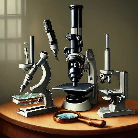
A Journey into the World of Microscopy: From Humble Beginnings to High-Tech Magnification
The science of looking into the hidden invisible Microscopy has transformed our understanding of the world around us. It can explore the universe beyond the reach of our naked eyes, with complex cellular structures, red blood cells, viruses and other viruses and microorganisms taking on amazing perspectives
The history of the microscope is a fascinating story of human curiosity, scientific genius, and relentless exploration. From the humble beginnings of simple magnifying glasses to the sophistication of modern electronic microscopes, the invention of microscopes has shaped our understanding of the microscopic world
In the 1600s, Dutch opticians such as Hans and Zachary Janssen are credited with inventing the first microscope. Known for this hybrid microscope, many lenses were used to magnify objects up to 30 times.At the end of the 17th century, Antony van Leeuwenhoek, Dutch draper some changed our perception of thumbnails. Armed with a well-made single-lens microscope, and explored the hidden reaches of nature. In 1674, Leeuwenhoek discovered microorganisms in lake water, which he aptly named “animalcules”. His discovery laid the foundations of biology and inspired generations of scientists. This incredible feat allowed him to uncover a hidden universe – the first sightings of bacteria, red blood cells, and other microorganisms.
Formation of the scientific environment (17th-19th centuries): Leeuwenhoek’s discoveries boosted scientific research. Robert Hooke, an English scientist, established these developments. In 1665, his book "Micrographia" recorded his observations with a compound microscope. Notably, the term "cell" was coined by Hooke when he examined cork tissue, laying the foundation for cell biology.Microscope systems flourished throughout the 18th and 19th centuries Joseph Lister and other scientists addressed the limitations of the early lenses, introducing improvements that reduced image distortion.
Beyond the Limits of Light: The Beginning of the New Age (19th-20th century): As the 19th century progressed, the limitations of optical microscopy became apparent and scientists yearned for a tool which can go deeper into cells. This research culminated in the development of the electron microscope in the 1930s. The 20th century was revolutionary with the invention of the electron microscope. Unlike light microscopes, which use visible light, electron microscopes use electron beams to achieve much higher magnification.Formation of the scientific environment (17th-19th centuries): Leeuwenhoek’s discoveries boosted scientific research. Robert Hooke, an English scientist, established these developments. In 1665, his book "Micrographia" recorded his observations with a compound microscope. Notably, the term "cell" was coined by Hooke when he examined cork tissue, laying the foundation for cell biology.Microscope systems flourished throughout the 18th and 19th centuries Joseph Lister and other scientists addressed the limitations of the early lenses, introducing improvements that reduced image distortion.
Beyond the Limits of Light: The Beginning of the New Age (19th-20th century): As the 19th century progressed, the limitations of optical microscopy became apparent and scientists yearned for a tool which can go deeper into cells. This research culminated in the development of the electron microscope in the 1930s. The 20th century was revolutionary with the invention of the electron microscope. Unlike light microscopes, which use visible light, electron microscopes use electron beams to achieve much higher magnification.
In the 1930s, German experts Max Knoll and Ernst Ruska made the first electron microscope. This tool let us see tiny things like cells and even atoms by using electron beams, not light, getting images many times bigger. This cool invention showed us the tiny parts inside cells, viruses, and stuff too small to see before. The 1900s brought even more cool microscopes. New kinds like phase-contrast and confocal microscopy let scientists look at live cells without using stuff that could hurt them. Now, the world of looking at tiny things is getting even better. Today, we have high-tech microscopes that use computers and lasers. These let us see and even change tiny things in ways we never could before.
Modern Microscopy's Diverse Arsenal - Today, the field of microscopy boasts a diverse range of specialized instruments, each tailored to address specific scientific needs. Here's a glimpse into some remarkable examples:
Scanning Electron Microscope (SEM): Imagine a high-tech camera that captures images using a beam of electrons instead of light. That's the essence of a SEM. By scanning the surface of a sample with a focused electron beam, SEMs generate detailed information about its topography and composition. This makes them ideal for studying the intricate structures of materials like insect wings, microchips, and even pollen grains.
Transmission Electron Microscope (TEM): While SEMs provide exceptional surface detail, TEMs take us a step further. They function by transmitting a beam of electrons through a very thin sample, allowing us to observe its internal structure. TEMs are the go-to instruments for visualizing the intricate world of viruses, organelles within cells, and macromolecules like proteins.
Confocal Microscopy: Ever wished to focus on a specific layer within a thick biological sample and blur out the rest? Confocal microscopy makes this possible. It utilizes a laser beam to precisely illuminate a chosen plane within the sample, effectively eliminating information from out-of-focus regions. This allows researchers to create sharp, three-dimensional images of cells, tissues, and even small organisms.
Atomic Force Microscopy (AFM): This technique takes a completely different approach, venturing into the realm of physical interaction. AFM employs a tiny cantilever, akin to a microscopic feeler, to physically scan the surface of a sample. By measuring the minute forces between the cantilever and the sample's surface, AFM can map its topography at an atomic level. This provides invaluable insights into the properties of materials at an unimaginable scale, making it crucial for research in fields like nanotechnology and surface science.
Fluorescence Microscopy: Imagine illuminating a sample with specific wavelengths of light and observing it glowing in response. That's the essence of fluorescence microscopy. This technique utilizes fluorescent molecules or tags that bind to specific structures within a cell or tissue. When excited by light, these tags emit their own light, highlighting the target structures with remarkable clarity. This allows researchers to visualize specific proteins, DNA, or even pathogens within biological samples.
Super-resolution Microscopy (SRM): Overcoming the limitations imposed by the wavelength of light, SRM techniques like STED (Stimulated Emission Depletion) and PALM (Photoactivated Localization Microscopy) achieve resolutions surpassing the diffraction limit. This allows researchers to visualize structures as small as 20 nanometers, enabling the observation of intricate cellular machinery and the dynamics of individual molecules within living cells.
Cryo-Electron Microscopy (Cryo-EM): This powerful technique takes a snapshot of biological samples in their near-life state. Samples are rapidly frozen at ultra-low temperatures, preserving their native structure and minimizing damage caused by traditional fixation methods. Cryo-EM has been instrumental in determining the three-dimensional structures of complex molecules like proteins and viruses, providing crucial insights into their function and potential drug targets.
Correlative Microscopy: Combining the strengths of multiple microscopy techniques, correlative microscopy offers a comprehensive view of biological samples. For instance, researchers can utilize fluorescence microscopy to identify specific structures within a cell and then switch to electron microscopy to examine those structures in high detail. This integrated approach provides a deeper understanding of cellular processes and their underlying mechanisms.
Light Sheet Microscopy (LSM): Imagine illuminating a thin slice of a sample within a living organism. LSM achieves this feat by focusing a laser beam into a thin sheet of light, minimizing photobleaching and phototoxicity – damaging effects caused by prolonged exposure to light. This allows researchers to observe dynamic processes within living organisms over extended periods, providing valuable insights into cellular behavior and development.
Expansion Microscopy (ExM): This innovative technique physically expands biological samples by several folds while preserving their structural integrity. This expansion allows for better resolution and visualization of intricate cellular structures that would otherwise be difficult to distinguish using traditional microscopy methods. ExM holds immense potential for studying the organization and function of organelles within cells.
Scanning Near-Field Optical Microscopy (SNOM): This innovative technique pushes the boundaries of resolution by utilizing a tiny probe that interacts with the sample at an extremely close range. SNOM can not only image the surface features of a sample with exceptional detail but also probe its optical properties at the nanoscale. This opens doors for research in areas like material science and photonics, allowing scientists to study the behavior of light at the interface between materials.
X-ray Microscopy: Stepping outside the realm of light and electrons, X-ray microscopy offers unique capabilities. By utilizing high-energy X-rays, this technique can penetrate deep into samples, making it ideal for studying the internal structure of dense materials like bones and minerals. Additionally, it allows for the visualization of elements within a sample, providing valuable information about their distribution and composition.
From revealing the building blocks of life to aiding in the development of new medicines, the microscope has played an undeniable role in shaping our scientific understanding. As technology continues to evolve, one can only imagine the future breakthroughs this remarkable invention holds in unveiling the secrets of our universe, both seen and unseen. These advancements hold the potential to revolutionize our understanding of biological processes, develop new materials with extraordinary properties, and ultimately pave the way for breakthroughs in medicine, nanotechnology, and countless other fields. As we continue to refine and develop novel microscopy techniques and the future holds immense promise for further groundbreaking discoveries that will undoubtedly revolutionize our perception of the world around us.
#science sculpt#life science#science#molecular biology#biology#biotechnology#artists on tumblr#microscopy#microscope#Scanning Electron Microscope#Transmission Electron Microscope#Confocal Microscopy#Atomic Force Microscopy#Fluorescence Microscopy#Expansion Microscopy#X-ray Microscopy#Super-resolution Microscopy#Light Sheet Microscopy#illustration#illustrator#illustrative art#education#educate yourself#techniques in biotechnology#scientific research#the glass scientists#scientific illustration#scientific advancements
6 notes
·
View notes
Text
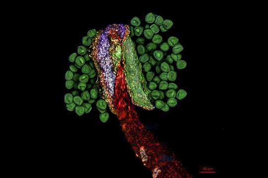
Edelweiss stamens, which were scanned and reconstructed in three dimensions using laser scanning confocal microscopy.
By Jiao Li (China)
Light Microscopy Awards
#jiao li#photographer#china#edelweiss stamens#laser scanning confocal microscopy#micro photography#light microscopy awards#nature
2 notes
·
View notes
Text

i cannot stop thinking about this tweet
#he’d fr stop mid battle and ask foo if he could have a few of their planktons on a microscope slide or smthn#he would’ve been 2 steps early just so he could go home n write a grant proposal to the speedwagon foundation asking 4 a confocal microscope#jjba#jotaro kujo
18 notes
·
View notes
Text

Победа Горячего Обеда
2 notes
·
View notes
Text
Gorgeous mature lesbian playgirl gets her shaved pussy toyed
Pleazure Belly Dancing in Skimpy Outfit
Japanese teen demolished in tight swimsuit
Royal Two Stunning Big Tit MILFS Share Two Huge Cock
Pecker riding makes captivating honey Rilynn Rae cum
Riley Reid Cum Tribute
Kendra Lust Let Me Purge Your Urge
Lesbian Cougar Mindi Mink Sensually Touched On Tight Asshole
Boy teen gay emos xxx Luke Shaw is back for his very first couple
My Stepsister Loves Sexy Games
#fingerroot#confocally#Ekwok#nonvisceral#coho#exegetically#unequitable#birrotch#Housum#waferwork#nonerrantly#raciest#Ampelosicyos#Swarthmore#lemon-faced#noodling#hardsalt#dea-nettle#tetrobolon#spaders
2 notes
·
View notes
Text
microscope time again !!
2 notes
·
View notes
Text
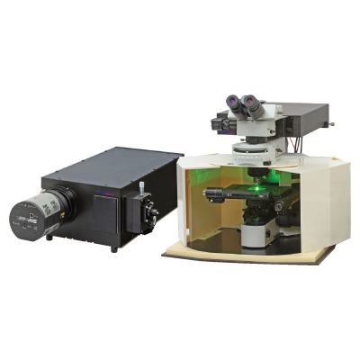
Confocal Laser Raman Spectrometer
Discover the wonders of science with the Confocal Laser Raman Spectrometer from Technos Photonics! This advanced tool uses laser light to analyze tiny samples, revealing their unique chemical fingerprints. Explore the world of molecules and compounds with precision and clarity, unlocking insights into materials and substances like never before. Dive into the world of scientific discovery with Technos Photonics. To find more information, you can contact us or visit our website.
0 notes
Text
youtube
The confocal microscope at Imperial College's Sir Alexander Fleming Building lab is used for imaging the interior of living plant and animal cells.
During my PhD project, I used the confocal microscope to view the interior of Nicotiana benthamiana plant cells which were expressing Green Fluorescent Protein (GFP) tagged genes of interest. I aimed to find out where the proteins encoded by the genes of interest were localised in the plant cell, which turned out to be in the cytoplasm.
From Wikipedia's entry on Confocal Microscopy: "Confocal microscopy, most frequently confocal laser scanning microscopy (CLSM) or laser scanning confocal microscopy (LSCM), is an optical imaging technique for increasing optical resolution and contrast of a micrograph by means of using a spatial pinhole to block out-of-focus light in image formation. Capturing multiple two-dimensional images at different depths in a sample enables the reconstruction of three-dimensional structures (a process known as optical sectioning) within an object. This technique is used extensively in the scientific and industrial communities and typical applications are in life sciences, semiconductor inspection and materials science. Light travels through the sample under a conventional microscope as far into the specimen as it can penetrate, while a confocal microscope only focuses a smaller beam of light at one narrow depth level at a time. The CLSM achieves a controlled and highly limited depth of field."
Music by the Fiechter Brothers
Images by Katia Hougaard & the Facility for Imaging by Light Microscopy at Imperial College London
#katia plant scientist#botany#plant biology#plants#plant science#biology#science#science and technology#confocal microscopy#microscopy#microscope#scientific instruments#laboratory#research#phdblr#phd#phd life#molecular biology#cell biology#green fluorescent protein#plant scientist#Youtube
1 note
·
View note
Text
Distance Measurement Sensors Market - Forecast(2025 - 2032)
Distance Measurement Sensors can actively check distances, position system parts and monitor other parameters in order to intelligently and independently initiate action and these sensors supply an analog signal representing the distance proportional to the selected sensor’s range. The global Distance Measurement Sensors market in 2017 is valued at $2.5 billion and is anticipated to $2.81 billion by 2023 growing at an estimated value of CAGR of more than 2% during the forecast period 2018 to 2023. North America will dominate the Distance Measurement Sensors Market in the upcoming years with a significant share due to several factors. The penetration of the market is quite high in this category, the presence of several automotive and manufacturing giants is a vital growth causing factors.
𝐑𝐞𝐩𝐨𝐫𝐭 𝐒𝐚𝐦𝐩𝐥𝐞 @ https://bitly.ws/3aerY
Distance Measurement Sensors Market Outlook:
Distance Measurement Sensors Market is segmented by sensing technology, measurement type, application and end user. On the basis of Sensing Technology, the Distance Measurement Sensors Market is segmented in to Laser Diodes, IR LED, Ultrasonic, Capacitive, Inductive, Photo electric, Draw wire, Image sensor and others. Ultrasonic sensors are very widely used in this industry along with laser diodes giving them the edge and making them the largest product segment in this market. On the basis of measurement type, the Distance Measurement Sensors market is categorized in to Laser Triangulation, TOF, Confocal chromatic imaging, Photoelectric Sensor, Others. On the basis of application, the Distance Measurement Sensors market is segmented in to Automation, Safety Systems, Robotics, Defense aerospace & intelligent, Packaging, Automatic identification, tracking position, automobiles Others. On the basis of end user, the Distance Measurement Sensors market is segmented in to Oil and Gas Industry, Automotive, Military & Defense, Packaging, Consumer Electronics. The global Distance Measurement Sensors market is set to experience a substantial growth owing to the strong demand from Oil & Gas segment across the worldwide.
The current price quoted for Infrared sensors of Distance Measurement Sensors is around $45 to $80 per piece by most manufacturers supplying these sensors.
0 notes
Text

Innervating Kidneys
Using a combination of light-sheet and confocal microscopy to generate anatomical maps of the nerve networks establishing throughout development of the mouse kidney from embryo to adult
Read the published research paper here
Image from work by Pierre-Emmanuel Y. N’Guetta and colleagues
Department of Cell Biology and Physiology, The University of North Carolina at Chapel Hill, Chapel Hill, NC, USA
Image originally published with a Creative Commons Attribution 4.0 International (CC BY NC 4.0)
Published in bioRxiv, November 2023 (not peer reviewed)
You can also follow BPoD on Instagram, Twitter and Facebook
#science#biomedicine#immunofluorescence#biology#renal#kidney#neuroscience#nerves#confocal microscopy#light-sheet microscopy#embryo development#developmental biology
7 notes
·
View notes
Text
Transforming Biopsy Procedures with Cutting-Edge Devices
A biopsy is a medical operation in which a small sample of tissue or cells from the body is removed for examination under a microscope. Biopsies can be performed for a variety of reasons, including cancer or other diseases diagnosis, determining the cause of abnormal growths or lumps, checking for infections or inflammation, and monitoring disease progression.
Biopsies come in several forms, and…

View On WordPress
#biopsy#biopsy devices#biopsy guns#biopsy systems#confocal microscopy#excisional biopsy#inspect disorders
0 notes
Text
The use of confocal laser scanning microscopy, which allows for the direct observation of translocation through living sieve elements, addresses the additional question of whether the sieve plate pores and sieve element lumen are open in intact, translocating tissues.
"Plant Physiology and Development" int'l 6e - Taiz, L., Zeiger, E., Møller, I.M., Murphy, A.
#book quote#plant physiology and development#nonfiction#textbook#confocal laser scanning microscopy#microscopy#observation#plant cells#sieve elements#sieve plates#pores#lumen#translocation
0 notes
Text
Sexy slut hooks up with guy she met online for rough anal
milf babe fucked with young lover vintage
Madura Hermosa Culona y lindos pies
Steamy sexy blowjob session with cute gals
Jaye Summers facialized in gangbang
White girl sucks swallows bbc cum
Teen Fucked From Pussy To Mouth
3D HENTAI neko girl masturbates and cums
Bachelorette squirts with strippers help
Little Piece of Shit
#cerebrations#apostatise#Matagalpan#saturatedness#gyros#boccias#merenchyma#interknotting#fingerroot#confocally#Ekwok#nonvisceral#coho#exegetically#unequitable#birrotch#Housum#waferwork#nonerrantly#raciest
0 notes
Text
I have experienced contact with modern western hardware engineering send help.
See, this is Nice Device.
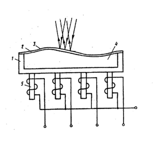
It hails from soviet patent for ferrofluid adaptive optics device with 10×10 actuators КВФ-74 of Leningrad Institute of Refrigerators.
This is done in 2006 in Toronto.
This is an affront to everything human. This design is not human at all. You are losing more magnetic field on resistance than you gain from WINDING MORE TURNS. This could have been a PCB with a soft magnetic screen. Fuck you.
1 note
·
View note
Last Seen Blogs

djdanialiasdanielhass
Dj Dani ( Daniel Haß )

jennyiz
Untitled

cassiopea-ornata
cassie
salvateure
𝒔𝒖𝒑𝒆𝒓𝒔𝒕𝒂𝒓 𝒔𝒉*𝒕 .

sarausaidesign
Creative Design