ioag
we all love red things
records of my disease
385 posts
Don't wanna be here? Send us removal request.
Last Seen Blogs

romancistass
L-ie biert

jaydoolez
Untitled

kanamacina-blog
Sans titre
manureboy1997
worker.97
cosywinged
you are safe here
Photo
Sometimes knowledge of microbiological media and microbes can have an unusual effect. Especially when it gets too boring or hectic in the lab. Every person who has ever been in contact with microbiology has a picture of their "masterpiece" created thanks to microbes and the reactions of these microbes with the substrate. Of course, some people do it better
(https://lnkd.in/d9tgYvgc
https://lnkd.in/d9vCBdZu (as in my case) worse. Such a springboard often saves the situation and (what's funny) can well systematize knowledge. After all, it's always better to remember with pleasant associations.
I know that the post fits the group on average, but I hope that the members will not be ashamed and will share their works as part of a rest over a quick coffee.
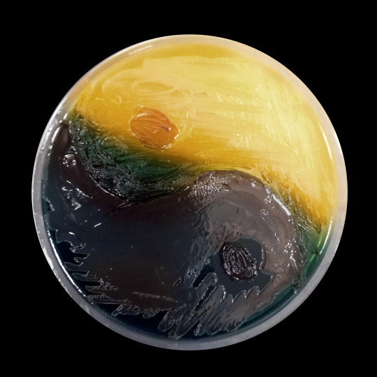
Today you will be able to see some of this microbiological - mushroom art FUNGAL UPDATE: MYCOLOGY 2023 at the QEII Conference Centre, London
In the photo: BiGGY agar + mix of yeast-like fungi; title:between, the (or i'll burn this place down with those @#$%! fungi)
#veterinary #veterinarymedicine #veterinaryjobs #laboratory #microbiology #microbiologist #jobapplication #mycology #examination #numbers #lookingforjob #microscope #phdjourney #phd #cryptococcus #animals #fungi #yeast #candida
#examination #teaching #chromogens

bittersweet
between, the
(BiGGY agar, Candida spp.)
#veterinary#veterinarymedicine#veterinaryjobs#laboratory#microbiology#microbiologist#jobapplication#mycology#examination#numbers#lookingforjob#microscope#phdjourney#phd#cryptococcus#animals#fungi#yeast#candida#teaching#chromogens#I.O.A.G.#veterinary medicine
5 notes
·
View notes
Text
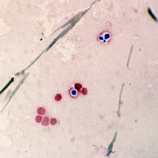
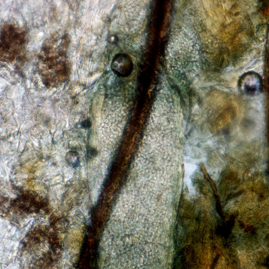
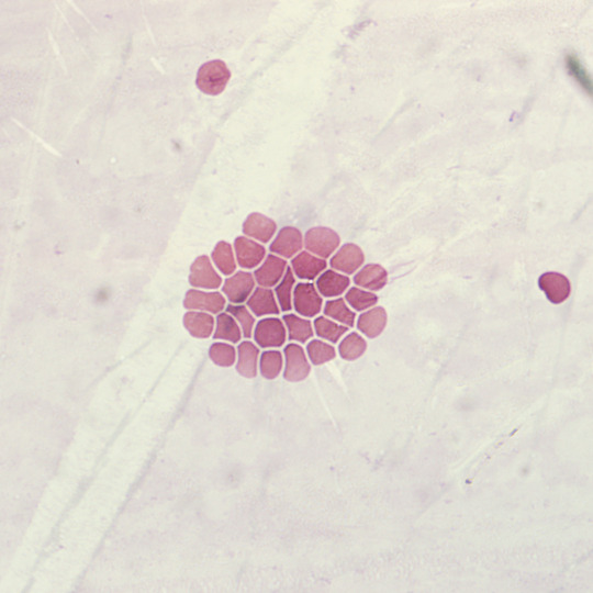
n veterinary medicine, in the case of many pathogens, staining of clinical material can narrow down the range of possible elements. Sometimes even pinpoint a specific pathogen (e.g. P. multocida and Loeffler's blue staining). Each type of staining has diagnostic significance depending on what we really need to assess and count or just see. Unfortunately, in the case of microbiology courses, you work with pure cultures, usually one pathogen per class. Apart from reference points in the form of clinical material. Another thing is that during the classes the preparations are made so that they look as similar as possible to those from atlases and books. Let's be honest - it's almost never possible to get such an effect when working with clinical material and then with a pathogen. This is a key element in quick diagnostics to remember that what we see under the microscope only resembles what is in the book and that it is a spectrum of forms, extreme cases and very often poor quality diagnostic material.
As in this case: blood smear stained with MGG. Visible erythrocytes and large dark blue cells with a pink halo (envelope) the preparation is nasty but that's how the blood was taken but it is still diagnostic. Cryptococcosis in a cat (which quite often occur pretending to be malignant squamous cell tumors around the mouth). In this case, additionally confirmed by histopathological examination. The size of an erythrocyte and a yeast cell are actually the same (reference scale), and in the case of a Titan cell (plus envelope) they may resemble lymphocytes.
return to
the last tears after what will never be again
(Blood sample, MGG staining; Cryptococcus spp.)
#veterinary#veterinarymedicine#veterinaryjobs#laboratory#microbiology#microbiologist#jobapplication#mycology#examination#numbers#lookingforjob#microscope#phdjourney#phd#cryptococcus#feline#fungi#MGG#teaching#veterinary medicine#i.o.a.g.#blood
4 notes
·
View notes
Text
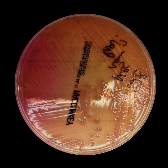
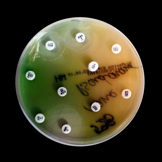
Quick and accurate diagnosis of bacteria is often a key element not only in identifying the etiological factor, but also in its fight against it. On a daily basis, whether in "human" or "veterinary" microbiology, we use a set of main microbiological media, which are a quick way to identify these main pathogens. Can we trust them implicitly? NO. Most pathogens will show a characteristic pattern on a given medium, but a few percent will look quite different. And so it was this time. What in macromorphology, gram staining resembled a little larger coccobacilli and there were forms of quite long threads, but the most important on MacConkey's medium were colorless colonies with a slightly pink center. At a glance: Providencia spp.
However, no. After careful identification panel it was lactose negative E. coli. Very rare in veterinary medicine.
Let's remember that all quick tests, identification media or even MALDI TOF are only tools that are supposed to help put a solution, not be the final result. As it is in nature, there are always extremes that we like to forget.
#veterinary#veterinarymedicine#veterinaryjobs#laboratory#microbiology#microbiologist#jobapplication#bacteriology#examination#numbers#lookingforjob#microscope#infection#antimicrobials#kirbybauer#ecoli#i.o.a.g.
4 notes
·
View notes
Text
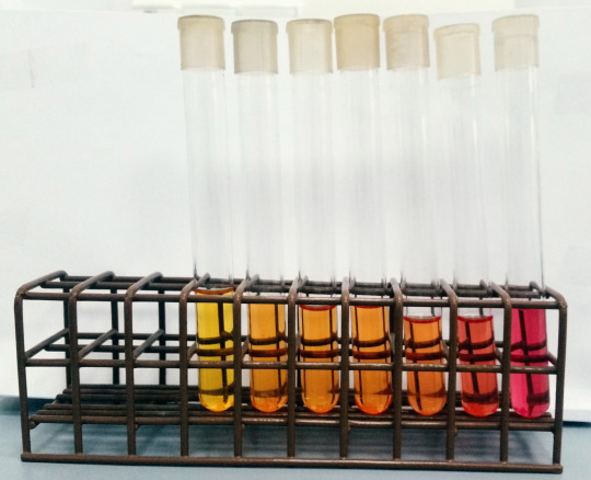
Very long time ago…
This is a photo from the beginning of my work as a future/would-be mycologist. By the color of the test tubes, you can easily see that we are dealing with a medium with urea. In bacteriology, the urea hydrolysis test is used routinely for almost all bacteria. In turn, in the case of dermatophytes, although it is a differentiating feature, it usually gives unexpected results. By accident I managed (them) to get quite a nice color gradient from yellow (which doesn't happen often) to very pink (fuchsia/bishop color). And the culprit was Trichophyton verrucosum, which according to the literature should be urea positive. This yellow color (i.e. the opposite of the intended effect) is due to the fact that this strain of T. verrucosum very quickly used up a small amount of dextrose in the medium, which acidified the medium so much that the phenol red from a slightly orange color turned yellow. In general, we all use the term Christensen's medium, but every lab's composition is a little different. Note that there is such a thing as Stuart's medium for detecting urease, and it is not much different from Christiansen's medium. Virtually every laboratory uses a combination of these media so as not to add dextrose but at the same time the medium is "rich" enough for other microbes to grow on it. Hence, there are often considerable discrepancies - especially in mycology. Believe it or not, the same strain that gives a typical positive result in lab A will give +/- or the opposite in lab B.
#veterinary#veterinarymedicine#veterinaryjobs#laboratory#microbiology#microbiologist#jobapplication#mycology#examination#numbers#lookingforjob#microscope#infection#biochemistry#urea#ureabroth#trichophyton
8 notes
·
View notes
Text
I am pleased to inform, that an article of which I am a co-author has just been accepted.
Thank you Dr. Marta Dec for the opportunity to cooperate. At the same time, this is my last article with the affiliation of the University of Life Sciences in Lublin. Fortunately, there will never be such cooperation again :)
This is a bit of a sentimental article for me - the strains used in it, I collected as a veterinary student, later as a doctor and employee of the University of Life Sciences in Lublin. It is an interesting end to a career.
It's also funny that I'm ending my career with the most veterinary bacteria.
#veterinary#veterinarymedicine#veterinaryjobs#laboratory#microbiology#microbiologist#jobapplication#bacteriology#examination#numbers#lookingforjob#microscope#infection#antimicrobials#kirbybauer#swine#mdpi#pathogens#i.o.a.g.#end
38 notes
·
View notes
Text

Bolesław Leśmian - The Girl
Twelve brothers who believed in dreams, scouted a wall among phantasms;
Beyond the wall there cried a voice - voice of a Girl long gone through chasms.
They fell in love with voice’s sound and with their own wishful believing,
And tried to guess shape of her lips from how her song died out in grieving.
They said “she cries therefore she is” - and nothing else they said but wondered,
They blessed the world with sign of cross - and then the world grew still and pondered.
The hammers held in hardened hands, they launched against the walls in clamor!
And night was blind, and couldn’t tell: which part was man and which - the hammer?
“Let’s hurry and undo cold stone, before in death the Girl’s enrusted!”
The youngest brother thus cried out - and in their hammers’ strength they trusted.
But all their efforts were in vain, their arms exertions and pain - futile!
They sacrificed their bodies to the dream enticing, yet so brutal!
Their chests caved in, their bones crushed down, decayed their hands and faded faces…
They died together in one day and shared one night’s eternal spaces.
But dead men’s shadows - my good Lord! - instead of stopping they persisted!
And they went on, in eerie time - the hammers’ sounds continued, twisted.
They clanged ahead! And back they clashed! And upwards in resounding clamor!
And night was blind, and couldn’t tell: which part was shade and which - the hammer?
“Let’s hurry and undo cold stone, before in death the Girl’s enrusted!”
The youngest shadow thus cried out - and in their hammers’ strength they trusted.
But suddenly their strength had waned, night came and they were overpowered!
And - since you never die enough - they died again, by dark devoured.
Never enough, never the way the moribund would want, departing!…
Their substance - lost without a trace, their story closed instead of starting!
But stalwart hammers - my good Lord! - didn’t surrender to bereavement!
And on their own they fought the wall, rumbling for naught but the achievement!
They rumbled forth through days and nights, sweating like humans do, through clamor!
And night was blind, and couldn’t tell: what’s hammer if not just a hammer?
“Let’s hurry and undo cold stone, before in death the Girl’s enrusted!”
The youngest hammer thus cried out - and in their own pure strength they trusted.
And the wall fell with booming crash, sounding through every nook and cranny!
Alas! Beyond they found no Girl, only the waiting void; uncanny.
There was no eyes! There was no lips! Nobody’s fate needed securing!
There was but voice - and only voice, nothing but voice tempting and luring!
Nothing but night, and cries, and grief, and loss in every uttered letter!
This is the world! Such awful world! Couldn’t it have been different, better?
Against the dreams that lied out loud, against the wish obliterated,
The hammers finally went to rest, relief deserved and so belated.
And there was silence all around! The emptiness reigning forever!
Why do you mock that emptiness although it doesn’t mock you ever?
Translation: Maria Gral
#Poland#Polska#linguistics#poetry#poems#wiersze#Leśmian#Lesman#translation#writing#Irian writes stuff#signal boosts are deeply appreciated btw#Leśmian deserves to be read by the whole wide world#i.o.a.g.#veterinary#art#uvlight#veterinaryjobs#sunset
86 notes
·
View notes
Text
Sometimes, even with pure bacterial cultures, unexpected "guests" can appear - especially when you need to give the result, stat.
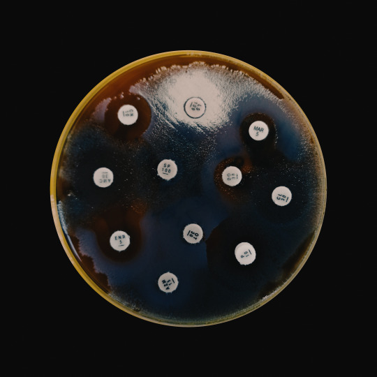
Disk diffusion test for strains of alpha hemolyzing Streptococcus spp. from the dog's vagina.
Streptococci are very often isolated from the genital tracts of companion animals. Very often, Proteus spp is also isolated, which in the first passage (after previous antibiotic therapy) can mask itself well in the culture of other bacteria and show all its features in the next passage. Unfortunately, that was the case here. In the case of Streptococci, at least medium supplemented with blood is needed, while Proteus plain MH is sufficient for the disc diffusion test.
Of course, the antibiogram had to be repeated...
#veterinary#veterinarymedicine#veterinaryjobs#laboratory#microbiology#microbiologist#jobapplication#bacteriology#examination#numbers#lookingforjob#microscope#infection#antimicrobials#kirbybauer#i.o.a.g.
2 notes
·
View notes
Text
Pseudomonas aeruginosa is one of the most pleasant bacteria to identify from a microbiologist's perspective. In turn, from the veterinarian's point of view - one of the worst to treat.
#microbiology#veterinarymedicine#veterinarian#veterinaryjobs#laboratory#diagnostics#bacteriology#jobhunt#pseudomonas#uvlight#antimicrobialresistance#antimicrobial#AST#i.o.a.g.
73 notes
·
View notes
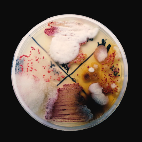
Text
It's not the best "microbiology" art, but it has a very interesting background. Two bacteria from two different clinical cases were inoculated on the TSCB medium. This metallic blue spilling bacterium is of course Pseudomonas aeruginosa. The yellow one (positive reaction on TSCB medium) is Vibrio metschnikovii isolated from chronic UTI in a dog. It was an unusual microbiological diagnosis. But what can you do when even your dog has a better holiday than you?
Problems with urination (in this dog) began just after returning from the Mediterranean, the owners and the dog intensively used the charms of warm and salty water.
#veterinary#veterinarymedicine#veterinaryjobs#laboratory#microbiology#microbiologist#jobapplication#bacteriology#examination#numbers#lookingforjob#microscope#urinarytractinfection#art#rarediseases#i.o.a.g.
172 notes
·
View notes
Text
return to
above the steel sky
(Arthrospores; CBE stained clinical material)
#microbiology#microscope#veterinary#laboratory#i.o.a.g.#veterinary medicine#fungi#spring#june 2022#alone#chlorazol black
2 notes
·
View notes
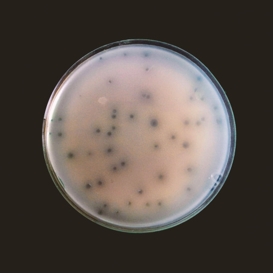
Text
return to
the wheel of unpredictability
((Pagano medium; clinical samples))
#microbiology#veterinary#veterinary medicine#fungi#laboratory#microscope#i.o.a.g.#red#pink#mold#yeast#alone
37 notes
·
View notes
Note
hey :)! i was wondering about your method of photographing agar plates. whats your trick to minimize reflections and keep a good contrast? theyre very neat your pics!
Hello:)
I use a matte black pad for the photos. It largely depends on the thickness of the agar. Usually I pour out thicker agar plates and then there is less shine. And I take the photos themselves with my Huawei P20 smartphone and I can recommend it for such things
0 notes
Text
return to
Bloob
6 notes
·
View notes
Text
return to
And they will dance drunk in front of us
(Negative nigrosin staining of a blood sample)
#microbiology#veterinary#laboratory#microscope#veterinary medicine#fungi#i.o.a.g.#alone#may 2022#negative
1 note
·
View note
Text
return to
the last tears after what will never be again
(Blood sample, MGG staining; Cryptococcus spp.)
#microbiology#veterinary#laboratory#veterinary medicine#microscope#fungi#i.o.a.g.#cryptococcus#blood#alone#may 2022
4 notes
·
View notes
Text
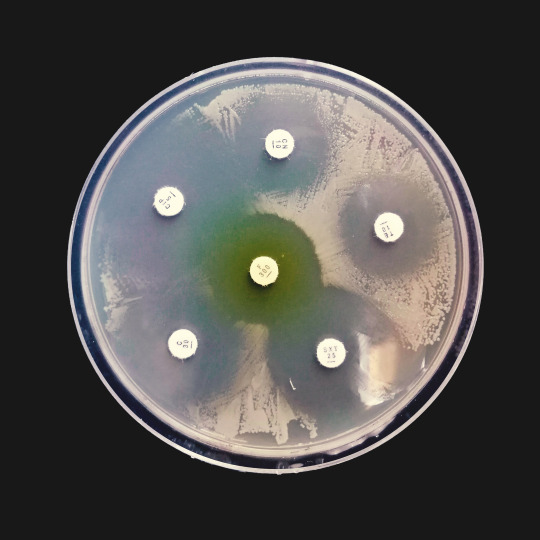
return to
inability to preserve the zone
(Kirby-Bauer Disk Diffusion test; E. coli from pigeon)
#microbiology#veterinary medicine#veterinary#i.o.a.g.#laboratory#bacteriology#antibiotic resistance#pigeon#escherichia coli#may 2022
0 notes
Text
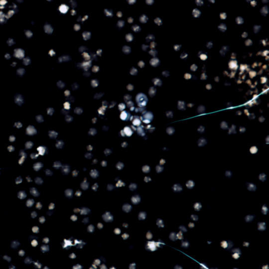
return to
we will never reach that limit
(Plaques and halos produced by phage T4)
3 notes
·
View notes